

 PDF
PDF ePub
ePub Citation
 Print
Print


Desmoid-type fibromatosis (DF) is a rare mesenchymal tumor, which was first described by MacFarlane in 18321) and termed desmoid tumor by Mueller in 1838. This neoplasm is considered benign but locally infiltrative and can occur in nearly any part of the body.2) It occurs sporadically but its association with familial adenomatous polyposis is well known.3) Despite being non-metastasizing, this neoplasm is reported to have a local recurrence rate of 20%–64% after surgical resection, and repeated surgery often results in substantial morbidity.45) Nonsurgical modalities such as chemotherapy, radiotherapy, and hormonal therapy have been considered an alternative treatment option.67891011) Some authors, on the other hand, advocate a more conservative approach, which is known as “wait-and-see” strategy, as an initial approach.4510111213141516)
The biological behavior of DF is unpredictable and varies widely from indolent to rapidly progressive.17) Meanwhile, spontaneous stabilization of this neoplasm has been reported in many studies.45181920) To the best of our knowledge, only one report19) has investigated possible predictors of spontaneous stabilization of sporadic extra-abdominal DF and the size of the cohort was small. In this study, we sought to assess the rate of spontaneous stabilization of DF treated with the frontline conservative approach in a single institute and to analyze the factors associated with spontaneous stabilization.
METHODS
Patient Cohort
We retrospectively reviewed 197 patients who were diagnosed as having sporadic extra-abdominal DF in our institute from 1995 to 2016. We conducted this study in compliance with the principles of the Declaration of Helsinki. The protocol of this study was reviewed and approved by the Institutional Review Board of Seoul National University Hospital (IRB No. 1610-008-795). The informed consent was waived. We included patients in whom (1) observation with a watchful waiting strategy was used as primary treatment, (2) regular follow-up with imaging modality was performed, and (3) follow-up period was more than 12 months. We excluded patients in whom (1) observation was less than 1 year, (2) medical or radiological information was insufficient to assess the extent of tumor, and (3) additional treatment such as chemotherapy and radiotherapy was required during conservative treatment. In total, 76 patients satisfied our inclusion criteria and were eligible for analysis.
Tumor Surveillance
Regular surveillance was performed with clinical examination and imaging modalities such as magnetic resonance imaging (MRI) and ultrasound. The extent of tumor was evaluated at the time of diagnosis and followed up with the same imaging modality every 3 to 6 months from the start of observation treatment except for four patients who were followed up with ultrasound. After the spontaneous stabilization of the disease, imaging assessment was continued with the same interval or annually. We defined stabilization of DF as radiological evidence of the size of the tumor unchanged or decreasing continuously for 6 months or more. The size of tumor was measured as the longest diameter of a measurable mass in any plane. Measurement was done by a radiologist (JYC) specializing in the musculoskeletal section and confirmed by three orthopedic oncologists (MSR, YK, and IH); in case of discrepancy, the three authors had a discussion and made a decision. During follow-up, date, symptoms including pain and neurological deficits, and tumor status including size and surveillance frequency were documented. The duration of our watchful waiting treatment was defined as the period from the date of diagnosis to the last follow-up unless the strategy was changed.
Data Collection
The following clinicopathological variables were collected from the institutional database: sex, age at first diagnosis, disease presentation, size and location of tumor, origin of tumor compartment, resection margin status, adjuvant chemotherapy, and adjuvant radiotherapy. The information is summarized in Table 1. Tumor location was dichotomized as axial or appendicular. The size of tumor was measured as the longest diameter of a measurable mass in any plane on MRI. Data on tumor size, compartment of origin, and tumor status at final follow-up were obtained from the MRI report written by the radiologist (JC) specializing in musculoskeletal oncology and were further confirmed in consensus by the three authors (MSR, YK, and IH). Histology was confirmed by an experienced pathologist (CL). In patients with recurrence after previous surgical treatment outside of our institute, we reviewed the original slides from the previous hospital.
Statistical Analysis
The primary endpoint was spontaneous stabilization of DF. Time to stabilization was calculated as the interval between the date of diagnosis of DF and the date when stabilization was initially noted by imaging modality. For recurrent DF, the time to stabilization was defined as the interval from the date of recurrence to the date when stabilization was confirmed. Evolving tumors were classified as censored at the last follow-up. Possible factors associated with disease stabilization of DF were analyzed with the Kaplan-Meier survival method and Cox proportional hazard method. Time to stabilization was estimated using the Kaplan-Meier survival method, and the log-rank test was used for univariate analysis. Multivariate analysis was performed by using the Cox proportional hazards model on variables in which p-values of < 0.05 were obtained after univariate analysis. All statistical analyses were performed by using IBM SPSS ver. 21.0 (IBM Corp., Armonk, NY, USA). A two-tailed p-value of less than 0.05 was considered statistically significant.
RESULTS
Stabilization Rate of DF
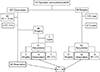
Of the total 197 patients with DF, the initial treatment strategy was frontline observation in 107 patients and upfront surgery in 90 patients (Fig. 1). Of the 107 patients with observation, 47 patients were excluded from the analysis because of the follow-up of less than 1 year (n = 45) and changes in treatment strategy to chemotherapy or radiotherapy during observation (n = 2). The observational therapy was continued to the final follow-up in 32 patients, whereas 28 patients underwent palliative surgery during the observational period (mostly because of neurologic symptoms and severe pain caused by mass effect). Among the 28 patients, seven were cured after complete excision without recurrence, whereas 21 patients had recurrence. Of the 21 patients, seven had additional chemotherapy or radiotherapy, and two continued to have surgical treatment; the remaining 12 had observational treatment after surgery and thus were included in the analysis. Of the 90 patients who underwent upfront surgery, 28 patients were excluded from the analysis: 27 with continuous disease-free status after surgery until the last follow-up (n = 27) and 1 with incomplete medical record (n = 1). The remaining 62 patients had recurrence after surgical intervention. Of those, 21 had repetitive surgery and 9 had additional chemotherapy or radiotherapy; the remaining 32 underwent observational treatment until the last follow-up. In total, 76 patients satisfied our inclusion criteria and were included in the analysis.
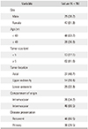
On patient demographic characteristics (Table 1) of the 76 included patients, 29 (38.2%) were men and 47 (61.8%) were women. The mean age of the patients was 30.2 years (range, 1 to 77 years), and 48 patients (63.2%) were younger than 40 years. Sixty-two patients (81.6%) had tumors larger than 5 cm. The location of the tumors was axial in 37 patients (48.7%) and appendicular in 39 (51.3%). Forty-six patients (60.5%) had intermuscular tumors regardless of the depth, and 26 (34.2%) had intramuscular tumors. The tumor was recurrent in 46 patients (60.5%) at the time of presentation, whereas it was primary in 30 patients (39.5%). The mean follow-up from the start of observation was 54.8 months (range, 12 to 226 months). Fifty-four (71.0 %) of 76 tumors showed spontaneous stabilization during observation treatment and were stable at the final follow-up. The mean time to stabilization was 30.4 months (range, 7 to 112 months). On Kaplan-Meier survival analysis, the spontaneous stabilization rate was 25.4% at 1 year, 52.7% at 2 years, and 70.9% at 3 years.
Predictors of Disease Stabilization
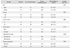
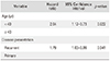
On univariate analysis, age and disease presentation were associated with stabilization of DF (Table 2). Patients who were younger than 40 years (p = 0.014) or had recurrence (p = 0.036) needed longer time for spontaneous stabilization. Tumor size had no correlation with spontaneous stabilization (p = 0.887). On multivariate analysis with the Cox proportional hazard method, age younger than 40 years (hazard ratio [HR], 2.04; p = 0.022) and recurrence (HR, 1.79; p = 0.041) were significant predictive factors of longer time to disease stabilization (Table 3).
DISCUSSION
DF is a benign, nonmetastasizing neoplasm marked by monoclonal fibroblastic proliferation of spindle cells with a locally infiltrative growth pattern.2) Although this neoplasm is considered intermediately malignant because of its inability to metastasize, its aggressive nature is mainly reflected in the high rate of local recurrence,567891011212223) which often leads to repetitive operations and entails substantial morbidity.24252627) After publication of many reports on spontaneous disease stabilization or even regression without treatment, the shift from early proactive treatment approach to conservative “wait-and-see” approach has emerged.451819) In this study, we confirmed the validity of this recent approach by assessing the spontaneous stabilization rate of DF and evaluating predictive factors.
The spontaneous stabilization rate of DF after non-surgical treatment varies from 50% to 97% in previous reports with a mean follow-up of 3 to 6 years.512181924) The moderate stabilization rate (71.0%) found in the present study falls within the described range. The potential of progression of DF seems to be limited after 3 years.512181924) On the other hand, the mean time to tumor stabilization in the present study (30.4 months) is longer than that in a report by Barbier et al. (13.2 months).19)
In the present study, recurrence was found to be an independent predictor of spontaneous stabilization of DF as reported by Barbier et al.19) Shin et al.28) have also reported that recurrence is significantly associated with poor recurrence-free survival in their 119 patients treated with surgical resection. Huang et al.15) also reported that patients with previous history of recurrence have shorter recurrence-free survival on the basis of the analysis of 214 patients who underwent surgical resection.
We found age younger than 40 years was associated with longer time to spontaneous stabilization of DF. Many studies71123) have also suggested that younger age be considered a predictive factor of shorter recurrence-free survival after surgical treatment. Therefore, age should be an important factor when a physician considers watchful waiting strategy in clinic.
Tumor size was variable and did not show significant correlation with stabilization in this study. Barbier et al.19) retrospectively reviewed 26 cases of extra-abdominal DF and reported that the tumor size was very variable and independent of location and evolutivity. Even though tumor size is a well-known independent prognostic factor,9152329) it did not appear to be predictive of the disease stabilization process. Fiore et al.12) reported that patients with tumors located in trunk or thoracic wall had worse progression-free survival. However, we found no significant correlation of the location of tumor with time to stabilization.
Among 54 patients whose tumor had stabilized, 13 patients showed transient spontaneous stabilization (the tumor size increased later). The increase in size was not significant in most patients. None of them required additional treatment and stayed with observation. The mean time to rebound was 31.3 months (range, 5 to 93 months). On the other hand, nine patients showed continuous shrinkage or decrease in tumor size. In particular, one patient with aggressive fibromatosis in the psoas muscle showed total regression during 15 months of observation. Further research on this issue is necessary.
The study is not without limitations. The shift towards a frontline conservative treatment approach is a relatively recent trend and surgical resection has been the gold standard for a long time. In our cohort, 90 patients had upfront surgery and 44 (48%) of them had surgical treatment before 2010. Also, given the retrospective nature of our study, there were certain cases in which we could not identify the clinical reference to determine either surgical resection or a frontline conservative strategy. Measuring the proper size or volume of the tumors and evaluating the effect of treatment are another issue that needs to be addressed in further studies. In the current study, we used only the longest diameter of the measurable mass in any plane. In a recent study by Villalobos et al.30) on the long-term follow-up of the effect of PF03084014 of DF, they used both Response evaluation criteria in solid tumors criteria and World Health Organization criteria for assessment of the response.
Kasper et al.31) suggested a consensus algorithm for the initial management of DF. According to the algorithm, there is a consensus that surgical treatment is no longer a gold standard for the initial treatment and the conservative “wait-and-see” approach should be the frontline treatment approach for patients with newly diagnosed DF, irrespective of symptoms. Our study supports this approach as an effective and reasonable initial option for the treatment of DF, and considering the identified predictive factors in treating DF can play a role in promoting disease stabilization. Frontline conservative treatment seems to be the optimal treatment for most patients who present with DF. Younger patients or those with recurrence may require longer time to spontaneous stabilization of the neoplasm.
 XML Download