

 PDF
PDF ePub
ePub Citation
 Print
Print


Abstract
Purpose
To report a case of non-glaucomatous retinal nerve fiber layer (RNFL) defect associated with paravascular inner retinal defect (PIRD) in a patient with idiopathic epiretinal membrane (ERM).
Case summary
A 70-year-old male who was diagnosed with ERM in his right eye and pseudoexfoliative glaucoma in his left eye visited our clinic. His intraocular pressure was 14 mmHg in both eyes while using topical hypotensive medications in both eyes. His right eye showed no glaucomatous change of the optic disc head, and also no glaucomatous visual field defect on standard automated perimetry. Red-free fundus photography and swept-source optical coherence tomography showed an ERM and wedge-shaped RNFL defect starting from the PIRD, not the optic disc head. He was diagnosed with non-glaucomatous RNFL defect in the right eye and was told to stop using topical hypotensive medication for the right eye. After 2 years of discontinuing the medication, the IOP was within the normal range, the RNFL defect showed no progression, and the visual field remained stationary.
Figures and Tables
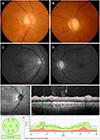 | Figure 1Ophthalmic examinations performed at the first visit. (A, B) Disc photography of the patient shows intact neuroretinal rim (NRR) in the right eye and narrowing of superotemporal and inferotemporal NRR in the left eye. (C, D) Red-free fundus photography shows nasal extrafoveal epiretinal membrane with multiple retinal nerve fiber layer (RNFL) defect in the right eye that are not connected to the optic disc head border, and diffuse RNFL thinning in the left eye. (E) Circumpapillary scan image of spectral-domain optical coherence tomography of the right eye shows focal narrow RNFL thinning (marked as yellow line) just nasal to the superotemporal retinal venous arcade, which corresponds to the most nasally located slit-like RNFL defect seen in the red-free fundus photography. Other RNFL defects seen in the red-free fundus photography are not detected on the scan circle of 3.5 mm diameter. TS = superotemporal; NS = superonasal; T = temporal; N = nasal; G = general; TI = temporal inner; NI = nasal inner; TMP = temporal; SUP = superior; NAS = nasal; INF = inferior.
|
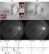 | Figure 2Comparisons between 2017 and 2019.(A) The en-face 52 µm slab image of swept-source optical coherence tomography (OCT) shows epiretinal membrane and paravascular inner retinal defect (PIRD) along the retinal vascular arcades. It also shows retinal nerve fiber layer (RNFL) defect connected to the PIRD, but not the optic disc head. There was no definite change in PIRD and RNFL defect between results of 2017 and 2019. (B) Red-free fundus photography also showed no change between 2017 and 2019. Note that locations of OCT B-scans in the en-face 52 µm slab image are also indicated as red horizontal lines in the red-free fundus photography. (C) Standard automated perimetry results did not show development of glaucomatous visual field defect between 2017 and 2019.
|
References
1. Oray M, Onal S, Bayraktar S, et al. Nonglaucomatous localized retinal nerve fiber layer defects in Behcet uveitis. Am J Ophthalmol. 2015; 159:475–481.

2. Jonas JB, Schiro D. Localized retinal nerve fiber layer defects in nonglaucomatous optic nerve atrophy. Graefes Arch Clin Exp Ophthalmol. 1994; 232:759–760.
3. Choi JA, Ko SH, Park YR, et al. Retinal nerve fiber layer loss is associated with urinary albumin excretion in patients with type 2 diabetes. Ophthalmology. 2015; 122:976–981.

4. Jeon SJ, Kwon JW, La TY, et al. Characteristics of retinal nerve fiber layer defect in nonglaucomatous eyes with type II diabetes. Invest Ophthalmol Vis Sci. 2016; 57:4008–4015.
5. Muraoka Y, Tsujikawa A, Hata M, et al. Paravascular inner retinal defect associated with high myopia or epiretinal membrane. JAMA Ophthalmol. 2015; 133:413–420.
6. Chihara E, Chihara K. Apparent cleavage of the retinal nerve fiber layer in asymptomatic eyes with high myopia. Graefes Arch Clin Exp Ophthalmol. 1992; 230:416–420.
7. Komeima K, Kikuchi M, Ito Y, et al. Paravascular inner retinal cleavage in a highly myopic eye. Arch Ophthalmol. 2005; 123:1449–1450.
8. Hood DC, Fortune B, Mavrommatis MA, et al. Details of glaucomatous damage are better seen on OCT en face images than on OCT retinal nerve fiber layer thickness maps. Invest Ophthalmol Vis Sci. 2015; 56:6208–6216.

9. Hood DC, De Cuir N, Mavrommatis MA, et al. Defects along blood vessels in glaucoma suspects and patients. Invest Ophthalmol Vis Sci. 2016; 57:1680–1686.
10. Kim JH, Kang SY, Kim NR, et al. Prevalence and characteristics of glaucoma among Korean adults. Korean J Ophthalmol. 2011; 25:110–115.
11. Lee SH, Kim GA, Lee W, et al. Vascular and metabolic comorbidities in open-angle glaucoma with low- and high-teen intraocular pressure: a cross-sectional study from South Korea. Acta Ophthalmol. 2017; 95:e564–e574.
12. Demer JL, Clark RA, Suh SY, et al. Magnetic resonance imaging of optic nerve traction during adduction in primary open-angle glaucoma with normal intraocular pressure. Invest Ophthalmol Vis Sci. 2017; 58:4114–4125.
13. Wang X, Teoh CKG, Chan ASY, et al. Biomechanical properties of Bruch's membrane-choroid complex and their influence on optic nerve head biomechanics. Invest Ophthalmol Vis Sci. 2018; 59:2808–2817.
14. Hwang YH, Kim YY, Kim HK, Sohn YH. Characteristics of eyes with inner retinal cleavage. Graefes Arch Clin Exp Ophthalmol. 2015; 253:215–220.
15. Jung KI, Kim SJ, Park CK. Systemic vascular risk factors for multiple retinal nerve fiber layer defects. Sci Rep. 2018; 8:7797.
16. Burgoyne CF, Downs JC, Bellezza AJ, et al. The optic nerve head as a biomechanical structure: a new paradigm for understanding the role of IOP-related stress and strain in the pathophysiology of glaucomatous optic nerve head damage. Prog Retin Eye Res. 2005; 24:39–73.
 XML Download