

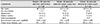
 PDF
PDF ePub
ePub Citation
 Print
Print


Abstract
Purpose
To evaluate clinical outcomes of idiopathic epiretinal membrane removal in patients ≥ 80 years of age.
Methods
A retrospective review of medical records was performed with 56 patients who underwent vitrectomy and removal of idiopathic epiretinal membrane. In the ≥ 80 years of age group (n = 28), the best-corrected visual acuity (BCVA) and central macular thickness (CMT) before surgery were compared with those at the final follow-up. The amount of change in the BCVA after surgery was also compared between the ≥ 80 years of age group and the < 80 years of age group (n = 28).
Results
In the ≥ 80 years of age group, the mean follow-up period was 19.1 ± 17.0 months. Before surgery, 11 eyes were pseudophakic and 17 eyes were phakic. Combined cataract surgery was performed with epiretinal membrane removal in all 17 phakic eyes. The mean logarithm of the minimal angle of resolution BCVA was 0.75 ± 0.30 before surgery, which improved to 0.50 ± 0.30 at the final follow-up (p < 0.001). The CMT was 458.0 ± 79.7 µm before surgery, which decreased to 367.2 ± 83.4 µm at the final follow-up (p < 0.001). There was no significant difference in the amount of change in the BCVA after the surgery between the ≥ 80 years of age group and the < 80 years of age group (p = 0.547).
Conclusions
In patients with idiopathic epiretinal membrane who were ≥ 80 years of age, the visual acuity was improved or maintained, and was accompanied with anatomical improvement after epiretinal membrane removal with or without cataract surgery. These results suggest the usefulness of epiretinal membrane removal in older patients.
Figures and Tables
 | Figure 1Horizontal (A) and vertical (B) optical coherence tomography scan images showing method to measure inner-retinal irregularity index. The yellow line indicates inferior border of inner plexiform layer (IPL) and the blue line indicates retinal pigment epithelium (RPE). The inner-retinal irregularity index was calculated as length of inferior border of IPL/length of RPE.
|
 | Figure 2Outcomes of 84-year-old patient who was diagnosed with epiretinal membrane and cataract. The best-corrected visual acuity (BCVA) was 0.2 (decimal) before surgery (A, B). The patients underwent combined epiretinal membrane removal and cataract surgery. Five months after the surgery (C, D), the BCVA improved to 0.4 (decimal).
|
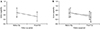 | Figure 3Changes in logarithm of minimal angle of resolution (logMAR) best-corrected visual acuity (BCVA) of patients who underwent epiretinal membrane removal with or without cataract surgery. There was a significant improvement in BCVA after surgery (p < 0.001). (A) Outcomes in all 28 patients. (B) Comparison of outcome between patients who underwent epiretinal membrane removal only (solid line, n = 11) and patients who underwent combined epiretinal membrane removal and cataract surgery (dotted line, n = 17). Op = operation; F/U = follow-up.
|
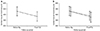 | Figure 4Changes in central macular thickness of patients who underwent epiretinal membrane removal with or without cataract surgery. There was a significant decrease in central macular thickness after surgery (p < 0.001). (A) Outcomes in all 28 patients. (B) Comparison of outcome between patients who underwent epiretinal membrane removal only (solid line, n = 11) and patients who underwent combined epiretinal membrane removal and cataract surgery (dotted line, n = 17).
|
References
1. Kim JM, Lee H, Shin JP, et al. Epiretinal membrane: prevalence and risk factors from the Korea National Health and Nutrition Examination Survey, 2008 through 2012. Korean J Ophthalmol. 2017; 31:514–523.


2. Fang IM, Hsu CC, Chen LL. Correlation between visual acuity changes and optical coherence tomography morphological findings in idiopathic epiretinal membranes. Graefes Arch Clin Exp Ophthalmol. 2016; 254:437–444.
3. Suh MH, Seo JM, Park KH, Yu HG. Associations between macular findings by optical coherence tomography and visual outcomes after epiretinal membrane removal. Am J Ophthalmol. 2009; 147:473–480.e3.
4. Lee JW, Kim IT. Outcomes of idiopathic macular epiretinal membrane removal with and without internal limiting membrane peeling: a comparative study. Jpn J Ophthalmol. 2010; 54:129–134.
5. Lee P, Lee TG, Kim MS, et al. Prognostic factors in vitrectomy for macular epiretinal membrane. J Korean Ophthalmol Soc. 2011; 52:1302–1307.
6. Kauffmann Y, Ramel JC, Lefebvre A, et al. Preoperative prognostic factors and predictive score in patients operated on for combined cataract and idiopathic epiretinal membrane. Am J Ophthalmol. 2015; 160:185–192.e5.
7. Spiteri Cornish K, Lois N, Scott NW, et al. Vitrectomy with internal limiting membrane peeling versus no peeling for idiopathic full-thickness macular hole. Ophthalmology. 2014; 121:649–655.
8. Cho KH, Park SJ, Cho JH, et al. Inner-retinal irregularity index predicts postoperative visual prognosis in idiopathic epiretinal membrane. Am J Ophthalmol. 2016; 168:139–149.
9. Bu SC, Kuijer R, Li XR, et al. Idiopathic epiretinal membrane. Retina. 2014; 34:2317–2335.
10. Yang HS, Hong JW, Kim YJ, et al. Characteristics of spontaneous idiopathic epiretinal membrane separation in spectral domain optical coherence tomography. Retina. 2014; 34:2079–2087.
11. Muto T, Ide T, Chikuda M, Machida S. Vitrectomy in patients over 90 years of age. Clin Ophthalmol. 2016; 10:239–242.
12. Park JC, Ramasamy B, Shaw S, et al. A prospective and nationwide study investigating endophthalmitis following pars plana vitrectomy: clinical presentation, microbiology, management and outcome. Br J Ophthalmol. 2014; 98:1080–1086.
13. Rizzo S, Belting C, Genovesi-Ebert F, di Bartolo E. Incidence of retinal detachment after small-incision, sutureless pars plana vitrectomy compared with conventional 20-gauge vitrectomy in macular hole and epiretinal membrane surgery. Retina. 2010; 30:1065–1071.
14. Chang S. LXII Edward Jackson lecture: open angle glaucoma after vitrectomy. Am J Ophthalmol. 2006; 141:1033–1043.
15. Christensen UC, Krøyer K, Sander B, et al. Value of internal limiting membrane peeling in surgery for idiopathic macular hole stage 2 and 3: a randomised clinical trial. Br J Ophthalmol. 2009; 93:1005–1015.
16. Miguel AI, Legris A. Prognostic factors of epiretinal membranes: a systematic review. J Fr Ophtalmol. 2017; 40:61–79.
17. Kim TN, Lee JE, Lee EJ, et al. Prevalence of and factors associated with lens opacities in a Korean adult population with and without diabetes: the 2008–2009 Korea National Health and Nutrition Examination Survey. PLoS One. 2014; 9:e94189.
18. Lee JJ, Kim KH, Shin MK, et al. Myopic shift and cataract change after lens sparing vitrectomy in patients with idiopathic epiretinal membrane in their 5th and 6th decade. J Korean Ophthalmol Soc. 2015; 56:1038–1043.
19. Jung KI, Song MH, Roh YJ. Combined clear corneal phacoemulsification and vitrectomy versus two-step surgery in Korean patients with idiopathic epiretinal membrane. J Korean Ophthalmol Soc. 2011; 52:203–209.
20. Shimozono M, Oishi A, Hata M, et al. The significance of cone outer segment tips as a prognostic factor in epiretinal membrane surgery. Am J Ophthalmol. 2012; 153:698–704.e1.
21. Kim J, Rhee KM, Woo SJ, et al. Long-term temporal changes of macular thickness and visual outcome after vitrectomy for idiopathic epiretinal membrane. Am J Ophthalmol. 2010; 150:701–709.
22. Okamoto F, Okamoto Y, Hiraoka T, Oshika T. Effect of vitrectomy for epiretinal membrane on visual function and vision-related quality of life. Am J Ophthalmol. 2009; 147:869–874.e1.
 XML Download