

 PDF
PDF ePub
ePub Citation
 Print
Print


Abstract
Methods
Results
Figures and Tables
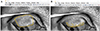 | Figure 1The area of the central two-thirds on upper tarsal conjunctiva, in which meibomian glands were best visible, was selected through drag and drop with mouse (A). Then, the area of meibomian gland dropout area within the aforementioned area was selected in the same manner (B). The ratio of the two area was calculated.
|
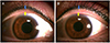 | Figure 2Distance between the tattoo pigment and the meibomian gland orifice was scored and was recorded as the tattoo score (mm). In each right (A) and left eye (B), the blue arrows on the top indicates tattoo pigment, and the yellow arrows at the bottom indicates meibomian gland orifice. In this case, the distance or tattoo score was measured as 1.0 mm in each eye.
|
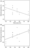 | Figure 3Meibomian gland (MG) loss of eyeliner tattoo group and tear film lipid layer thickness (LLT) of eyeliner tattoo group. (A) MG loss was significantly correlated with the tattoo score (rs = −0.563, p = 0.023; Spearman correlation test). (B) LLT was significantly correlated with the tattoo score (rs = 0.567, p = 0.022; Spearman correlation test).
|
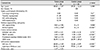
Table 1
Demographics and clinical data of the study subjects
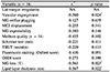
Values are presented as mean ± standard deviation unless otherwise indicated.
NA = not available; MG = meibomian gland; MCJ = mucocutaneous junction; TBUT = tear film break-up time; OSDI = ocular surface disease index.
*Fisher's exact test is not able since the lid margin of all subjects is normal in both groups; †Fisher's exact test was used. p < 0.05 were considered statistically significant; ‡Mann-Whitney U test was used. p < 0.05 were considered statistically significant; §for meibum quality, the number of the subjects of tattoo group was 14, not 16, because of omitted record.
![]()
Table 2
Correlation between upper eyeliner tattoo score and other indices
NA = not able; MG = meibomian gland; MCJ = mucocutaneous junction; TBUT = tear film break-up time; OSDI = ocular surface disease index.
*rs = Spearman correlation coefficient; †NA = Spearman correlation test is not able since the lid margin of all subjects is normal in tattoo group; ‡p < 0.05, which were considered statistically significant.
![]()
 XML Download