

 PDF
PDF ePub
ePub Citation
 Print
Print


Abstract
Purpose
The Ocular Surface Disease Index (OSDI) and the Standardized Patient Evaluation of Eye Dryness (SPEED) which are standard questionnaires of dry eye syndrome were used to determine the associations between clinical dry eye tests and meibomian gland dysfunctions (MGD).
Methods
Forty-one patients with MGD were enrolled in this study. The score of the dry eye syndrome questionnaire and the degree of blepharitis (score: 0–4), Schirmer test results, degree of fluorescence staining of cornea (Oxford Grading System), tear break-up time (TBUT), Pentacam imaging, and anterior segment optical coherence tomography results were used to compare and analyze the results of each test for possible correlations with the dry eye questionnaire answers.
Results
There was a significant correlation between OSDI and SPEED (R = 0.278, p = 0.011). SPEED was correlated with the Oxford grade (R = 0.478, p < 0.001) and MGD grade (R = 0.280, p = 0.011) while there was no significant correlation with corneal aberrations, tear meniscus height, tear meniscus area, Schirmer test results, or TBUT. The OSDI correlated with the MGD grade (R = 0.651, p < 0.001), TBUT (R = −0.360, p = 0.001), and age (R = −0.230, p = 0.037). Using multiple regression analyses, the MGD grade affected the OSDI (β = 0.580, p < 0.001) and the Oxford grade significantly influenced the SPEED (β = 0.447, p < 0.001).
Figures and Tables
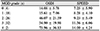
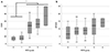 | Figure 1Mean and standard deviation of (A) Ocular Surface Disease Index (OSDI) and (B) Standardized Patient Evaluation of Eye Dryness (SPEED) sorted by meibomian gland dysfunction (MGD) grading. Based on one-way analysis of variance (ANOVA). *Statistically significant (p < 0.05). One way ANOVA with Tukey post-hoc test.
|
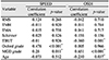
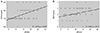 | Figure 2Spearman correlation between Standardized Patient Evaluation of Eye Dryness (SPEED) and dry eye signs. Correlation graphs between SPEED and (A) Oxford grade, (B) meibomian gland dysfunction (MGD) grade. ‘R’ means correlation coefficient. Statistical significance was calculated by Spearman correlation analysis.
|
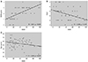 | Figure 3Spearman correlation between Ocular Surface Disease Index (OSDI) and dry eye signs. Correlation graphs between OSDI and (A) meibomian gland dysfunction (MGD) grade, (B) tear break-up time (TBUT), and (C) age. ‘R’ means correlation coefficient. Statistical significance was calculated by Spearman correlation analysis.
|
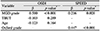
Table 1
Demographic and dry eye sign characteristics by classification according to severity of MGD
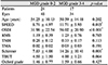
Values are presented as mean ± standard deviation or number. MGD = meibomian gland dysfunction; SPEED = Standardized Patient Evaluation of Eye Dryness; OSDI = Ocular Surface Disease Index; RMS = root mean square; TMH = tear meniscus height; TMA = tear meniscus area; TBUT = tear break up time. *Statistically significant t-test (p < 0.05).
![]()
Notes
References
1. Arita R, Fukuoka S, Morishige N. Meibomian gland dysfunction and contact lens discomfort. Eye Contact Lens. 2017; 43:17–22.


2. Goto E, Monden Y, Takano Y, et al. Treatment of non-inflamed obstructive meibomian gland dysfunction by an infrared warm compression device. Br J Ophthalmol. 2002; 86:1403–1407.

3. Mathers WD. Ocular evaporation in meibomian gland dysfunction and dry eye. Ophthalmology. 1993; 100:347–351.
4. McDonald JE. Surface phenomena of the tear film. Am J Ophthalmol. 1969; 67:56–64.
5. Zeev MS, Miller DD, Latkany R. Diagnosis of dry eye disease and emerging technologies. Clin Ophthalmol. 2014; 8:581–590.
6. McMonnies C, Ho A, Wakefield D. Optimum dry eye classification using questionnaire responses. Adv Exp Med Biol. 1998; 438:835–838.
7. Walt JG, Rowe MM, Stern KL. Evaluating the functional impact of dry eye: the Ocular Surface Disease Index. Drug Inf J. 1997; 31:1436.
8. Narayanan S, Miller WL, Prager TC, et al. The diagnosis and characteristics of moderate dry eye in non-contact lens wearers. Eye Contact Lens. 2005; 31:96–104.
9. Oden NL, Lilienfeld DE, Lemp MA, et al. Sensitivity and Specificity of a Screening Questionnaire for Dry Eye. Adv Exp Med Biol. 1998; 438:807–820.
10. Ngo W, Situ P, Keir N, et al. Psychometric properties and validation of the Standard Patient Evaluation of Eye Dryness questionnaire. Cornea. 2013; 32:1204–1210.
11. Schiffman RM, Christianson MD, Jacobsen G, et al. Reliability and validity of the Ocular Surface Disease Index. Arch Ophthalmol. 2000; 118:615–621.
12. Hosaka E, Kawamorita T, Ogasawara Y, et al. Interferometry in the evaluation of precorneal tear film thickness in dry eye. Am J Ophthalmol. 2011; 151:18–23.
13. Koh S, Tung C, Aquavella J, et al. Simultaneous measurement of tear film dynamics using wavefront sensor and optical coherence tomography. Invest Ophthalmol Vis Sci. 2010; 51:3441–3448.
14. Nguyen P, Huang D, Li Y, et al. Correlation between optical coherence tomography-derived assessments of lower tear meniscus parameters and clinical features of dry eye disease. Cornea. 2012; 31:680–685.
15. Jung NY, Baek JW, Shin SJ, Chung SK. Tear meniscus evaluation using optical coherence tomography in dry eye patients. J Korean Ophthalmol Soc. 2015; 56:323–330.
16. Bron AJ. The doyne lecture. Reflections on the tears. Eye (Lond). 1997; 11 (Pt 5):583–602.
17. Bron AJ, Benjamin L, Snibson GR. Meibomian gland disease. Classification and grading of lid changes. Eye (Lond). 1991; 5 (Pt 4):395–411.
18. Wang L, Shirayama M, Koch DD. Repeatability of corneal power and wavefront aberration measurements with a dual-Scheimpflug Placido corneal topographer. J Cataract Refract Surg. 2010; 36:425–430.
19. Baudouin C, Aragona P, Van Setten G, et al. Diagnosing the severity of dry eye: a clear and practical algorithm. Br J Ophthalmol. 2014; 98:1168–1176.
20. Nichols KK, Nichols JJ, Mitchell GL. The lack of association between signs and symptoms in patients with dry eye disease. Cornea. 2004; 23:762–770.
21. Pult H, Purslow C, Murphy PJ. The relationship between clinical signs and dry eye symptoms. Eye (Lond). 2011; 25:502–510.
22. Unlü C, Güney E, Akçay BI, et al. Comparison of ocular-surface disease index questionnaire, tearfilm break-up time, and Schirmer tests for the evaluation of the tearfilm in computer users with and without dry-eye symptomatology. Clin Ophthalmol. 2012; 6:1303–1306.
23. Finis D, Pischel N, König C, et al. Comparison of the OSDI and SPEED questionnaires for the evaluation of dry eye disease in clinical routine. Ophthalmologe. 2014; 111:1050–1056.
24. Moon IH, Kim TI, Seo KY, et al. The relationship between subjective ocular discomfort and blepharitis severity in dry eye patients. J Korean Ophthalmol Soc. 2016; 57:1507–1513.
25. Seo MH, Shin JY, Lee DH, Kim JH. Objective parameters associated with subjective symptom severity in dry eye syndrome patients. J Korean Ophthalmol Soc. 2017; 58:259–267.
26. Vehof J, Kozareva D, Hysi PG, et al. Relationship between dry eye symptoms and pain sensitivity. JAMA Ophthalmol. 2013; 131:1304–1308.
27. Schein OD, Tielsch JM, Munõz B, et al. Relation between signs and symptoms of dry eye in the elderly. A population-based perspective. Ophthalmology. 1997; 104:1395–1401.
28. Kim JH, Ro JW, Yi K, et al. Changes of the meibomian gland according to age in the normal Korean population. J Korean Ophthalmol Soc. 2015; 56:13–18.
29. Asiedu K, Kyei S, Mensah SN, et al. Ocular Surface Disease Index (OSDI) Versus the Standard Patient Evaluation of Eye Dryness (SPEED): A Study of a Nonclinical Sample. Cornea. 2016; 35:175–180.
30. Finis D, Hayajneh J, König C, et al. Evaluation of an automated thermodynamic treatment (LipiFlow®) system for meibomian gland dysfunction: a prospective, randomized, observer-masked trial. Ocul Surf. 2014; 12:146–154.
31. Pult H, Riede-Pult B. Non-contact meibography: keep it simple but effective. Cont Lens Anterior Eye. 2012; 35:77–80.
32. Osae EA, Ablorddepey RK, Horstmann J, et al. Assessment of meibomian glands using a custom–made meibographer in dry eye patients in Ghana. BMC Ophthalmol. 2018; 18:201.
 XML Download