

 PDF
PDF Citation
 Print
Print


Abstract
The replacement of missing teeth, especially in the anterior region, is an essential part of dental practice. Fiber-reinforced composite resin bridges are a conservative alternative to conventional fixed dental prostheses or implants. It is a minimally invasive, reversible technique that can be completed in a single visit. The two cases presented herein exemplify the treatment of root-fractured anterior teeth with a natural pontic immediately after extraction.
Go to : 
References
1. Kim H, Song MJ, Shin SJ, Lee Y, Park JW. Esthetic rehabilitation of single anterior edentulous space using fiber-reinforced composite. Restor Dent Endod. 2014; 39:220–225.

2. Kumar KP, Nujella SK, Gopal SS, Roy KK. Immediate esthetic rehabilitation of periodontally compromised anterior tooth using natural tooth as pontic. Case Rep Dent. 2016; 2016:8130352.
3. Kermanshah H, Motevasselian F. Immediate tooth replacement using fiber-reinforced composite and natural tooth pontic. Oper Dent. 2010; 35:238–245.
4. Khetarpal A, Talwar S, Verma M. Creating a single-visit, fibre-reinforced, composite resin bridge by using a natural tooth pontic: a viable alternative to a PFM bridge. J Clin Diagn Res. 2013; 7:772–775.
5. Vallittu PK, Shinya A, Baraba A, Kerr I, Keulemans F, Kreulen C, Lassila L, Malmstrom H, Novotny R, Peumans M, Van Rensburg J, Wolff D, Özcan M. Fiber-reinforced composites in fixed prosthodontics-Quo vadis? Dent Mater. 2017; 33:877–879.
6. Frese C, Schiller P, Staehle HJ, Wolff D. Fiber-reinforced composite fixed dental prostheses in the anterior area: a 4.5-year follow-up. J Prosthet Dent. 2014; 112:143–149.
7. Ahmed KE, Li KY, Murray CA. Longevity of fiber-reinforced composite fixed partial dentures (FRC FPD)-systematic review. J Dent. 2017; 61:1–11.
8. Kumbuloglu O, Özcan M. Clinical survival of indirect, anterior 3-unit surface-retained fibre-reinforced composite fixed dental prosthesis: up to 7.5-years follow-up. J Dent. 2015; 43:656–663.
9. Van der Weijden F, Dell'Acqua F, Slot DE. Alveolar bone dimensional changes of post-extraction sockets in humans: a systematic review. J Clin Periodontol. 2009; 36:1048–1058.
10. Spear FM, Cooney JP. Restorative interrelationships. Newman M, Takei H, Klokkevold P, Carranza F, editors. editors.Carranza's clinical periodontology. 12th ed.St. Louis, MO: Saunders Elsevier;2014. Chapter 67.
11. Steigmann M, Cooke J, Wang HL. Use of the natural tooth for soft tissue development: a case series. Int J Periodontics Restorative Dent. 2007; 27:603–608.
12. Perea-Lowery L, Vallittu PK. Framework design and pontics of fiber-reinforced composite fixed dental prostheses – an overview. J Prosthodont Res. 2018; 62:281–286.
13. Xie Q, Lassila LV, Vallittu PK. Comparison of load-bearing capacity of direct resin-bonded fiber-reinforced composite FPDs with four framework designs. J Dent. 2007; 35:578–582.
14. Malmstrom H, Dellanzo-Savu A, Xiao J, Feng C, Jabeen A, Romero M, Huang J, Ren Y, Yunker MA. Success, clinical performance and patient satisfaction of direct fibre-reinforced composite fixed partial dentures – a two-year clinical study. J Oral Rehabil. 2015; 42:906–913.
15. Perea L, Matinlinna JP, Tolvanen M, Lassila LV, Vallittu PK. Fiber-reinforced composite fixed dental prostheses with various pontics. J Adhes Dent. 2014; 16:161–168.
16. Aktas G, Basara EG, Sahin E, Uctasli S, Vallittu PK, Lassila LV. Effects of different cavity designs on fracture load of fiber-reinforced adhesive fixed dental prostheses in the anterior region. J Adhes Dent. 2013; 15:131–135.
Go to :
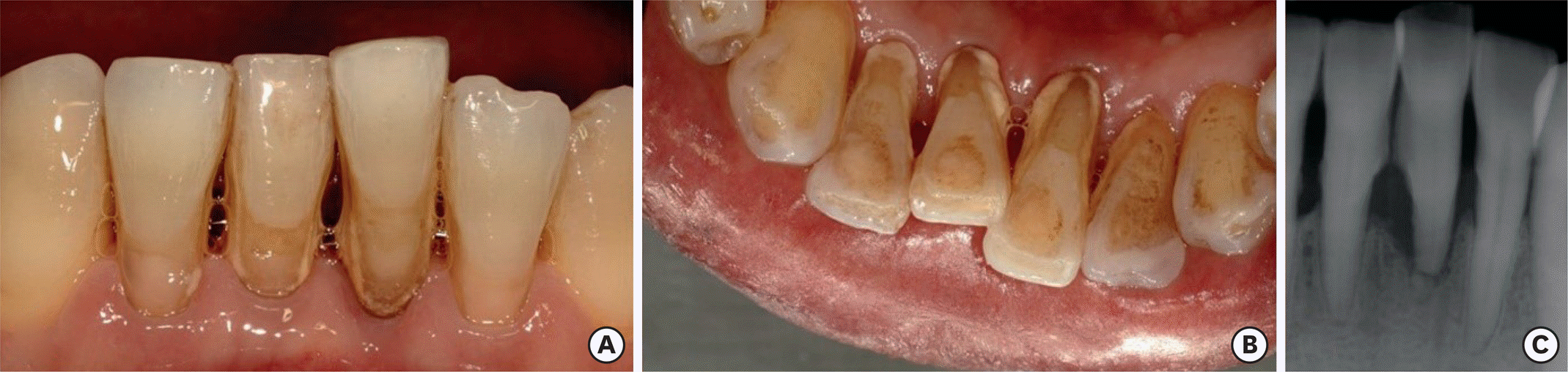
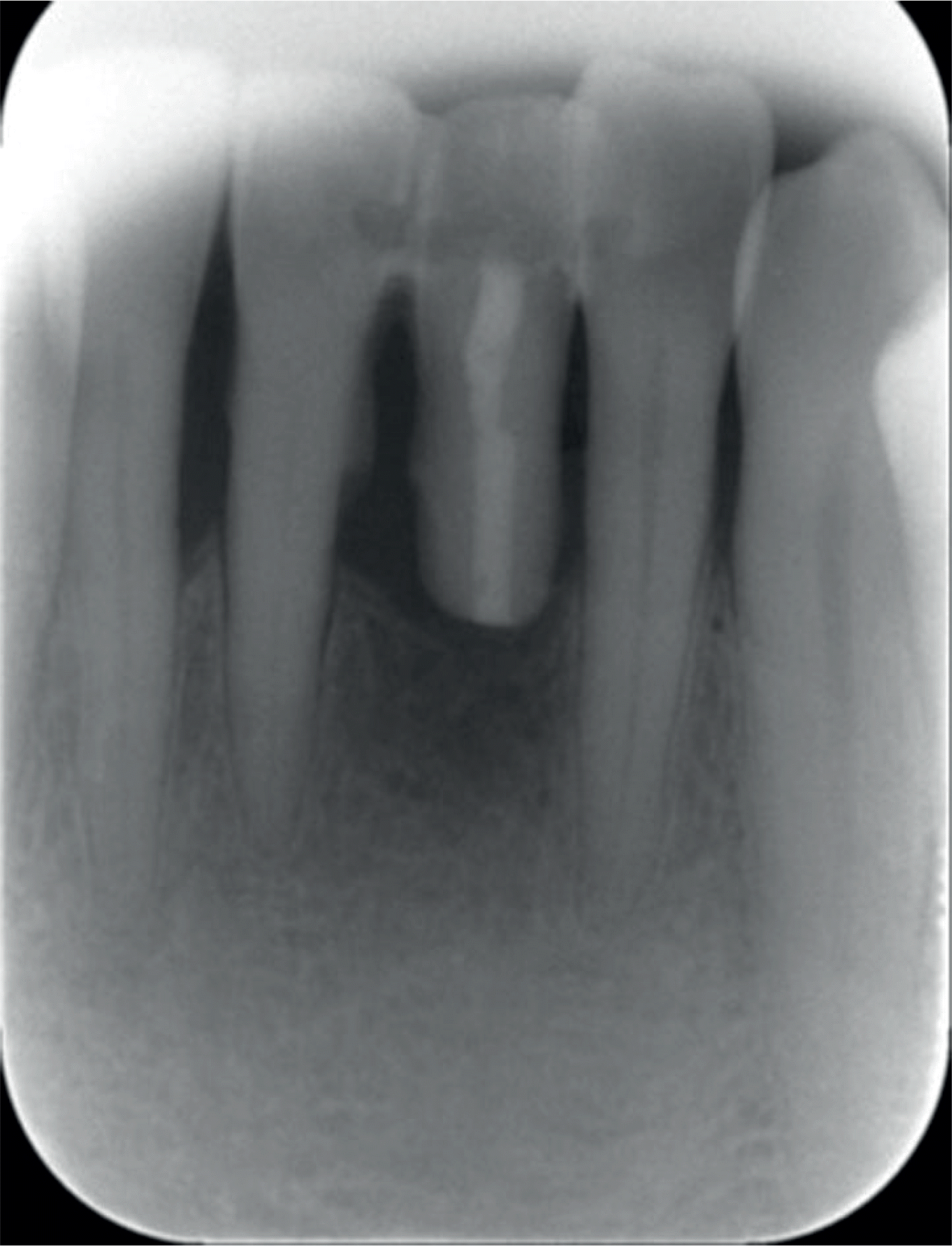
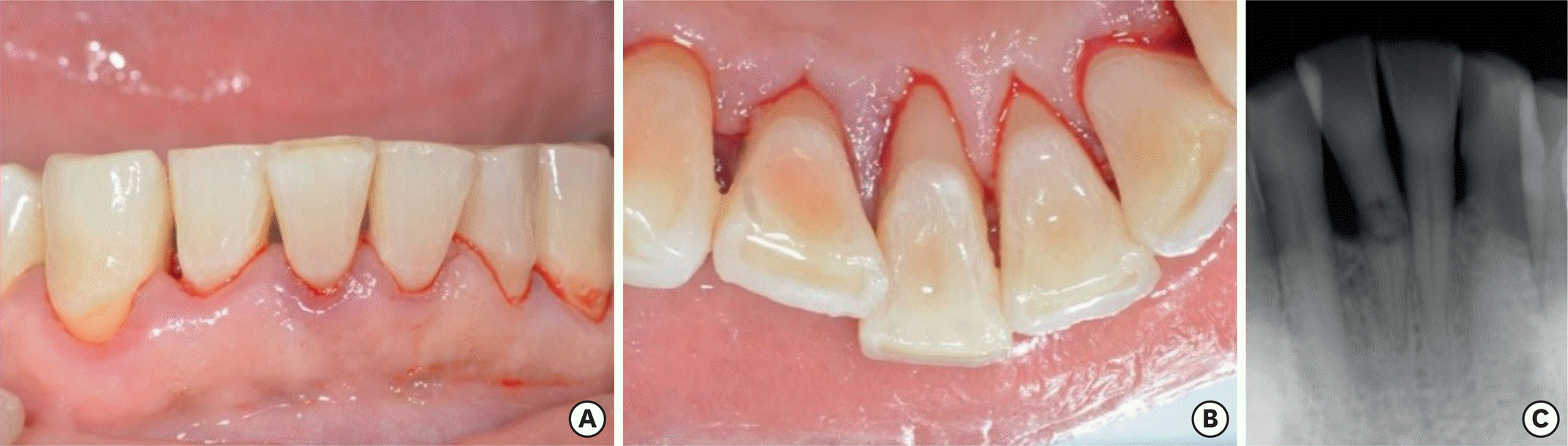 | Figure 1.Preoperative view. (A) Intraoral photograph (labial view); (B) intraoral photograph (lingual view); (C) periapical view. |
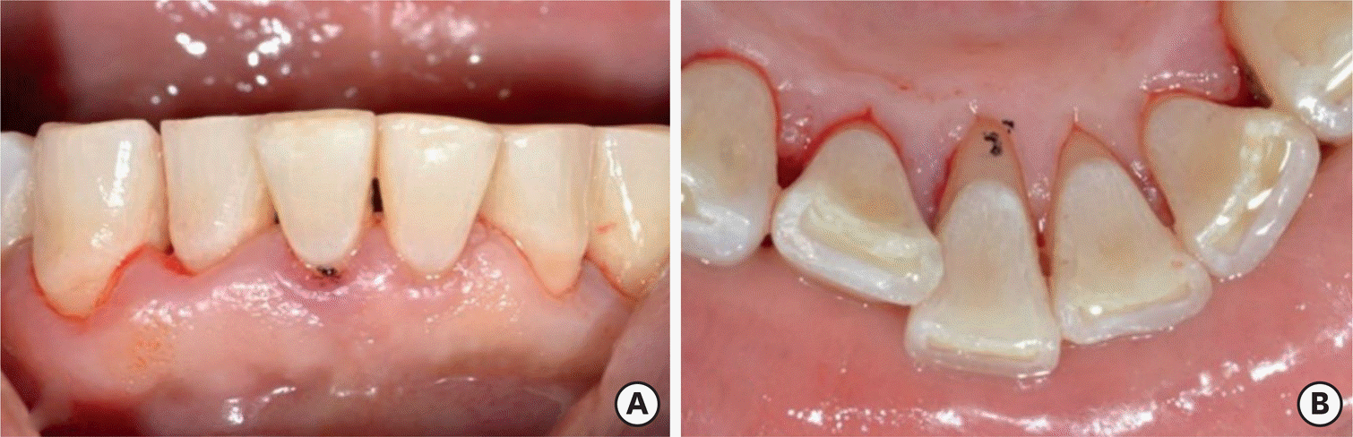 | Figure 2.A black dot was made to mark the gingiva level. (A) Intraoral photograph (labial view); (B) intraoral photograph (lingual view). |
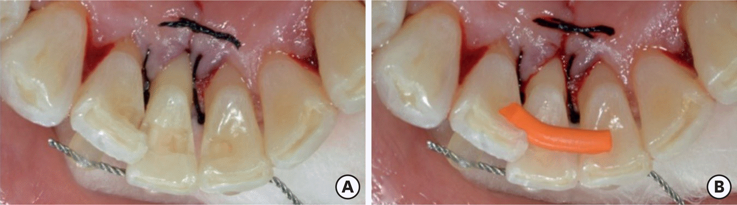
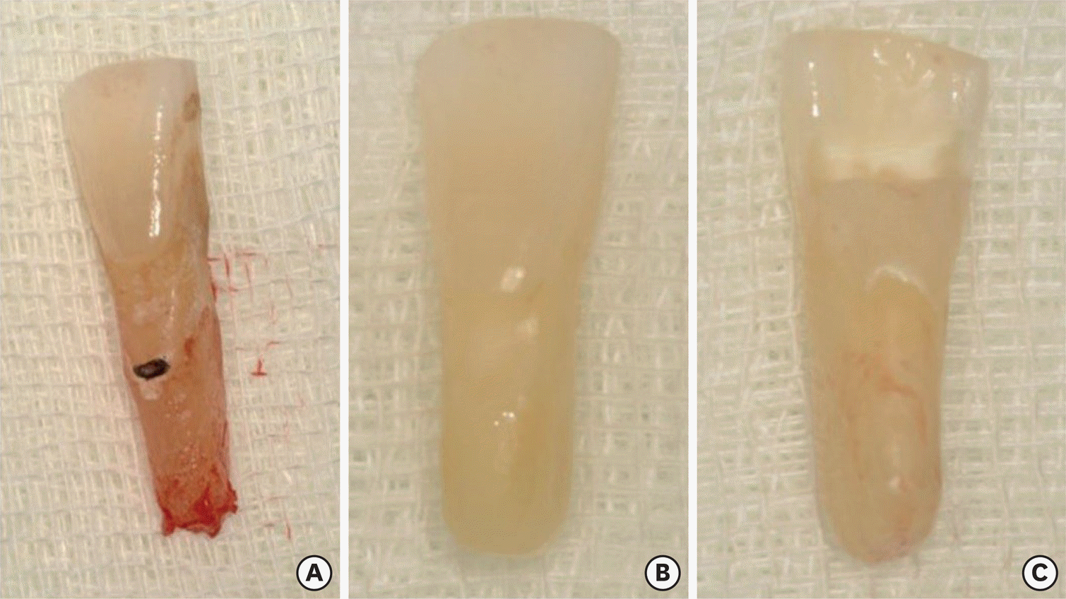
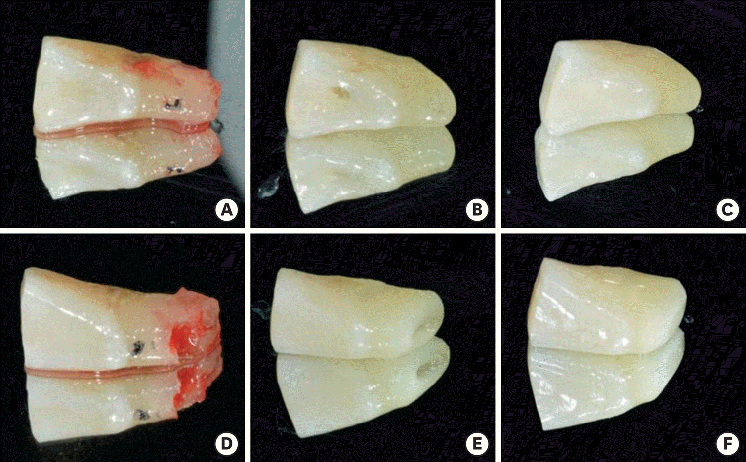 | Figure 4.Coronal fragment of tooth #41 as a natural tooth pontic. (A, D) Extracted coronal fragment; (B, E) prepared coronal fragment; (C, F) polished coronal fragment; (A-C) lingual view; (D-F) labial view. |
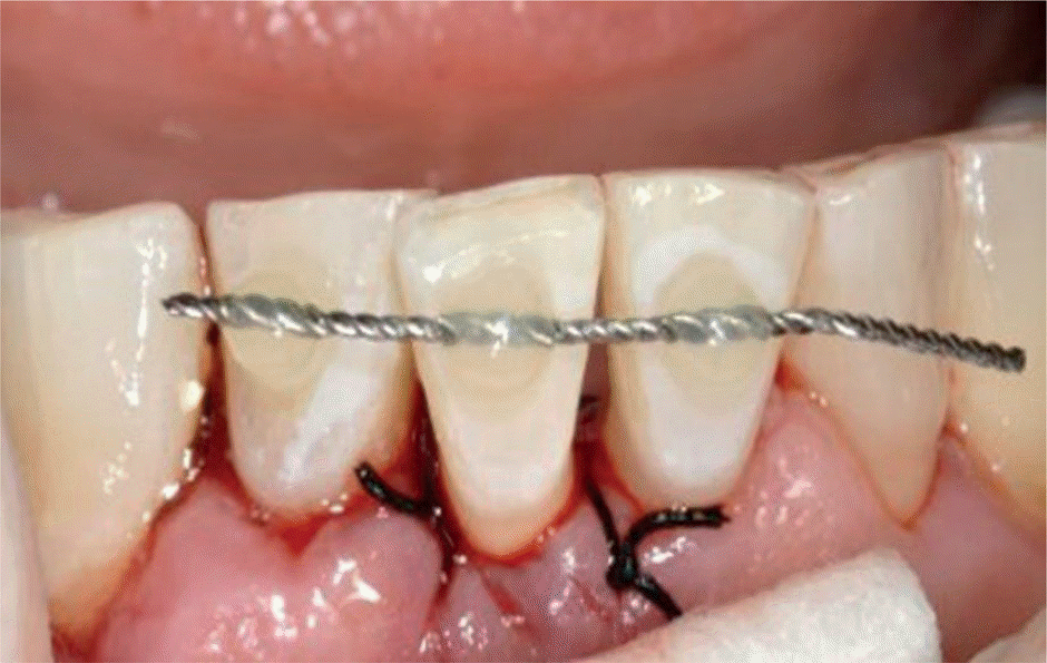
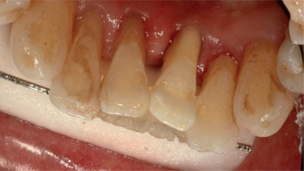
 | Figure 6.Lingual view of tooth preparation. (A) Preparation of space for fiber extending from the mesial aspect of tooth #42 to the distal aspect of tooth #31 across tooth #41; (B) measurement of the span using a Wedjet. |
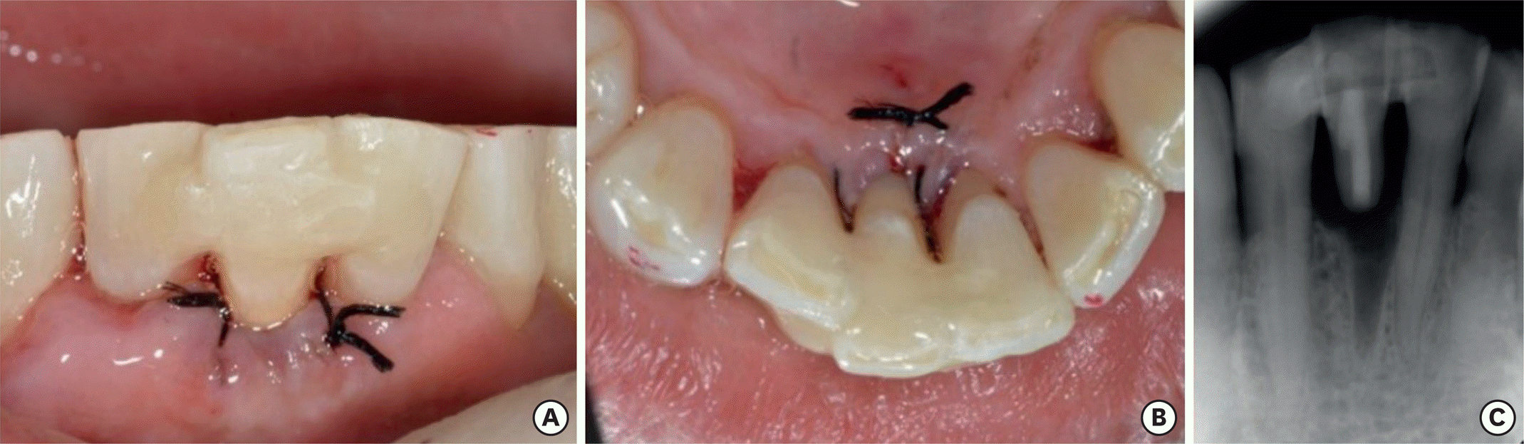 | Figure 7.Postoperative view. (A) Intraoral photograph (labial view); (B) intraoral photograph (lingual view); (C) periapical view. |
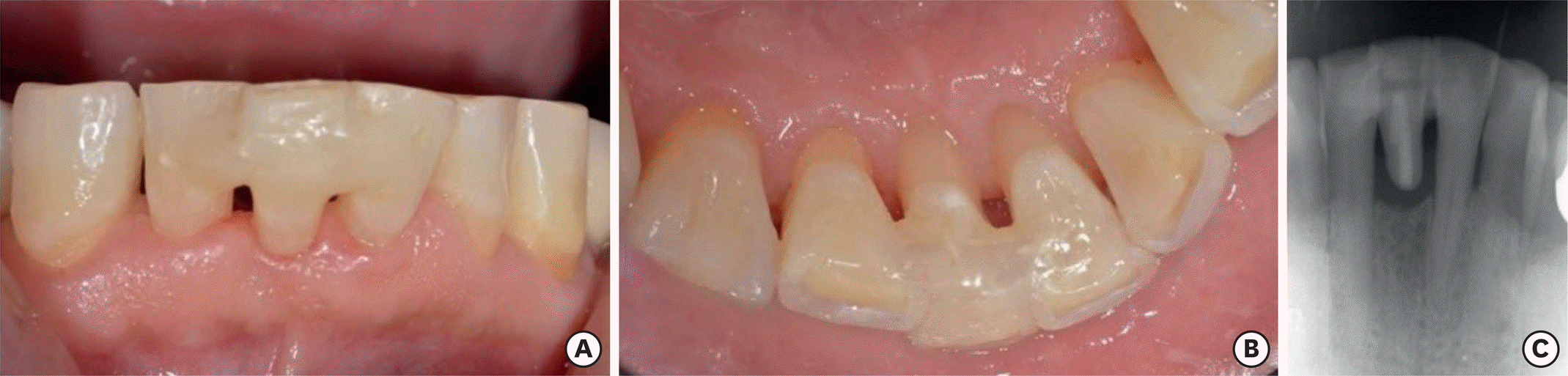 | Figure 8.At a 10-month recall check. (A) Intraoral photograph (labial view); (B) intraoral photograph (lingual view); (C) periapical view. |
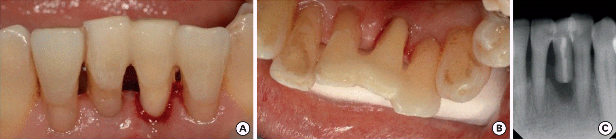
 | Figure 9.Preoperative view. (A) Intraoral photograph (labial view); (B) intraoral photograph (lingual view); (C) periapical view. |
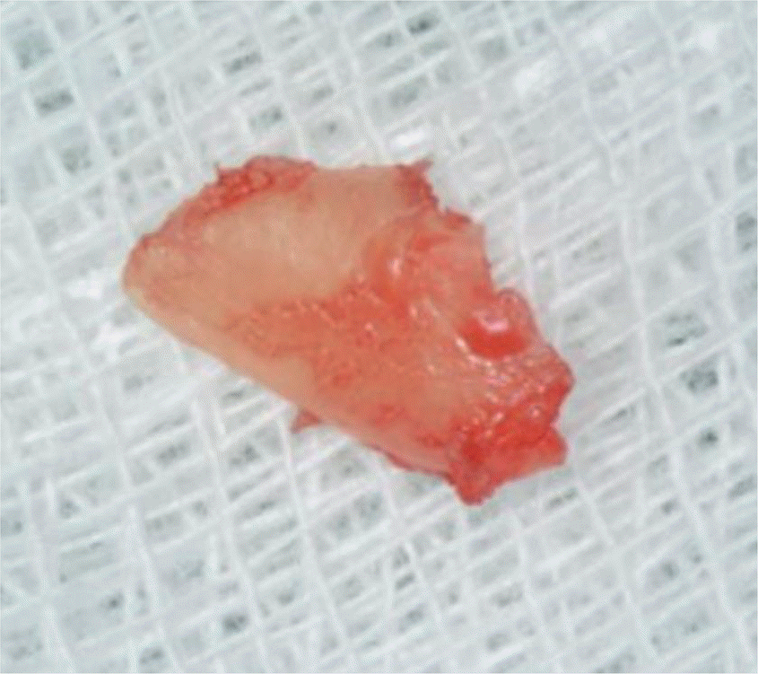
 | Figure 10.#31 Coronal fragment as a natural tooth pontic. (A) Extracted coronal fragment; (B) polished coronal fragment (labial view); (C) polished coronal fragment (lingual view). |
 XML Download