

 PDF
PDF Citation
 Print
Print


Abstract
Objectives
The obturation quality of MTA, Biodentine, Total Fill BC root canal sealer (RCS), and warm gutta-percha (WGP) in teeth with simulated internal root resorption (IRR) was evaluated by using micro-computed tomography.
Materials and Methods
Standardized IRR cavities were created using 40 extracted maxillary central incisor teeth and randomly assigned into 4 groups (n = 10). IRR cavities were filled with MTA, Biodentine, Total Fill BC RCS (bulk-fill form) and WGP + Total Fill BC RCS. Percentage of voids between resorptive cavity walls and obturation material (external void), and inside the filling materials (internal voids) were measured.
Results
Total Fill BC sealer in the bulk-fill form presented significantly highest values of external and internal void percentages (p < 0.05). Biodentine showed a significantly lowest external void percentage (p < 0.05). WGP + Total Fill BC RCS presented significantly lower values of internal void percentages than all groups (p < 0.05), except Biodentine (p > 0.05).
References
1. Patel S, Ricucci D, Durak C, Tay F. Internal root resorption: a review. J Endod. 2010; 36:1107–1121.

2. Rodríguez-Lozano FJ, García-Bernal D, Oñate-Sánchez RE, Ortolani-Seltenerich PS, Forner L, Moraleda JM. Evaluation of cytocompatibility of calcium silicate-based endodontic sealers and their effects on the biological responses of mesenchymal dental stem cells. Int Endod J. 2017; 50:67–76.
3. Ersahan S, Aydin C. Dislocation resistance of iRoot SP, a calcium silicate-based sealer, from radicular dentine. J Endod. 2010; 36:2000–2002.
4. Zhang W, Li Z, Peng B. Assessment of a new root canal sealer's apical sealing ability. Oral Surg Oral Med Oral Pathol Oral Radiol Endod. 2009; 107:e79–e82.
5. Huffman BP, Mai S, Pinna L, Weller RN, Primus CM, Gutmann JL, Pashley DH, Tay FR. Dislocation resistance of ProRoot Endo Sealer, a calcium silicate-based root canal sealer, from radicular dentine. Int Endod J. 2009; 42:34–46.
6. Jainaen A, Palamara JE, Messer HH. Push-out bond strengths of the dentine-sealer interface with and without a main cone. Int Endod J. 2007; 40:882–890.
7. Nagas E, Cehreli Z, Uyanik MO, Durmaz V. Bond strength of a calcium silicate-based sealer tested in bulk or with different main core materials. Braz Oral Res. 2014; 28:1–7.
8. Topçuoğ lu HS, Düzgün S, Ceyhanlı KT, Aktı A, Pala K, Kesim B. Efficacy of different irrigation techniques in the removal of calcium hydroxide from a simulated internal root resorption cavity. Int Endod J. 2015; 48:309–316.
9. Huang Y, Celikten B, de Faria Vasconcelos K, Ferreira Pinheiro Nicolielo L, Lippiatt N, Buyuksungur A, Jacobs R, Orhan K. Micro-CT and nano-CT analysis of filling quality of three different endodontic sealers. Dentomaxillofac Radiol. 2017; 46:20170223.
10. Silveira FF, Nunes E, Soares JA, Ferreira CL, Rotstein I. Double ‘pink tooth'associated with extensive internal root resorption after orthodontic treatment: a case report. Dent Traumatol. 2009; 25:e43–e47.
11. El-Ma'aita AM, Qualtrough AJ, Watts DC. A microcomputed tomography evaluation of mineral trioxide aggregate root canal fillings. J Endod. 2012; 38:670–672.
12. Sisli SN, Ozbas H. Comparative microcomputed tomographic evaluation of the sealing quality of ProRoot MTA and MTA Angelus apical plugs placed with various techniques. J Endod. 2017; 43:147–151.
13. Küçükkaya Eren S, Aksel H, Askerbeyli Örs S, Serper A, Koçak Y, Ocak M, Çelik HH. Obturation quality of calcium silicate-based cements placed with different techniques in teeth with perforating internal root resorption: a microcomputed tomographic study. Clin Oral Investig. 2019; 23:805–811.
14. Ozturk TY, Guneser MB, Taschieri S, Maddalone M, Dincer AN, Venino PM, Del Fabbro M. Do the intracanal medicaments affect the marginal adaptation of calcium silicate-based materials to dentin? J Dent Sci. 2019; 14:157–162.
15. Setbon HM, Devaux J, Iserentant A, Leloup G, Leprince JG. Influence of composition on setting kinetics of new injectable and/or fast setting tricalcium silicate cements. Dent Mater. 2014; 30:1291–1303.
16. Grech L, Mallia B, Camilleri J. Investigation of the physical properties of tricalcium silicate cement-based root-end filling materials. Dent Mater. 2013; 29:e20–e28.
17. Komabayashi T, Spångberg LS. Comparative analysis of the particle size and shape of commercially available mineral trioxide aggregates and Portland cement: a study with a flow particle image analyzer. J Endod. 2008; 34:94–98.
18. Bioč anin V, Antonijević Đ, Poštić S, Ilić D, Vuković Z, Milić M, Fan Y, Li Z, Brković B, Đurić M. Marginal gaps between 2 calcium silicate and glass ionomer cements and apical root dentin. J Endod. 2018; 44:816–821.
19. Gencoglu N, Yildirim T, Garip Y, Karagenc B, Yilmaz H. Effectiveness of different gutta-percha techniques when filling experimental internal resorptive cavities. Int Endod J. 2008; 41:836–842.
20. Keles A, Ahmetoglu F, Uzun I. Quality of different gutta-percha techniques when filling experimental internal resorptive cavities: a microcomputed tomography study. Aust Endod J. 2014; 40:131–135.
21. Lottanti S, Tauböck TT, Zehnder M. Shrinkage of backfill gutta-percha upon cooling. J Endod. 2014; 40:721–724.
22. Keleş A, Alçin H, Kamalak A, Versiani MA. Micro-CT evaluation of root filling quality in oval-shaped canals. Int Endod J. 2014; 47:1177–1184.
23. Ng YL, Mann V, Rahbaran S, Lewsey J, Gulabivala K. Outcome of primary root canal treatment: systematic review of the literature – Part 2. Influence of clinical factors. Int Endod J. 2008; 41:6–31.
24. Bogen G, Kuttler S. Mineral trioxide aggregate obturation: a review and case series. J Endod. 2009; 35:777–790.
25. Schilder H. Filling root canals in three dimensions. 1967. J Endod. 2006; 32:281–290.
Figure 1.
Experimental design. (A) Semi-circular cavities were created on each half of the roots; (B) Individual molds in Eppendorf tubes; (C) Periapical radiography; (D) Micro-computed tomography (µCT) images for obturation of resorption cavity.
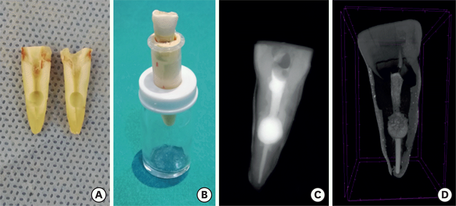
Figure 2.
Micro-computed tomography (µCT) analysis stages. (A) Measurement of percentage voids on CTAn software (Bruker microCT); (B). Selection of the region of interest; (C) Threshold selection from gray-level histogram.
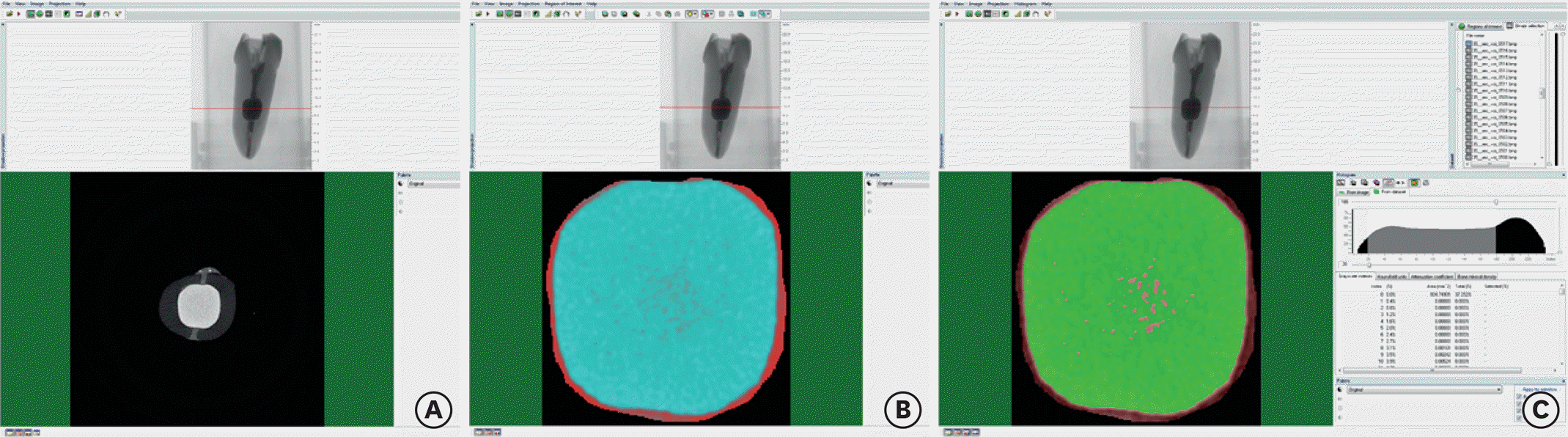
Table 1.
Median and range percentage values of experimental groups (%)
 XML Download