

 PDF
PDF Citation
 Print
Print


Abstract
Objectives
The purpose of this study was to determine the incidence of radix molaris (RM) (entomolaris and paramolaris) in the mandibular first permanent molars of a sample Saudi Arabian subpopulation using cone-beam computed tomography (CBCT).
Materials and Methods
A total of 884 CBCT images of 427 male and 457 female Saudi citizens (age 16 to 70 years) were collected from the radiology department archives of 4 dental centers. A total of 450 CBCT images of 741 mature mandibular first molars that met the inclusion criteria were reviewed. The images were viewed at high resolution by 3 examiners and were analyzed with Planmeca Romexis software (version 5.2).
Results
Thirty-three (4.5%) mandibular first permanent molars had RM, mostly on the distal side. The incidence of radix entomolaris (EM) was 4.3%, while that of radix paramolaris was 0.3%. The RM roots had one canal and occurred more unilaterally. No significant difference in root configuration was found between males and females (p > 0.05). Types I and III EM root canal configurations were most common, while type B was the only RP configuration observed.
Go to : 
References
1. Segura-Egea JJ, Jiménez-Pinzón A, Ríos-Santos JV. Endodontic therapy in a 3-rooted mandibular first molar: importance of a thorough radiographic examination. J Can Dent Assoc. 2002; 68:541–544.
2. Slowey RR. Radiographic aids in the detection of extra root canals. Oral Surg Oral Med Oral Pathol. 1974; 37:762–772.

3. Vertucci FJ. Root canal anatomy of the human permanent teeth. Oral Surg Oral Med Oral Pathol. 1984; 58:589–599.
4. Curzon ME, Curzon JA. Three-rooted mandibular molars in the Keewatin Eskimo. J Can Dent Assoc (Tor). 1971; 37:71–72.
5. Reichart PA, Metah D. Three-rooted permanent mandibular first molars in the Thai. Community Dent Oral Epidemiol. 1981; 9:191–192.
6. Walker RT. Root form and canal anatomy of mandibular first molars in a southern Chinese population. Endod Dent Traumatol. 1988; 4:19–22.
7. Younes SA, al-Shammery AR, el-Angbawi MF. Three-rooted permanent mandibular first molars of Asian and black groups in the Middle East. Oral Surg Oral Med Oral Pathol. 1990; 69:102–105.
8. Zaatar EI, al-Kandari AM, Alhomaidah S, al-Yasin IM. Frequency of endodontic treatment in Kuwait: radiographic evaluation of 846 endodontically treated teeth. J Endod. 1997; 23:453–456.
9. Sperber GH, Moreau JL. Study of the number of roots and canals in Senegalese first permanent mandibular molars. Int Endod J. 1998; 31:117–122.
10. al-Nazhan S. Incidence of four canals in root-canal-treated mandibular first molars in a Saudi Arabian sub-population. Int Endod J. 1999; 32:49–52.
11. Ahmed HA, Abu-bakr NH, Yahia NA, Ibrahim YE. Root and canal morphology of permanent mandibular molars in a Sudanese population. Int Endod J. 2007; 40:766–771.
12. Schäfer E, Breuer D, Janzen S. The prevalence of three-rooted mandibular permanent first molars in a German population. J Endod. 2009; 35:202–205.
13. Al-Qudah AA, Awawdeh LA. Root and canal morphology of mandibular first and second molar teeth in a Jordanian population. Int Endod J. 2009; 42:775–784.
14. Song JS, Kim SO, Choi BJ, Choi HJ, Son HK, Lee JH. Incidence and relationship of an additional root in the mandibular first permanent molar and primary molars. Oral Surg Oral Med Oral Pathol Oral Radiol Endod. 2009; 107:e56–e60.
15. Zhang R, Wang H, Tian YY, Yu X, Hu T, Dummer PM. Use of cone-beam computed tomography to evaluate root and canal morphology of mandibular molars in Chinese individuals. Int Endod J. 2011; 44:990–999.
16. Demirbuga S, Sekerci AE, Dinçer AN, Cayabatmaz M, Zorba YO. Use of cone-beam computed tomography to evaluate root and canal morphology of mandibular first and second molars in Turkish individuals. Med Oral Patol Oral Cir Bucal. 2013; 18:e737–e744.
17. Mukhaimer R, Azizi Z. Incidence of radix entomolaris in mandibular first molars in Palestinian population: a clinical investigation. Int Sch Res Notices. 2014; 2014:405601.
18. Rodrigues CT, Oliveira-Santos C, Bernardineli N, Duarte MA, Bramante CM, Minotti-Bonfante PG, Ordinola-Zapata R. Prevalence and morphometric analysis of three-rooted mandibular first molars in a Brazilian subpopulation. J Appl Oral Sci. 2016; 24:535–542.
19. Rahimi S, Mokhtari H, Ranjkesh B, Johari M, Frough Reyhani M, Shahi S, Seif Reyhani S. Prevalence of extra roots in permanent mandibular first molar s in Iranian population: a CBCT analysis. Iran Endod J. 2017; 12:70–73.
20. Gupta A, Duhan J, Wadhwa J. Prevalence of three rooted permanent mandibular first molars in Haryana (North Indian) population. Contemp Clin Dent. 2017; 8:38–41.
21. Walker RT, Quackenbush LE. Three-rooted lower first permanent molars in Hong Kong Chinese. Br Dent J. 1985; 159:298–299.
22. Gulabivala K, Opasanon A, Ng YL, Alavi A. Root and canal morphology of Thai mandibular molars. Int Endod J. 2002; 35:56–62.
23. Sert S, Aslanalp V, Tanalp J. Investigation of the root canal configurations of mandibular permanent teeth in the Turkish population. Int Endod J. 2004; 37:494–499.
24. Bahammam LA, Bahammam HA. The incidence of radix entomolaris in mandibular first permanent molars in a Saudi Arabian sub-population. JKAU Med Sci. 2011; 18:83–90.
25. Wu DM, Wu YN, Guo W, Sameer S. Accuracy of direct digital radiography in the study of the root canal type. Dentomaxillofac Radiol. 2006; 35:263–265.
26. Omer OE, Al Shalabi RM, Jennings M, Glennon J, Claffey NM. A comparison between clearing and radiographic techniques in the study of the root-canal anatomy of maxillary first and second molars. Int Endod J. 2004; 37:291–296.
27. Matherne RP, Angelopoulos C, Kulild JC, Tira D. Use of cone-beam computed tomography to identify root canal systems in vitro. J Endod. 2008; 34:87–89.
28. Al-Shehri S, Al-Nazhan S, Shoukry S, Al-Shwaimi E, Al-Sadhan R, Al-Shemmery B. Root and canal configuration of the maxillary first molar in a Saudi subpopulation: a cone-beam computed tomography study. Saudi Endod J. 2017; 2:69–76.
29. Carlsen O, Alexandersen V. Radix paramolaris in permanent mandibular molars: identification and morphology. Scand J Dent Res. 1991; 99:189–195.
30. Song JS, Choi HJ, Jung IY, Jung HS, Kim SO. The prevalence and morphologic classification of distolingual roots in the mandibular molars in a Korean population. J Endod. 2010; 36:653–657.
31. Wang Y, Zheng QH, Zhou XD, Tang L, Wang Q, Zheng GN, Huang DM. Evaluation of the root and canal morphology of mandibular first permanent molars in a western Chinese population by cone-beam computed tomography. J Endod. 2010; 36:1786–1789.
32. Quackenbush LE. Mandibular molar with three distal root canals. Endod Dent Traumatol. 1986; 2:48–49.
33. Loh HS. Incidence and features of three-rooted permanent mandibular molars. Aust Dent J. 1990; 35:434–437.
34. Salarpour M, Farhad Mollashahi N, Mousavi E, Salarpour E. Evaluation of the effect of tooth type and canal configuration on crown size in mandibular premolars by cone-beam computed tomography. Iran Endod J. 2013; 8:153–156.
35. Patel S, Dawood A, Ford TP, Whaites E. The potential applications of cone beam computed tomography in the management of endodontic problems. Int Endod J. 2007; 40:818–830.
36. Cotton TP, Geisler TM, Holden DT, Schwartz SA, Schindler WG. Endodontic applications of cone-beam volumetric tomography. J Endod. 2007; 33:1121–1132.
37. Neelakantan P, Subbarao C, Subbarao CV. Comparative evaluation of modified canal staining and clearing technique, cone-beam computed tomography, peripheral quantitative computed tomography, spiral computed tomography, and plain and contrast medium-enhanced digital radiography in studying root canal morphology. J Endod. 2010; 36:1547–1551.
38. Gu Y, Lu Q, Wang H, Ding Y, Wang P, Ni L. Root canal morphology of permanent three-rooted mandibular first molars–part I: pulp floor and root canal system. J Endod. 2010; 36:990–994.
39. Fabra-Campos H. Unusual root anatomy of mandibular first molars. J Endod. 1985; 11:568–572.
40. Wasti F, Shearer AC, Wilson NH. Root canal systems of the mandibular and maxillary first permanent molar teeth of south Asian Pakistanis. Int Endod J. 2001; 34:263–266.
41. Rwenyonyi CM, Kutesa A, Muwazi LM, Buwembo W. Root and canal morphology of mandibular first and second permanent molar teeth in a Ugandan population. Odontology. 2009; 97:92–96.
42. Scott GR, Turner CG. The anthropology of modern human teeth: dental morphology and its variation in recent human populations. Cambridge, NY: Cambridge University Press;1997. p74–130.
43. De Moor RJ, Deroose CA, Calberson FL. The radix entomolaris in mandibular first molars: an endodontic challenge. Int Endod J. 2004; 37:789–799.
44. Tinelli ME. Ethnic variations in the topography of the root canals. Electronic J Endod Rosario. 2011; 2:558–562.
45. Kim KR, Song JS, Kim SO, Kim SH, Park W, Son HK. Morphological changes in the crown of mandibular molars with an additional distolingual root. Arch Oral Biol. 2013; 58:248–253.
46. Kim HH, Jo HH, Min JB, Hwang HK. CBCT study of mandibular first molars with a distolingual root in Koreans. Restor Dent Endod. 2018; 43:e33.
Go to :
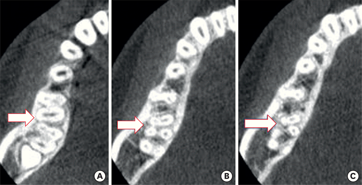 | Figure 1.(A) Cone-beam computed tomography (CBCT) images of mandibular first molar showing 4 canals (arrow). (B) CBCT showing disto-buccal root with one canal (arrow). (C) Separated distal roots with one canal each (arrow). |
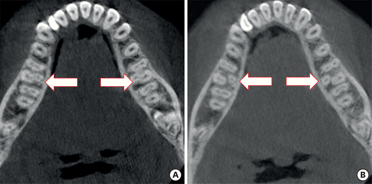 | Figure 2.(A) Cone-beam computed tomography (CBCT) images of mandibular first molar showing disto-lingual root bilateral entomolaris (arrow). (B) Bilateral separated roots with one canal (arrow). |
Table 1.
Prevalence of supernumerary root in mandibular first molar
Table 2.
Total number of evaluated cone-beam computed tomography images
Table 3.
Number of roots of mandibular first molar in relation to sex and jaw side
Table 4.
Number and percentages of patients with entomolaris (distolingual root) and paramolaris (mesiobuccal root) in mandibular first molars according to sex and jaw side (n = 741)
Table 5.
Morphology of the distolingual root (entomolaris) based on Song et al. [30] classification. (n = 741)
 XML Download