

 PDF
PDF ePub
ePub Citation
 Print
Print


INTRODUCTION
As a progressive neurodegenerative disease, Alzheimer's disease (AD) is the most common form of dementia worldwide, affecting more than 30 million people worldwide [1]. The main pathological characteristics of AD are extracellular amyloid β (Aβ) plaque deposition and neurofibrillary tangles (NFT) [2]. Aβ is derived from the amyloid protein precursor (APP) and NFT are mainly composed of hyperphosphorylated tau [3]. The production and clearance rates of Aβ maintain a dynamic balance in normal individuals [4]. The imbalance between the production and removal of Aβ results in the formation of Aβ oligomers, which eventually accumulate into senile plaques in the brain [3]. The accumulation of this peptide in AD patients leads to synaptic dysfunction, inflammation stress, apoptosis of neurons and cognitive deficits [56]. Significant progress has been made in therapies for AD in recent years; immunotherapy, targeting Aβ, is considered to be the most promising treatment due to its specific activity to limit the production, accumulation and deposition of Aβ [7]. However, serious side effects limit wide clinical application. There is an urgent need for complementary therapies, such as naturally derived drugs.
Drugs made from plant compounds have important clinical potential for the treatment of AD [8]. Natural compounds extracted from plants, such as catechins [9] and curcumin [10], have shown beneficial effects in AD. As a compound with a structure similar to cholesterol, β-Sitosterol is one of the most widely distributed plant sterols existing in plants [11]. β-Sitosterol has been widely investigated in medical research [12]. Dietary phytosterols can accumulate in brain tissue via the blood-brain barrier (BBB) and may affect brain function [13]. β-Sitosterol changes the shear pattern of APP [14], suggesting that it may be a potential therapy for AD. However, whether it has a neuroprotective effect on AD in vivo is still unclear. APP/presenilin 1 (PS1) transgenic mice show spatial memory impairment and imbalanced amyloid production and clearance [1516]. In the present study, we simulated the pathogenesis of AD in APP/PS1 mice and used β-Sitosterol as an intervention method, to explore the effect and mechanism on cognitive function in these mice. We report, for the first time, that administration of β-Sitosterol in APP/PS1 mice can reduce Aβ deposition and improve cognitive impairment. These results strongly support that β-Sitosterol is a promising therapeutic agent for AD.
METHODS
Animals and drug administration
Age matched, gender mixed APP/PS1 and wild-type (WT) littermates were purchased from Shanghai Nanfang Research Center for Model Organisms (Shanghai, China). Mice were housed no more than five per cage, with free access to food and water, under standard specific-pathogen-free laboratory conditions. The mice were handled to adapt to the experimenter before behavior testing. All animal procedures were performed in strict accordance with the guidelines for the care and use of laboratory animals of the Institute for Experimental Animals at Hebei Medical University and with the laws of China. All experiments were performed in accordance with the guidelines for Animal Care and Use of China, and the experimental schemes were approved by the animal ethics committee of Hebei Medical University (approval No. 2018011201A). The 30-week-old mice were divided into three groups: WT littermate controls (WT), APP/PS1 vehicle-treated controls (APP/PS1) and APP/PS1 treated with β-Sitosterol (APP/PS1 + BSS). β-Sitosterol was purchased from Sigma-Aldrich (Sigma, St. Louis, MO, USA) and dissolved in dimethyl sulfoxide (DMSO) as a stock solution (20 mg/kg). Control groups were treated with 0.1% DMSO. All groups were treated once per day by gavage for continuously 28 days.
Behavioral testing
Three days after β-Sitosterol administration, spatial learning and recognition memory testing was performed during the daylight hours.
The Morris water maze (MWM) test was conducted following a previously described method [17]. Briefly, A stainless steel pool (110 cm in diameter) was divided into four quadrants, containing a submerged escape platform (10 cm in diameter) located 1 cm under the water surface. The water temperature was maintained at 23 ± 1℃. The acquisition period consisted of 5 consecutive days of training with four trials/day. The time taken for the mouse to swim onto the hidden platform was recorded for all mice. Each mouse was allowed up to 60 sec to find the hidden platforms. If a mouse could not find a platform within 60 sec, the training was interrupted and the mouse was guided to a platform, to remain for 10 sec. The time that mice spent in the target quadrant and the number of target platform crossings, were recorded. On the 6th day, the platform was removed and spatial probe trials were conducted. Mice were given 60 sec to perform their search bias.
The novel object recognition test was conducted in a self-made open-field arena (58 × 58 × 31 cm) as previously described [18]. Mice were placed in the arena for 10 min, 24 h prior to the test, to acclimatize them to the apparatus and environment. Seventy percent ethanol was used to remove olfactory cues. Exploratory objects were red cubes (1.5 × 1.5 × 1.5 cm) or white cubes (2 × 2 × 2 cm) and mice were allowed 10 min for exploration. After a 3-h retention interval, mice were returned to the arena and exposed to the objects, in addition to a novel object, for a 10-min recognition trial. Object location and type were randomized throughout the trial. Total time spent exploring the familiar objects (TF = time spent exploring the familiar object) or the novel object (TN = time spent exploring the novel object) was recorded by SMART 3.0 software (Panlab, Barcelona, Spain). A recognition index was used to measure recognition memory: [TF or TN / (TF + TN)] × 100. The discrimination index could be get by TN minus TF. All behavioral parameters of mice were tracked, recorded and analyzed using SMART ver. 3.0 (Harvard Apparatus, Holliston, MA, USA).
ELISA
Brain tissue was homogenized and supernatant samples containing soluble Aβ, or pellets containing insoluble Aβ, were collected. The hippocampus and cortex Aβ1–40 and Aβ1–42 levels were measured by ELISA according to the manufacturer's instructions (ELISA; BioSource International, Camarillo, CA, USA). The values of Aβ concentrations were acquired with reference to the standard curve.
Thioflavin S staining
Four percent paraformaldehyde (pH 7.4) fixed brain tissues were collected and dehydrated in 30% sucrose, and 35 µm frozen section slices were used for Thioflavin S (ThS) (1%, 8 min) staining. Coverslips were mounted using depex polystyrene (Electron Microscopy Sciences, Hatfield, PA, USA). Hippocampal images were taken with a Nikon digital camera (Coolpix 4500; Nikon, Tokyo, Japan). The Aβ plaques were counted in five representative sections from each animal.
Western blotting
Protein extraction from the hippocampus was performed as previously described [19]. Protein concentration was determined using the BCA protein assay (Tiangen Biotech Co. Ltd., Beijing, China). 10 µg of protein from each sample was separated by 10% SDS-PAGE and electrotransferred to polyvinylidene fluoride membranes (Millipore, Billerica, MA, USA) for immunoblotting. The following primary antibodies were used: anti-Aβ (6E10, 1:1000; Covance, Emeryville, CA, USA), anti-BACE1 (1:500, ab2077; Abcam, Cambridge, MA, USA), and anti-β-Actin (1:500, ab2077; Abcam). Band intensities were semi-quantified using ImageJ ver. 1.6 (http://imagej.nih.gov/ij/) software.
Dendritic spine density analysis
200 nl AAV-IRES-GFP (2.45E+13 vg/ml; OBiO, Shanghai, China) viral vector were stereotactically injected in the hippocampus to label the DG neurons. Fourteen days after the infection, the mice were perfused and brain were extracted. Forty µm frozen sections were used for quantification of dendritic spines which was evaluated by fluorescent confocal imaging (LSM 510; Carl Zeiss, Jena, Germany) with identical settings for all samples. Spines were counted on 30–40 µm segments of secondary dendrites extending at least 60 µm beyond the cell body. Five segments from each neuron were quantified.
Whole-cell patch clamp studies
Whole cell voltage-clamp was performed at 37℃ with a 60-channel voltage amplifier (MEA1060-Inv-BC; Multi-Channel Systems, Reutlingen, Germany). All recordings were monitored in all experiments. Simultaneous whole-cell current and voltage clamps were held at −68 mV during recording. Patch pipettes had an identical resistance of 3–4 MΩ. The frequency and amplitude of the miniature excitatory postsynaptic currents (mEPSC) were sampled at 10 kHz and low-pass filtered at 3 kHz (Axon Digidata 1440A; Molecular Devices, Union City, CA, USA). The noise level was below 4 pA, and 7 pA and was typically used as the threshold for mEPSC events. Five min of representative mEPSC recordings were used to generate the cumulative distribution plot.
Statistical analyses
All values were expressed as the mean ± the standard error of the mean. Data management and statistical analyses were performed using GraphPad Prism ver 7.0 (GraphPad Software, Inc., San Diego, CA, USA). All statistical assessments were two-sided and the corresponding p-values were calculated. Statistical significance was determined at the α-level of significance of p = 0.05. Statistical analyses were carried out according to the experimental design used and are listed in figure legends.
RESULTS
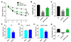
β-Sitosterol treatment rescues spatial and recognition memory deficits in APP/PS1 mice
To investigate whether β-Sitosterol treatment could attenuate hippocampus-dependent learning and memory impairment in APP/PS1 mice, age-matched WT, APP/PS1 and β-Sitosterol treated mice were assessed by the MWM and novel object recognition (NOR) tests. Our results showed that the β-Sitosterol treated APP/PS1 mice showed reduced escape latency (Fig. 1A). In the probe trial phase, the β-Sitosterol treated mice performed better than the APP/PS1 controls, as reflected by the significantly greater time spent in the target quadrant and more crossovers from the former platform (Fig. 1B, C). These observations indicate that β-Sitosterol treatment ameliorated spatial learning deficits in APP/PS1 mice. In the object recognition memory test, β-Sitosterol treated APP/PS1 mice and WT mice showed similar significant recognition of the novel object (Fig. 1D, F), but the APP/PS1 control mice did not (Fig. 1E). APP/PS1 vehicle-treated mice attained a significantly impaired score for novel object recognition (Fig. 1G).
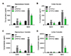
β-Sitosterol treatment rescues Aβ pathology in APP/PS1 mice
An association has been observed between Aβ deposition in the brains of both Alzheimer's disease patients and transgenic mouse models. After behavioural test, hippocampal and cortical tissues were isolated from each group. Total protein was extracted and quantified by BCA, the concentration of soluble and insoluble Aβ40 and Aβ42 in the brain was determined by using Aβ ELISA kit. Total soluble and insoluble brain Aβ42 or Aβ40 levels were significantly increased in the control APP/PS1 mice conpared with the WT control in both brain regions (Fig. 2). Insoluble tissue fractions showed the most prominent increases in Aβ42 (Fig. 2). However, the Aβ40 and Aβ42 were markly reduced in APP/PS1 mice treated withβ-Sitosterol (Fig. 2).
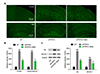
β-Sitosterol treatment attenuated Aβ accumulation and BACE1 expression
We further determined Aβ accumulation in the brain, by thioflavin-S staining and immunoblotting. Aβ staining result showed that β-Sitosterol administration results in a decrease in Aβ density in the hippocampus and cortex of APP/PS1 mice (Fig. 3A, B). Similarly, immunoblotting for Aβ and BACE1 expression showed that β-Sitosterol treatment significantly decreased their expression compared with the control APP/PS1 mice (Fig. 3C).
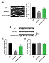
β-Sitosterol treatment restored the synaptic dysfunction in APP/PS1 mice
Dendritic spine and synaptic loss are well documented in APP/PS1 mice. We used AAV-IRES-GFP to label the hippocampal neurons of APP/PS1 mice. The dendritic spine densities of hippocampal neurons were analyzed for each group. β-Sitosterol treatment caused up-regulation of dendritic spine density compared with control APP/PS1 mice (Fig. 4A, B). Next, we used a single whole-cell recording method to record mEPSCs, to quantify synaptic transmission. The mEPSC frequency of APP/PS1 mice hippocampal neurons was significantly decreased compared with WT mice. However, in mice treated with β-Sitosterol, the mEPSC frequency showed a reversal to control levels. The mEPSC amplitude was not significantly different between any of the groups (Fig. 4C). These results showed that β-Sitosterol treatment rescued the synaptic transmission in APP/PS1 mice.
DISCUSSION
AD is a progressive neurodegenerative disorder characterized by Aβ plaque deposits, NFT, and neuronal loss, leading to learning and memory impairments [20]. Increased Aβ plaque deposition and decreased neuronal function in the hippocampus is implicated in the loss of memory performance in AD mice [21]. β-Sitosterol has been reported to cross the BBB and inhibit the production of Aβ [2223] and we speculate that this may contribute to a delay in the pathological development of AD. Therefore, we treated APP/PS1 mice with β-Sitosterol and evaluated cognitive dysfunction and extent of AD pathology. The APP/PS1 transgenetic mouse model has been widely used to explore the cognitive deficits related to AD. APP/PS1 mice show progressive accumulation of amyloid plaques and cognitive deficits after seven months [24]. Therefore, we initiated drug treatment in 30-week-old APP/PS1 mice, and continued for 4 weeks. We investigated the underlying role of β-Sitosterol in the protection against AD progression. The pathological characteristics of APP/PS1 mice, represented by memory and recognition deficits, impaired Aβ metabolism and synaptic loss, were evaluated to investigate the efficacy of β-Sitosterol in alleviating AD-related pathologies. Herein, we found that APP/PS1 transgenic mice treated with β-Sitosterol had significantly better working memory and visual recognition memory in the MWM and NOR test, compared with the AD control group. The behavioral results showed an observable decrease in escape latency and an increase in time spent on the target platform in the MWM test, compared with vehicle treated APP/PS1 mice. We suggest that β-Sitosterol plays a protective role in cognitive dysfunction during AD occurrence.
Imbalanced Aβ metabolism is a pathological characteristic of AD [25]. Accumulative Aβ production and aggregation in the brain has been associated with neuronal dysfunction and memory disorders [2627]. Inhibiting Aβ production or facilitating Aβ degradation are effective strategies for addressing AD-like pathologies [28]. Here, we provide in vivo evidence that β-Sitosterol is implicated in the mechanisms of Aβ production and deposition in mice. We identified roles of β-Sitosterol in the regulation of Aβ metabolism, providing a potential mechanism for the effects observed in the behavior test. We assessed amyloid plaque metabolism and found markedly reduced amyloid plaque deposits in the hippocampus and cortex. Beta-secretase 1 serves as the initiating enzyme in APP processing and Aβ production. The inhibition of BACE1 has been shown to be an effective strategy for Aβ clearance. The decreased expression of BACE1 in the brains of β-Sitosterol treated APP/PS1 mice is supported by the ELISA and Western blot analysis of Aβ. Our findings showed that the expression of BACE1 was down-regulated in the hippocampus after treatment with β-Sitosterol. This result implies that β-Sitosterol could also affect BACE1 to reduce the generation of Aβ in the AD brain.
Synaptic loss is widely observed in human and mice AD brains [2930]. Disconnections between neurons are responsible for the loss of cognitive function in AD patients. Through quantification of synapse numbers in the hippocampus, we demonstrated that β-Sitosterol treatment partly rescued the reduction in dendritic spine density in hippocampal neurons from APP/PS1 mice. In addition, the decreased frequency of mEPSCs was ameliorated. Synaptic spine density and neural electrical activity is believed to be a synaptic mechanism underlying recognition and memories in the brain [3132]. Therefore, maintenance of hippocampal spine and neural spiking explains the improved behavioral outcomes in β-Sitosterol-treated APP/PS1 mice.
In conclusion, the results presented in this study suggest that β-Sitosterol has therapeutic effects and reduces Alzheimer's disease progression in the aged APP/PS1 mouse model, which mimics the later stages of AD, raising hopes that β-Sitosterol may show positive effects, even in the later stages, for the AD patient.
Based on the encouraging effects that we found with β-Sitosterol, the neuroprotective effects of β-Sitosterol on other AD mouse models should also be examined, before commencement of clinical trials.
Issues remaining for further study include identifying the molecular mechanisms underlying Aβ clearance and accumulation, and determining whether this is due to the promotion of Aβ clearance.
 XML Download