

 PDF
PDF ePub
ePub Citation
 Print
Print


Introduction
The gradual wear of the occlusal surfaces of teeth is considered normal due to aging. However, pathological and physiological wear of teeth, if not treated appropriately and at the right time, can lead to decreased occlusal vertical dimension and functional and esthetic problems such as reduced chewing efficiency and disharmonious occlusal plane, respectively.1 Alteration of the vertical dimension of occlusion (VDO) requires accurate diagnosis, careful case analysis, and determination of a predictive treatment plan. Although, a minimalistic approach is almost always recommended while restoring the occlusal vertical dimension.2 Alteration in the VDO should be followed by an assessment of the adaptive responses of the temporomandibular joint (TMJ) and the periodontium, and the occlusal morphology, which is usually performed by clinically evaluating the diagnostic splint and/or the provisional restorations.3 Provisional restorations can provide critical information, which can prove to be of considerable help in determining various characteristics of the definitive restorations, such as VDO and tooth shape and position.
Following the introduction of computer-aided design and computer-aided manufacturing (CAD/CAM) technology in dentistry in the 1980s, the clinical and laboratory practices have evolved considerably, especially with respect to the fabrication of fixed dental prostheses.4 In clinical practice, early intraoral scanners had several issues such as lower precision as compared to the conventional impression techniques and problems associated with intraoral conditions. The recent advancements in technology have rendered the newer intraoral scanners capable of scanning complete dental arches and of fabricating multi-unit restorations, implant surgical guides, and clear aligners.567 In laboratory practice, zirconia is being widely used due to the development of zirconia materials, such as multi-layered zirconia and cubic phase zirconia, which show improved esthetic and optical properties. Due to these developments in the properties of zirconia materials, the use of the CAD/CAM technology has shown a steady increase.89 Several recent case reports have documented the use of CAD/CAM double scanning technique. The CAD/CAM double scanning technique can adopt the characteristics of provisional restorations to definitive restorations through the superimposition of provisional restorations on existing teeth abutments. The CAD/CAM double scanning technique can simplify the fabrication procedures and can easily refabricate the restorations from the stored CAD data.1011
This case report describes the full-mouth rehabilitation of a patient with esthetically compromised anterior teeth, generalized severe attrition of teeth, and reduced vertical dimension. The use of an intraoral scanner and of the CAD/CAM double scanning technique to fabricate provisional restorations and definitive monolithic zirconia restorations is described.
Case report
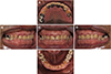
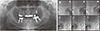
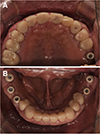
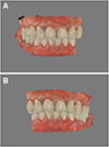
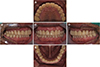
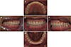
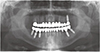
A 67-year-old patient was referred to the Department of Prosthodontics for the esthetic and functional restoration of severely-worn dentition. The patient was on anticoagulant therapy for hypertension. The patient did not report any painful symptoms related to the TMJ, musculature, or the associated dental structures, but gave a history of clenching the teeth during daytime. Clinical examination revealed generalized loss of dental structure, uneven occlusal plane, and insufficient space for the restoration of maxillary posterior teeth. Mandibular anterior teeth revealed attrition to the gingival level, and the mandibular anterior and premolar teeth showed sharp enamel edges and cupping, and the pulp was visible through the dentin (Fig. 1). The patient showed hypertrophy of the masseter muscle and thin lips with drooping commissures. No TMJ disorders were evident on radiographic examination (Fig. 2).
The following parameters were evaluated prior to treatment, to determine whether the VDO had been altered: loss of posterior support, history of wear, phonetics, interocclusal rest space, facial appearance, and space for restoration.1 A loss of posterior support had been suspected. Because, the mandibular posterior teeth were restored by implants. Parafunctional habits must have led to the pathologic wear of teeth. The closest speaking space (/s/ sound) was about 3 mm, and vertical dimension at rest (VDR) was approximately 7 mm greater than the VDO, which is greater than the normal range (2 – 4 mm). Wrinkles and drooping commissures around the mouth and an insufficient space for the restoration of maxillary posterior teeth were observed. Based on these findings, the patient's VDO was determined to be decreased due to mechanical wear. Hence, treatment options were full mouth rehabilitation with or without the crown lengthening procedure (CLP).
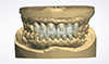
The patient's diagnostic casts were mounted on a semi-adjustable articulator (Hanau Modular Articulator, Whip Mix Corp., Louisville, KY, USA) using a facebow and a centric relation record to determine the amount of VDO that needed to be altered. The amount of VDO alteration was determined considering the width/length ratios of the anterior teeth and minimum restoration space. The vertical length between the maxillary central incisor and the mandibular central incisor of the gingival zenith was 8 mm in centric occlusion. The maxillary central incisor length and the mandibular central incisor length were set to 10 mm and 8 mm respectively based on the average crown length of Korean adults with a 2 mm overbite.12 As a result, the altered vertical length between the maxillary central incisor and the mandibular central incisor of the gingival zenith was 16 mm, the required vertical length was 8 mm. To solve uneven gingival contours of anterior teeth and to obtain an additional vertical length, the crown lengthening procedure was planned. The maxillary anterior teeth were planned based on the gingival zenith of right canine (tooth no. 13). And the mandibular premolar and anterior teeth were planned based on the gingival zenith of left second molar (tooth no. 35). The vertical length of maxilla and mandible were gained 2 mm respectively. Eventually, the required vertical length was 4 mm. An arbitrary opening of the articulator of 4 mm at the incisal pin revealed a sufficient space for the restorations of maxillary posterior teeth. Therefore, full mouth rehabilitation of the patient with re-establishment of the lost VDO through CLP was planned. The treatment plan included implant placement in the mandibular right second molar area and root canal therapy (RCT) of the mandibular premolars and of the anterior teeth for the placement of post and core. The new VDO was set by a 4 mm increase in the height of the incisal guidance pin of the articulator. An occlusal splint was fabricated at the new VDO. The splint was designed to achieve bilateral contacts in all the posterior teeth in centric relation and guides of the anterior teeth in excursive movements. The anterior guidance discluded the posterior teeth in all jaw positions except the centric relation. The patient used this occlusal splint for two months to verify the TMJ adaptation to the new VDO (Fig. 3). No muscle tenderness and discomfort in TMJ was noted. The diagnostic wax-up was completed at the new VDO with canine guidance in lateral movement and anterior guidance in protrusive movement (Fig. 4).
The patient was referred to the Department of Periodontology for CLP and implant placement. A surgical stent was fabricated for the CLP from the diagnostic wax-up. The required amount of gingival recontouring and ostectomy was guided by the surgical stent. The implant placement in the mandibular right second molar area was completed, and the patient was referred to the Department of Conservative dentistry for the RCT.
After the pre-prosthodontic treatment procedures, the diagnostic wax-up was digitized using a dental laboratory scanner (D1000, 3Shape Dental System, Copenhagen, Denmark) for the fabrication of provisional restorations, and based on the scanned data, poly methyl methacrylate blocks (PMMA block, Huge Dental Material Co., Shandong, China) were milled. The fiber-reinforced post and resin core treatment was performed on the mandibular premolar and anterior teeth due to the insufficient crown height. The teeth were prepared for restoration with complete zirconia crowns. Initially, provisional restorations were fabricated and cemented with noneugenol zinc oxide cement (TempBond NE, Kerr Corp., Orange, CA, USA). Next, an intraoral scanner (CS3600, Carestream, Rochester, NY, USA) and the scan bodies (Scanbody, Dentium, Seoul, Korea) were used to fabricate provisional restorations for the implant (Fig. 5, Fig. 6). Customized titanium abutments and PMMA provisional restorations were designed with a software (3Shape's CAD Design software, 3shape, Copenhagen K, Denmark) (Fig. 7). The abutments were milled in a 5-axis milling machine (Arum 5X-200, Doowon, Daejeon, Korea) using a titanium block (CMFit, GeoMedi Co., Uiwang, Korea), and the restorations were printed using a 3D printer (Bio3D W11, NextDent, Soesterberg, The Netherlands) and PMMA based 3D printing materials (NextDent C&B, NextDent, Soesterberg, The Netherlands). The customized titanium abutments were tightened at a torque of 30 Ncm with a torque wrench. The provisional restorations were cemented with noneugenol zinc oxide cement.
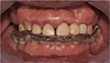
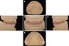
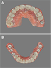
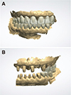
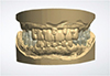
Patient's adaptation to the new VDO was monitored for three months. No muscle tenderness and/or discomfort in the TMJ were evident. The intraoral scanner was used to fabricate the second provisional restorations which had a better aesthetic and occlusion. Two-thirds of the arch with the prepared teeth and the titanium abutments were scanned separately for the left and right sides (Fig. 8). Two-thirds of the arch with the provisional restorations was also scanned separately (Fig. 9). A lateral scan in maximum intercuspation was recorded as the interocclusal record. The second set of provisional restorations was fabricated with the recorded data superimposed on each other (Fig. 10). The second provisional restorations were cemented with noneugenol zinc oxide cement. The canine guidance in lateral movements and anterior guidance in protrusive movements were established and verified (Fig. 11).
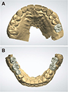
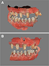
Patient's adaptation was monitored for two months. Improvement in mastication and esthetics was confirmed. For the fabrication of definitive restorations, final impressions were divided into two stages: anterior and posterior. First, final impressions of the anterior teeth abutments and second provisional restorations were made using polyvinylsiloxane impression material (Honigum light- and heavybody impression material; DMG, Hamburg, Germany) (Fig. 12). Bite registration was performed using occlusal registration material (Blue-Mousse, Parkell, Farmingdale, NY, USA). Working casts of the teeth abutments and second provisional restorations were fabricated, and the working casts were scanned by the dental laboratory scanner. The scanned data of the anterior teeth abutments and the second provisional restorations were superimposed (Fig. 13). Monolithic zirconia restorations of the anterior teeth were fabricated using the recorded superimposed data with multi-layered zirconia blocks (KATANA Zirconia Multi-Layered Disc (STML), Noritake Dental Supply Co., Ltd., Miyoshi, Japan). The canine guidance in lateral movements and anterior guidance in protrusive movements were confirmed. Definitive restorations of the anterior teeth were cemented using a dual-cure resin cement (Multilink N, Ivoclar Vivadent, Amherst, NY, USA) (Fig. 14). Next, definitive restorations of the posterior teeth were also fabricated using the CAD/CAM double scanning technique and cemented using the dual-cure resin cement on the teeth abutments and using the temporary resin cement (Premier implant cement, Premier dental products Co., Plymouth meeting, PA, USA) on titanium abutments (Fig. 15, Fig. 16. Fig. 17).
For the maintenance period, a centric relation splint was delivered. After 6 months, at the follow-up appointment, the patient was satisfied with the prosthesis, esthetically and functionally, and no radiographic abnormalities were detected (Fig. 18).
Discussion
Assessment of the adaptive responses of the TMJ and periodontium, and of the occlusal morphology of the provisional restorations is essential to alter the VDO.3 Therefore, in cases of full mouth rehabilitation requiring an increase in the VDO, patient-specific characteristics of the provisional restorations should be reproduced in the definitive restorations. The CAD/CAM double scanning technique can adopt the characteristics of the provisional restorations to the definitive restorations through superimposition of provisional restorations and the existing teeth abutments.10 However, limitations of this technique include inaccuracies in superimposition and the necessity of additional occlusal adjustments. In this case report, these limitations were minimized by separately fabricating the anterior and posterior teeth restorations during the fabrication of the definitive restorations.
Monolithic yttria-stabilized tetragonal zirconia (Y-TZP) has an excellent biocompatibility, high strength and fracture toughness. However, monochromatic and opaque optical characteristics of zirconia are a clinical drawback for esthetic restoration. Currently, monolithic zirconia with improved or high translucency, such as multi-layered zirconia and cubic phase zirconia, has been developed and indicated for use in anterior esthetic restoration. In this case, the definitive restorations fabricated using multi-layered zirconia block showed improved esthetic and function.
The intraoral scanner was used to scan the provisional restorations to evaluate the possibility of digital impressions in full mouth rehabilitation. The technology of intraoral scanners is continuously evolving, and with the introduction of newer intraoral scanners, digital impressions have become easier and accurate. And, intraoral scanner can minimize impression time, cost and potential clinical/laboratory errors, such as expansion, shrinkage, and distortion of impression materials and gypsum models.13 However, some limitations should be overcome in the recording of intraoral digital impressions. Unlike the laboratory scanner, which has been proved steady and accurate, intraoral scanners are facing concerns regarding the accuracy and precision of full-arch scans. According to Su and Sun14 precision of intraoral digital impression is clinically acceptable if the scanning scope is less than half of the arch. Nevertheless, in this case report, two-thirds of the arch were scanned separately for the left and right sides. The purpose of scanning the canine in the contralateral side was to digitize the lingual surface of the maxillary anterior teeth. By scanning beyond the midline, error could be obtained in the intraoral digital impression. So, the intraoral scanning data were used only to fabricate the provisional restorations, and error could be corrected at chairside. However, adaptations of the second provisional restorations were excellent and negligible occlusal adjustment was required.
In order to support practical use of intraoral scanner and completely digital workflow for full arch restorations, more evidences should be provided about accuracy, reliability and time requirement of the intraoral scanners for full arch impressions. Thus, additional studies of experimental and clinical research are needed in this area.
Conclusion
This case report described the full mouth rehabilitation of a patient with monolithic zirconia crowns and provisional restorations fabricated using an intraoral scanner and a CAD/CAM double scan technique. The patient was satisfied with the prosthesis esthetically and functionally. However, further studies and technological developments in intraoral scanners are required to make full-arch intraoral scan practical.


 XML Download