

 PDF
PDF ePub
ePub Citation
 Print
Print


INTRODUCTION
Endobiliary radiofrequency ablation (RFA) is performed widely to induce locoregional tumor control by inducing tumor necrosis through the transfer of thermal energy.1 Endobiliary RFA was performed prior to biliary stenting in a patient with an inoperable common bile duct (CBD) cancer. Cholecystectomy was required to remove the percutaneous transhepatic gallbladder drainage (PTGBD) catheter, but the surgical procedure could not be performed due to cystic ductal invasion of the tumor. Although gallbladder (GB) ablation is not a standard treatment, it could be an alternative option for cholecystectomy such as in inoperable circumstances.2 Pure ethanol was used to breakdown the GB mucosa. This paper reports a case, in which the procedures mentioned above were performed successfully without significant side effects for 3 months.
CASE REPORT
A 69-year-old male was admitted for 1 week owing to the complaints of right upper quadrant pain and chills. The patient had hypertension and type 2 diabetes. His initial laboratory findings were as follows: white blood cell count, 7,130/mm3; hemoglobin level, 8.6 g/dL; platelet count, 164,000/mm3; erythrocyte sedimentation rate, 43 mm/h; CRP, 14.25 mg/dL; AST, 91 U/L; ALT, 78 U/L; total bilirubin, 4.14 mg/dL; and direct bilirubin, 3.55 mg/dL. Abdominal ultrasound showed that his GB was distended. Abdominal CT scans revealed high-density nodules in the distal CBD, which indicated a CBD stone with a distended GB and dilated peripheral intrahepatic duct. ERCP, performed on the day of admission, revealed some filling defects and the presence of several black-pigmented stones. The stones were removed through endoscopic sphincterotomy and balloon sweeping. Cholecystectomy was not considered at this time owing to a lack of evidence of combined cholecystitis and the presence of a GB stone.
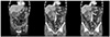
Three years after the last discharge, the patient was admitted again because of complaints of right upper quadrant pain and chills. His laboratory test results were as follows: white blood cell count, 13,640/mm3; hemoglobin level, 13.2 g/dL; platelet count, 246,000/mm3; CRP level, 23.10 mg/dL; AST value, 35 U/L; ALT value, 51 U/L; total bilirubin level, 1.62 mg/dL; direct bilirubin level, 1.28 mg/dL; and CA 19-9, 187.64. The abdominal CT scans showed a perforated GB and abscesses in the lesser sac and ligament teres, which indicated acute perforated cholecystitis and also showed a diffusely thickened CBD and intrahepatic duct dilatation with a stricture (Fig. 1). The ERCP cholangiogram showed that the upstream bile duct was dilated with focal segmental narrowing at the proximal CBD. A plastic biliary stent (double pigtail typed C-flex, 7 Fr×10 cm) was placed into the upstream duct and PTGBD catheter. An endobiliary biopsy revealed chronic inflammation. The endoscopic retrograde biliary drainage could be removed after several days of antibiotic treatment, but the PTGBD catheter could not be removed.
Two weeks after the last discharge, he was admitted to the hospital for the third time owing to fever and nausea. His laboratory results revealed the following: white blood cell count, 10,390/mm3; hemoglobin, 11.3/mm3; platelet count, 202,000/mm3; erythrocyte sedimentation rate, 59 mm/h; CRP level, 1.05 mg/dL; AST value, 599 U/L; ALT value, 323 U/L; ALP value, 541 U/L; GGT value, 1249 U/L; total bilirubin level, 2.81 mg/dL; and direct bilirubin level, 2.46 mg/dL. An abdominal CT scan showed that the intrahepatic and extrahepatic ducts were markedly dilated. Percutaneous transhepatic biliary drainage (PTBD) was performed at the distal CBD, which had been occluded completely. An endoscopic ultrasound scan showed that the biliary duct was dilated and that there was an internal echogenic lesion in the distal CBD. The PTBD cholangiogram showed that the biliary duct was dilated with a focal segmental narrowing at the proximal CBD. A histology examination after the endobiliary biopsy confirmed the presence of atypical epithelial cells, which were indicative of adenocarcinoma.
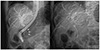
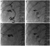
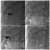
Tumor removal was attempted but was unsuccessful because it was challenging to dissect the tissue owing to its adhesion beginning at the liver hilum and extending up to the GB. In addition, multiple metastatic nodules were already present in the small bowel mesentery, anterior abdominal wall, and pelvic cavity. These abdominal wall and small bowel mesentery nodules were diagnosed as adenocarcinoma after analyzing the frozen biopsy tissue samples. Consequently, a decision was made to perform endobiliary RFA for such an unresectable CBD cancer. RFA was performed under seven watts using an 22 mm RFA catheter (ELRA® RF catheter, STARmed, Goyang, Korea) for 2 minutes. The partially covered biliary stent was placed against the narrowed CBD after performing endobiliary RFA (Fig. 2). Subsequently, the PTBD catheter was removed (Fig. 3A, B). After performing two cycles of chemical ablation of the GB using 5 mL of pure ethanol at one-week intervals (Fig. 4), the PTGBD catheter was also removed (Fig. 3C, D). Since then, he did not have any complications, such as jaundice or cholecystitis, for 3 months.
DISCUSSION
In Western countries, the most common cause of biliary stenosis is malignant diseases, such as pancreatic head cancer, extrahepatic cholangiocarcinoma, GB cancer, malignant hilar lymphadenopathy, and hepatocellular carcinoma.3 Because many of these cancers are often inoperable at the time of diagnosis, palliative care, particularly with regard to the resolution of a biliary obstruction, is of primary importance. Continual biliary decompression is achieved through biliary stenting; for this purpose, maintaining the patency of the stent becomes a key task. Recent studies suggested that performing RFA prior to biliary stenting yields better results in terms of stent patency. Endobiliary RFA is believed to induce locoregional tumor control by inducing tumor necrosis and transferring the thermal energy through the active electrode of a probe tip.
According to a recently published randomized trial by Yang et al.4 involving 19 patients with a Klatskin tumor and 46 with a distal cholangiocarcinoma, 32 patients were treated with RFA combined with plastic stent placement and 33 patients were treated only with plastic stent placement. The occurrence of side effects were similar between the two groups, whereas stent patency was significantly higher in the RFA and subsequent biliary stent inserted group compared to the only biliary stent inserted group; the results were 6.8 months and 3.4 months, respectively (p=0.02). Moreover, the mean overall survival period was also significantly longer in the RFA group (13.2 vs. 8.3 months, p<0.001). Sharaiha et al.5 reported favorable results regarding the bile duct diameter and survival rate in the RFA combined with self-expandable metal stent group compared to the results of the self-expandable metal stent only group. The study included 66 patients with a biliary obstruction complicated by cholangiocarcinoma and pancreatic cancer, and showed that there was no significant difference in adverse effects. According to a meta-analysis of reviewing nine investigations, RFA showed substantial prolongation of the stent patency and survival among patients with a biliary stricture caused by a malignancy. Regarding the adverse events, the frequency of abdominal pain was significantly higher in the RFA group; the frequency of other adverse events did not show notable differences between the two groups.6
The side effects after biliary stent placement include cholangitis, cholecystitis, and pancreatitis as well as stent obstruction. Recently, the frequency of such side effects has also increased because there has been an increase in the use of stents during biliary obstruction. Isayama et al.7 reported that cholecystitis occurred at a rate of 5.3% (13 out of 246 patients) after the insertion of a covered or an uncovered metallic stent. In addition, as in the case of this study, an obstruction of the cystic duct owing to a malignancy is one of the risk factors for cholecystitis occurring after biliary stent placement. Suk et al.8 reported nine cystic duct malignant involvements among a total of 15 patients with cholecystitis, who underwent the placement of a metal stent owing to a malignant biliary obstruction. In this case, the patient had no scope for undergoing cholecystectomy because of the cystic duct invasion of cancer. Cholecystostomy is also a safe option for cholecystectomy. On the other hand, GB ablation was performed because the PTGBD could not be removed in the present case. Chemical ablation of the GB could be an alternative option for patients in whom cholecystectomy cannot be performed. Many studies have been conducted on the selection of a sclerosant. Lee et al.9 reported successful treatment with GB sclerosis using pure ethanol in a case of a patient with a suspicious cystic ductal invasion of cholangiocarcinoma. Xu et al.10 performed the chemical ablation of GB in 34 patients who underwent minicholecystectomy for the treatment of cholelithiasis and/or acute cholecystitis. As a side effect of the chemical ablation of GB, five out of 34 patients developed a mucocele 2 to 5 years after the procedure.
RFA can be beneficial to the stent patency and survival and does not cause a significant increase in side effects in cases of biliary obstruction caused by malignancy. Accordingly, endoscopic RFA was performed before placing the biliary stent in the present case of an inoperable CBD cancer complicated by a biliary obstruction. Therefore, chemical ablation of GB was performed using pure ethanol for removing PTBD and because the patient had a prior history of acute cholecystitis with a GB rupture. This paper reports this case, where palliative treatment for biliary obstruction was achieved successfully. The PTGBD catheter was removed successfully through ethanol ablation of the GB with no significant side effects for 3 months. Consequently, PTGBD removal was performed, which brought about an improvement in the patient's quality of life.
 XML Download