

 PDF
PDF ePub
ePub Citation
 Print
Print


Introduction
The two canine adenovirus serotypes, CAV-1 and CAV-2, are distinguished by neutralization using canine antisera and via molecular analysis [1]. CAV-1 targets the digestive tract and causes infectious hepatitis; CAV-2 infects respiratory tissues and induces infectious laryngotracheitis in dogs, raccoon dogs, foxes, and wolves [2]. CAV-2, a member of the genus Mastadenovirus of the family Adenoviridae, is a non-enveloped icosahedral virus that replicates in the nucleus [3]. The CAV-2 genome is double-stranded DNA 35 kb in size with approximately 30 open reading frames; the virus is 70–90 nm in dimensions. The three major structural proteins form the hexon, the penton base, and the fiber [3]. The hexon (a major component of the viral capsid) is key in terms of inducing an immune response. The penton base stabilizes the capsid by interacting with the hexon, the capsomeres, and hexon-associated protein IIIa. The fiber featuring a tail, a shaft, and a knob interacts with the coxsackie adenovirus receptor (CAR); variable sequences on the fiber knob modulate hemagglutination with red cells of various species [4]. CAV infects a wide range of animals including dogs worldwide [56]. In Korea, the first CAV-1 infection was reported in a Eurasian rover otter in 2007, and the second in a fennec fox in 2014 [78]. The first CAV-2 infections were reported in stray dogs in 2010 [9]. The Korean CAV-2 isolate was obtained from a naturally infected dog in 2018 and its biological properties characterized [10]. Typical symptoms of CAV-2 infection in dogs include a cough, fever, a runny nose, and red watery eyes. Dogs with strong defense mechanisms exhibit relatively mild respiratory symptoms. However, dogs infected with bacteria such as Bordetella bronchiseptica, or that are immunocompromised, may die. In Korea, combination canine vaccines including CAV-1 and -2 antigens have been used to prevent dog infections since 1987. Although CAV-infected animals typically show moderate clinical symptoms, the prevalence of such infections among Korean animals is unclear. We previously performed a sero-surveillance of CAV-2 status in several animal species [11]. Although live attenuated CAV vaccines have been given to Korean dogs, they are sourced from the United States or Canada. A vaccine based on the Korean CAV-2 strain would be preferable. Recently, we isolated a novel CAV-2 strain termed APQA1701 from a naturally infected Korean dog and sequentially passaged it 40 times in MDCK cell culture. The complete sequence showed that APQA1701 was a CAV-2 strain. Various adjuvants added to inactivated, subunit, and DNA vaccines enhanced dendritic cell activation and thus induced strong immune responses [12]. New adjuvants such as Carbopol and the IMS gel are safe and efficient [1314]. Here, we prepare an inactivated CAV-2 vaccine and assess its safety and immunogenicity in guinea pigs and dogs.
Materials and Methods
Virus propagation
A Korean CAV-2 strain, APQA1701, isolated from a naturally infected dog, was passaged 40 times in Madin and Darby canine kidney (MDCK) cells (ATCC CCL-43; ATCC, Manassas, VA, USA); termed APQA1701-40P; and used as a vaccine. In brief, confluent monolayers of MDCK cells were washed with phosphate-buffered saline (PBS) and inoculated with APQA1701 suspended in Dulbecco's modified Eagle's medium (DMEM; Invitrogen, Carlsbad, CA, USA) with 5 mM urea (to induce mutations). After 1 hour at 37℃ under 5% (volume per volume, v/v) CO2, the culture medium was replaced to remove non-infectious viruses. The APQA1701-infected cells were maintained at 37℃ under 5% (v/v) CO2 and cytopathic effects (CPEs) evaluated daily under a microscope. When over 90% of cells evidenced CPEs, the flask was subjected to three freeze/thaw cycles and the clarified virus-containing fluid collected by centrifugation at 1,000×g for 15 minutes. The virus was serially passaged in six-well plates using the decimal limit dilution method. Viruses from each passage were stored at 70℃. To assess growth of the APQA1701 parent, APQA1701-40P, and APQA1601, MDCK and Vero cells (ATCC CCL1586; ATCC) grown in 25-cm2 flasks were inoculated with viruses at 50% tissue culture infectious doses (TCID50 values)/mL and harvested daily to 4 days post-inoculation. After several freeze-thaw cycles, 10-fold serial dilutions were titrated in 96-well microplates. Titers were determined using the method of Reed and Muench and expressed as TCID50 units/mL.
Next-generation sequencing
Viral DNAs were extracted from 200-µL aliquots of APQA1701 and APQA1701-40P suspensions using an AccuPrep DNA extraction kit (Bioneer, Daejeon, Korea) according to the manufacturer's instructions. The complete genomes of the two viruses were sequenced externally (Macrogen Inc., Seoul, Korea) using next-generation technology. Nucleotide sequence alignments were generated using Clone Manager ver. 10.0, basic edition (Sci-Ed Software, Denver, CO, USA).
Vaccine preparation
The titers of APQA1701-40P propagated in MDCK cells were measured using the method of Reed and Muench; a suspension of >107.5 TCID50/mL was inactivated with 0.05% (v/v) formaldehyde at 37℃ overnight. Inactivation was verified by the absence of viral growth on an MDCK monolayer over 5 days. The two adjuvants tested were the Montanid Gel (also termed IMS gel; SEPPIC, Paris, France) and Cabopol 974 PNF (Lubrizol, Wickliffe, OH, USA). The vaccine containing IMS gel was prepared by adding the adjuvant to a suspension of inactivated virus to a final concentration of 10% (v/v), followed by blending at room temperature for 30 minutes. Cabopol 974 PNF powder (2 g) was added to 94 mL distilled water, blended for 30 minutes, and the temperature then raised to 70℃ to melt the adjuvant. NaOH (6 mL of a 1 N solution) was then added to form a gel that was sterilized for 15 minutes at 121℃ (Cabopol concentration, 20 mg/mL). This Cabopol gel was added to a suspension of inactivated virus at a volume ratio of 9:1, followed by blending at room temperature for 30 minutes. Both vaccines were aseptically transferred to sterile vaccine vials and stored at 4℃.
Polymerase chain reaction
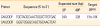
DNAs were extracted from APQA1701 strains propagated in MDCK cells, using a DNA Extraction Kit (Bioneer, Daejeon, Korea) according to the manufacturer's instructions; the DNAs were eluted into 50-µL of the elution buffer supplied with the kit. Portions of the E1b-19K genes were polymerase chain reaction (PCR)-amplified using specific primers (Table 1). Each reaction (in AccuPure PCR premix; Bioneer) contained 10 µL denatured DNA, 1 µL of each primer (10 pmol/µL), and 38 µL of distilled water in a final volume of 50 µL. Denaturation at 95℃ for 5 minutes was followed by 35 cycles of denaturation at 95℃ for 30 seconds, annealing at 55℃ for 30 seconds, and extension at 72℃ for 1 minute, with a final extension at 72℃ for 5 minutes. The products were visualized via 2.0% (weight per volume, w/v) agarose gel electrophoresis and application of Redsafe Nucleic Acid-Staining Solution (iNtRON, Seongnam, Korea).
Animal experiments
All animal experiments were performed in the Animal and Plant Quarantine Agency of Korea using protocols approved by the experimental animal ethics committee (approval no., 2019-160). Eight-week old guinea pigs were divided into three groups. Group 1 (five) was inoculated with inactivated CAV-2 vaccine containing IMS gel adjuvant; group 2 (five) with vaccine containing Cabopol adjuvant, and group 3 (two) was not vaccinated (control group). Vaccines (in 1 mL) were given intramuscularly, twice, with a 2-week interval. At 0, 2, 4, and 11 weeks post-vaccination (WPV), blood was collected from all guinea pigs.
Five 4-month-old dogs seronegative for CAV-2 were divided into two groups. Group 1 (three dogs) was subcutaneously immunized with one dose (2 mL) of inactivated CAV-2 vaccine with the Cabopol adjuvant, twice, with a 2-week interval. Group 2 (two) did not receive the vaccine (control group). Blood for immunoserological analysis was collected at 0, 2, and 4 WPV. The serum antibody titers were measured using the virus neutralization (VN) test and indirect enzyme-linked immunosorbent assay (I-ELISA).
Virus neutralization test and indirect enzyme-linked immunosorbent assay
The virus neutralizing antibody (VNA) titers in serum samples were determined using the VN test employing 96-well microplates. Briefly, serum samples collected before and after vaccination were subjected to successive two-fold dilutions (1:2 to 1:2,048) in DMEM and mixed with equal volumes of a virus suspension containing approximately 100 TCID50/0.1 mL of APQA1701-40P or the Korean CAV1V vaccine strain [15]. After 1-hour incubation at 37℃, 100 µL of an MDCK cell suspension (4×10⁵ cells/mL) was added to each well and the microplates incubated for 5 days at 37℃ under 5% (v/v) CO2. The CAV-1 and CAV-2 VNA titers were the reciprocals of the highest serum dilutions inhibiting CAV-specific CPEs.
The levels of CAV-2-specific immunoglobulin G (IgG) antibodies were measured via I-ELISA in serum samples. For I-ELISA, 100-µL serum samples diluted 100-fold in 5% (w/v) skim milk solution were added to a 96-well microplate (Maxisorp; NUNC, Roskilde, Denmark) coated with CAV-2 (APQA1701-40P) antigen. Following incubation at 37℃ for 1 hour, the plates were washed with PBS with 0.05% (v/v) Tween-20 and incubated with 100 µL/well anti-dog or anti-guinea pig horseradish peroxidase-conjugated IgG (KPL, Gaithersburg, MD, USA) for 1 hour at 37℃. After washing, 2'2-azino-di-3-ethylbenzthiazoline substrate solution and a stop solution (1.0% [w/v] sodium dodecyl sulfate) were added to all wells, in line with the manufacturer's instructions. Absorbances were measured at 450 nm using an ELISA plate reader (Sunrise; Tecan, Mannedorf, Switzerland).
Results
Molecular characterization of the APQA1701-strain
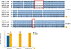
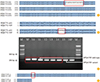
The complete genome sequences of the APQA1701 parent and APQA1701-40P were compared to those of the APQA1601 and Toronto-A26/61 strains. The APQA1701 genome contained 31,071 nucleotides with defects in genes encoding two early region (E1A) proteins and the precursor terminal protein (Fig. 1A, B). APQA1701 proliferated well in MDCK cells, but not in Vero cells (Fig. 1C). To explore whether APQA1701-40P had suffered a deletion after sequential passage, the complete genome sequence of the APQA1701-40P strain was compared to that of APQA1701. APQA1701-40P was >99% homologous with APQA1701. However, 18 nucleotides of the E1b-19K gene (encoding a non-structural protein) were lacking in the sequentially passaged strain (Fig. 2A). APQA1701-40P exhibited a six-amino acid deletion of the E1b-19K protein (amino acids 122–127) (Fig. 2B). Viral DNAs isolated after each passage were PCR-amplified using E1b-19K-specific primers; the deletion was evident at passage 11 and then persisted. Viruses of passages 11–13 contained both parental and mutant viruses. PCR showed that the CAV-2 mutant alone existed after 14 passages (Fig. 2C). Also, alanine 21 of the E312.5K gene was replaced by glutamine in APQA1701-40P (Fig. 2D).
Safety and immunogenicity of canine adenovirus type 2 vaccines in animals
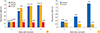
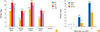
No vaccinated guinea pig died. The humoral antibody responses were assessed via I-ELISA and VN testing. I-ELISA revealed that both vaccines induced significant IgG antibody responses to CAV-2 in guinea pigs. The mean IgG levels of groups 1 and 2 increased to 11 weeks after vaccination (Fig. 3A). The IgG antibody levels in guinea pigs given either vaccine did not differ greatly; the Cabopol-adjuvanted vaccine induced a slightly better (1.03-fold) IgG antibody response 2 weeks after the first inoculation, compared to the IMS gel-adjuvanted vaccine; controls lacked any response. We thus tested the Cabopol-adjuvanted vaccine in dogs. No dog developed any illness to 4 weeks; all developed elevated (1.44 absorbance value) levels of anti-CAV2 IgG antibody 4 weeks after the second vaccination (Fig. 3B); the vaccine thus induced a significant response. The geometric mean (GM) VNA titers gradually attained 2,352.5 (11.2 log2) to 2,702.4 (11.4 log2) by 4 weeks in vaccinated guinea pigs, but fell slightly to 955.4 (9.9 log2) by 11 weeks in both groups (Fig. 4A). The maximum VNA titers were attained in both vaccinated groups by 4 WPV. Although the differences were not significant (p>0.05), group 2 given the Cabopol-adjuvanted vaccine developed a slightly higher GM VNA titer than did the group given the IMS gel-adjuvanted vaccine at 2, 4, and 11 weeks, reflected in the levels of CAV-2 specific IgG antibodies. Based on the VNA titers in guinea pigs, we tested the Cabopol-adjuvanted vaccine in dogs; the animals exhibited a mean GM VNA titer of 315.2 (8.3 log2) at 4 weeks (Fig. 4B), significantly higher than that of the control group (p<0.05). We explored cross-VNA titers. The differences in the GM VNA titers (CAV-1 and CAV-2) ranged from 41.06 (5.36 log2) to 519.15 (9.02 log2) in guinea pigs and dogs inoculated with the CAV-2 vaccine (Table 2). The VNA titers did not rise in unvaccinated animals (Fig. 4A, B).
Discussion
A CAV-1 based vaccine was introduced in Korea in the 1980s; since 2000, a steady decline in the number of CAV-infected dogs is evident [11]. Recently, CAV-2 infections have been reported in both domestic dogs and wild animals [910], suggesting that a new CAV-2 vaccine is required. Mutant canine adenoviruses feature multiple repeats in the inverted terminal repeat [16]; one such CAV-2 virus exhibited a duplicated E1A region [17]. Repeat CAV passage under stressful culture conditions may lead to the emergence of new mutants [17]. APQA1701-40P exhibited a new deletion in the E1b-19K gene; the gene product suppresses apoptosis induced by tumor necrosis factor and DNA-damaging agents such as ultraviolet irradiation during viral infection, and mitigates the cytotoxicity of the E1A protein [181920]. We passaged APQP1701 in MDCK cells in medium containing 5 mM urea, triggering mutations including the APQA1701-40P six-amino deletion in E1b-19K; this mutation may critically distinguish parent or field CAV-2 strains from the mutant; the mutation is a ‘marker’ of the vaccine.
Both Cabopol and the IMS gel are safe and effective adjuvants [131421]. Selection of an optimal adjuvant is essential when seeking to enhance the immune response to vaccination. We compared the immunogenicities of CAV-2 vaccines featuring adjuvants developed by SEPPIC (France) and Lubrizol (USA). The Cabopol-adjuvanted vaccine afforded slightly higher specific IgG antibody and VNA titers in guinea pigs, as was also the case for the PRRS MLV and atrophic rhinitis vaccines; Cabopol induces rapid differentiation of IFN-r-producing cells (NK and CD4+ T cells) and also drives T cell differentiation [1321]. The Korean veterinary standard states that a CAV vaccine should induce a VNA titer of at least 120 in dogs. Our Cabopol-adjuvanted vaccine induced a GM VNA titer of 315.2 (8.3 log2) at 4 WPV, and a CAV-1 VNA titer of 32 (5 log2); CAV-1/-2 cross-neutralization was in play. The VNA titer of 315.2 was identical to that in dogs given a live CAV-2 vaccine protective against challenge with virulent CAV-2 [22]. Thus, our novel, inactivated CAV-2 vaccine should be protective in dogs.
In summary, we created a new inactivated CAV-2 vaccine, APQA1701-40P, featuring a six-amino acid deletion in the E1b-19K protein; the strain was isolated after 40 consecutive passages in MDCK cells. The inactivated CAV-2 vaccine given with the Cabopol adjuvant was safe and slightly more immunogenic in guinea pigs than was the same vaccine with the IMS gel. The vaccine was safe and probably protective in dogs; we did not challenge dogs with virulent virus. More studies are required.

 XML Download