

 PDF
PDF Citation
 Print
Print


INTRODUCTION
Stroke is a major cause of serious long-term disability in adults, and post-stroke motor impairment is the primary cause of disability [1]. Hence, stroke burden is likely to increase due to its disabling impact. However, effective therapies for patients with stroke have been limited especially in upper limb motor impairment.
Repetitive transcranial magnetic stimulation (rTMS) has been known as an effective therapy for motor impairment by modulating cortical excitability and enhancing neural plasticity [2].
Previous research has shown that rTMS can improve the motor function in patients with acute and chronic stroke [345678]. However, these studies only investigated patients with mild or moderate motor deficits who were able to move fingers individually [3], tap the fingers [578], and grip the hands [6] or those with Motricity index range 72–76 [4]. It is still unclear whether rTMS can be an effective treatment for post-stroke patients with severe upper-limb motor impairment. Boggio et al. [9] reported only one patient with stroke and severe motor deficits who had improved motor function after low-frequency rTMS. Moreover, it remains unclear which stimulation method is more effective either high-frequency stimulation on lesional primary motor cortex (M1) or low-frequency stimulation on contralesional M1 for patients with stroke. Khedr et al. [5] have suggested that low-frequency rTMS was more effective than high-frequency rTMS. In contrast, Sasaki et al. [10] observed that high-frequency rTMS was more effective than low-frequency rTMS in improving the hand grip and finger tapping frequency of the affected hand in patients with early-stage stroke.
This study aimed to investigate whether rTMS improved the motor function in post-stroke hemiplegic patients with severe upper-limb motor impairment and to compare the effect of high-frequency stimulation on lesional M1 and low-frequency stimulation on contralesional M1.
MATERIALS AND METHODS
Subjects
Patients were included based on the following criteria: 1) first-ever stroke confirmed by magnetic resonance imaging or computed tomography, 2) the location of the stroke lesion is within the supratentorial area, 3) severely impaired hemiplegic patients who scored below 25 in the upper extremity motor scale of the Fugl-Meyer Assessment (FMA) [11], and 4) ability to maintain a sitting posture and proper cognitive function to cooperate during the procedure. Patients were excluded if they had 1) a seizure history or ictal waveform on electroencephalogram, 2) an intracranial metallic implantation, 3) a cardiac pacemaker, 4) impaired upper or lower extremity motor function before a stroke attack, 5) evidence of peripheral neuropathy by diabetic mellitus or focal peripheral neural injury, and 6) coexisting neurological and psychiatric diseases. Sixty patients who were admitted in the department of rehabilitation medicine in our hospital were enrolled in the study. However, 2 patients in low-frequency stimulation group (premature discharge: 1, transferred to another department: 1) were excluded. Thus, a total of 58 post-stroke patients with severe upper-limb motor impairment (38 men, 20 women) with a mean age of 62.9 years were analyzed in our study. Fifty patients had infarction while 8 patients had hemorrhage. The duration from stroke onset was 8.2 ± 5.9 weeks. The patients' characteristics are summarized in Table 1. No statistically significant differences were observed in the age, sex, duration from stroke onset, and other characteristics among the 3 groups.
Table 1
Baseline characteristics of subjects

Values are mean ± standard deviations. Duration from stroke onset to experimental day (week).
M, male; F, female; C, cortical; S, subcortical; R, right; L, left.
![]()
The study was approved by the local ethics committee. Informed consent was obtained from each patient prior to participation in the study.
Study design
This was a single-blind randomized controlled study. The subjects were randomly assigned into 3 groups: patients with 1 Hz rTMS (low-frequency stimulation group), those with 20 Hz rTMS (high-frequency stimulation group), and those with sham stimulation (control group). All groups received 10 stimulation sessions 5 days per week for 2 weeks. In the low-frequency rTMS group, 1 Hz rTMS was applied on the contralesional M1. In the high-frequency rTMS group, 20 Hz rTMS was applied on the lesional M1. The control group received sham stimulation on the primary motor hotspot of the contralesional hemisphere of the cerebral cortex. The FMA of the involved side and cortical excitability on the unaffected hemisphere were measured before and after the rTMS in all groups. Clinical assessment was conducted by an experienced physician blinded to patients' group allocation and rTMS was performed by another investigator who was not involved in clinical assessment, patient follow up, or data analysis.
rTMS
The subjects were comfortably seated in the chair with both hands supinated on the pillow. TMS of the motor cortex was performed with a figure-of-eight coil connected to a Magstim rapid stimulator (Magstim Co., Wales, UK). Prior to the rTMS intervention, the motor hotspot of the abductor digiti minimi (ADM) muscle was evaluated according to the recommendations of the International Federation of Clinical Neurophysiology [12]. The electromyography (EMG) was recorded from the ADM muscle of the hand via silver-silver chloride surface electrodes using a muscle belly-tendon set-up via a Synergy EMG system (Medelec Co., Oxford, UK). The hotspot was defined as the location where stimulation of a slightly suprathreshold intensity elicited the largest motor evoked potential (MEP) in the ADM muscle. If no lesional MEP response was observed, the hotspot was obtained on the contralesional hemisphere. Once motor hotspot was identified, the resting motor threshold (RMT) was evaluated. RMT was defined as the lowest stimulus intensity to elicit MEPs ≥50 uV peak-to-peak amplitude in 5 of 10 consecutive trials in a relaxed muscle [3]. The EMG activity was amplified in every rTMS trial. Each motor hotspot and RMT were reevaluated just before the stimulation; then, rTMS was delivered. One session of rTMS per day was performed on each subject for a total of 10 sessions for 2 weeks (5 days/week).
Patients in the low-frequency stimulation group received rTMS as follow; 1 Hz, total of 1,500 pulses and 90% RMT were applied on the motor hotspot of the ADM muscle in the contralesional hemisphere. For the high-frequency stimulation group, patients received rTMS as follows; 20 Hz, 5 seconds, intertrain interval 55 seconds, 15 trains, total 1,500 pulses and 90% RMT were applied on the motor hotspot of the ADM muscle in the lesional hemisphere. If no lesional MEP response was observed, the location for stimulation was decided to the equal point of scalp in the lesional hemisphere from the contralesional motor hotspot and 90% RMT of contralesional M1 was applied. In the sham stimulation group, the stimulation was delivered to the contra-lesional motor hotspot area with the coil held perpendicular to the scalp with 1 Hz and 1,500 pulses. All patients received the same conventional physical and occupational therapy to maintain optimal passive and active range of motion of all affected upper extremity joints for 30 minutes respectively after each rTMS session, which were conducted by well-trained therapists.
Measurement of motor function and cortical excitability
The FMA was used to assess the changes in motor function on the involved side. It is a well-known assessment tool to measure post-stroke physical impairments related to functional recovery. Furthermore, it is a reliable and valid measurement tool to assess the ability to isolate movements at each joint and the influence of unwanted synergies on movement [11]. Of its 5 domains (motor, sensory, balance, range of motion, joint pain), the motor scale of upper extremity FMA (UE-FMA) and lower extremity FMA (LE-FMA) are used to measure voluntary movement and include 33 items (score range, 0–66) and 17 items (score range, 0–34), respectively. Additionally, motor scale of UE-FMA has been subscaled into shoulder/elbow/forearm (score range, 0–36), wrist (score range, 0–10), and hand (score range, 0–14).
The MEP on the contralesional hemisphere was measured by single pulse TMS with 120% RMT on the primary motor hotspot of hand ADM area. Ten successive stimulations with more than 5 seconds of interval were performed, and MEP was calculated as the average of peak-to-peak amplitude of collected response. To assess whether the rTMS treatments cause changes in intrinsic cortical excitability, intercortical inhibition (ICI) and intercortical facilitation (ICF) were evaluated at baseline and after 10 sessions of rTMS treatment. Additionally, the ICI and ICF were examined in the fully relaxed ADM muscle using a paired conditioning-test pulse paradigm [13]. The intensity of the test stimulus was 120% RMT and that of the conditioned stimulus was 80% RMT. The interstimulus interval (ISI) were 3 ms and 5 ms for ICI and 10 ms and 15 ms for ICF. For each ISI, 5 trials were performed and the average value was calculated and divided by the mean MEP amplitude. Therefore, the amplitudes of the ICI and ICF were expressed as a ratio of the amplitude. All measurements were performed before and after 10 stimulation sessions by the examiner who was blinded to the stimulation methods used.
Statistical analysis
Statistical analysis was performed using the SPSS 20.0 software (SPSS Inc., Chicago, IL, USA). Sex, laterality, and location of the lesion among the different groups were compared using the χ2 test. Meanwhile, age and duration from stroke onset to experimental day were compared using a one-way analysis of variance (ANOVA) at baseline. Repeated measures ANOVA was used to evaluate the effects of TIME before, after and GROUP low, high, control to compare the FMA and cortical excitability among the 3 groups. Comparison of the changes of cortical excitability and FMA among the 3 groups were performed using a one-way ANOVA. A post hoc analysis was performed with Bonferroni's correction. The level of significance was set at p < 0.05. All data are expressed as mean ± standard deviations.
RESULTS
Motor function
No significant difference was noted in the UE-FMA measured at baseline among the 3 groups. Repeated measures ANOVA revealed a significant interaction between TIME before, after and GROUP low, high, control on the total UE-FMA (F = 8.36; p = 0.001). On the contrary, no significant difference was found in the total LE-FMA (F = 2.58; p = 0.085). UE-FMA significantly improved in both high- and low-frequency rTMS groups following the intervention (p < 0.05), but not in the control group (Fig. 1A). Changes in the UE-FMA after stimulation were significantly more improved in both low- and high-frequency rTMS groups compared to the control group (p < 0.05). However, no significant difference was noted between low- and high-frequency rTMS groups (p > 0.05, Table 2).
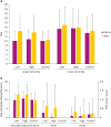 | Fig. 1Comparison of changes of FMA after stimulation according to stimulation methods. (A) UE-FMA scores were improved by low and high frequency rTMS. Repeated measures ANOVA showed a significant TIME × GROUP factor interaction (p < 0.05). (B) UE-FMA scores of shoulder/elbow/forearm were improved by low and high frequency rTMS. ANOVA showed a significant TIME × GROUP factor interaction (p < 0.05).FMA, Fugl-Meyer Assessment; UE-FMA, upper extremity Fugl-Meyer Assessment; rTMS, repetitive transcranial magnetic stimulation; ANOVA, analysis of variance; Low, low frequency group; High, high frequency group; Control, control group; Before, before stimulation; After, After stimulation.
*Significant at p < 0.05 vs. before stimulation.
|
Table 2
Comparison of changes of FMA after stimulation according to stimulation methods
Values are mean ± standard deviations.
FMA, Fugl-Meyer Assessment; UE-FMA, upper extremity Fugl-Meyer Assessment; LE-FMA, lower extremity Fugl-Meyer Assessment; Before, before stimulation; After, after stimulation.
*p < 0.05 vs. before stimulation; †p < 0.05 vs. control group.
![]()
Moreover, repeated measures ANOVA revealed a significant interaction between TIME before, after and GROUP low, high, control on the shoulder/elbow/forearm subscore of the UE-FMA (F = 5.29; p = 0.008), but not on the wrist and hand subscore of the FMA (F = 1.83; p = 0.171, F = 0.82; p = 0.446). The shoulder/elbow/forearm subscale of the UE-FMA significantly increased in both high- and low-frequency rTMS groups (p < 0.05), but not in the control group (Fig. 1B). Changes after the stimulation were significantly more improved in both low- and high-frequency rTMS groups compared to the control group (p < 0.05), but we did not find any significant difference between low- and high-frequency rTMS groups (p > 0.05). Detailed information of FMA after the stimulation are summarized in Table 2.
Cortical excitability
No lesional MEP response was observed in 54 of 58 patients in this study. Of these, 16 were in low-frequency stimulation group, 19 were in high-frequency stimulation group and 19 were in control group. There were no significant differences in response of lesional MEP among the 3 groups. No significant differences were found in the amplitude of MEP and MEP with ISI (3 ms, 5 ms, 10 ms, 15 ms) measured at baseline when stimulating the contralesional hemisphere among the 3 groups. Repeated measures ANOVA revealed a significant interaction between TIME before, after and GROUP low, high, control on the amplitude of MEP (F = 6.15; p = 0.004). However, no significant difference was observed in the amplitude of MEP with ISI (3 ms, 5 ms, 10 ms, 15 ms) (F = 0.95; p = 0.392, F = 0.87; p = 0.426, F = 2.45; p = 0.096, F = 1.12; p = 0.334, respectively). The amplitude of MEP significantly decreased in both high- and low-frequency rTMS groups (p < 0.05). A significant difference was noted in the changes after the stimulation in both low- and high-frequency rTMS groups compared to the control group (p < 0.05); however, no significant difference was observed between low- and high-frequency rTMS groups (p > 0.05). Detailed information on cortical excitability after stimulation are summarized in Table 3.
Table 3
Comparison of changes of contralesional cortical excitability after stimulation according to stimulation methods

Values are mean ± standard deviations.
MEP, motor evoked potential; ISI, interstimulus interval; Before, before stimulation; After, after stimulation.
*p < 0.05 vs before stimulation; †p < 0.05 vs control group.
![]()
DISCUSSION
The findings in this study indicated that low- and high-frequency rTMS improved the upper extremity motor function, especially in the proximal upper limb, compared with the sham stimulation in post-stroke patients with severe upper-limb motor impairment. Additionally, the improvement was not statistically significant between low- and high-frequency rTMS.
These positive findings after rTMS were consistent with most randomized controlled trials documenting the effects of rTMS on the upper limb function in post-stroke patients [61415161718192021]. However, the upper limb function of their subjects was heterogeneous or mild to moderate. Khedr et al. [5] reported a significant positive correlation between pre rTMS MEP amplitude of the affected hemisphere and percent improvement in National Institutes of Health Stroke Scale and modified Rankin Scale. Carey et al. [22] reported that the responders showed a significantly greater baseline paretic hand function and preservation volume of the ipsilesional posterior limb of the internal capsule than the non-responders. Futhermore, Lee et al. [23] reported that the presence of an MEP response and high Motricity index arm score were significantly associated with a response to rTMS. Therefore, few studies have been reported on the effects of rTMS in post-stroke patients with severe upper-limb motor impairment. Boggio et al. [9] reported only one patient with stroke with a severe motor deficit whose motor function improved following low-frequency rTMS. Demirtas-Tatlidede et al. [24] reported motor improvement in post-stroke patients with severe upper-limb motor impairment after 1 Hz rTMS on the contralesional M1. However, this study was a single open-label trial. To our knowledge, this is the first randomized controlled study on post-stroke patients with severe upper-limb motor impairment.
This study revealed that rTMS was also effective even in post-stroke patients with severe upper-limb motor impairment. Interestingly, this study showed improvement of the FMA in the shoulder/elbow/forearm subscore rather than the wrist or hand subscore, although the hotspot of the ADM muscle was targeted. It is possible to explain that the rTMS induced rapid changes in the distorted cortical representation after extensive neuronal damage and improved the motor function. Although it was not clearly understood that the improvement of motor function mechanism was associated with rTMS, it has been thought that rTMS modulates cortical excitability [25]. After stroke, the abnormal interhemispheric inhibition is increased and may impede motor function [42126]. Modulation of cortical excitability using rTMS would result in a decrease in the transcallosal inhibition to the affected hemisphere and improve motor function [421]. Animal studies showed that one strategy for recovery after stroke is for the surrounding surviving cortex to regain control over the function of the damaged motor area through somatotopic reorganization, a phenomenon called vicariation [272829]. However, when extensive neuronal damage has occurred, there may not be enough surviving cortical neurons for representation in the perilesional area [30]. Extensive neuronal damage induces reorganization of perilesional and secondary motor area in functional recovery after stroke [30]. Nudo et al. [31] demonstrated that focal neuroplastic changes occurred following intracortical stimulation to the motor region representing a rat forelimb. This kind of reorganization, placed in the motor region, modifies the motor cortical representational map, and this process can be induced with rTMS [32]. Another hypothesis is that rTMS stimulate the extrapyramidal tract as well as pyramidal tract. Extrapyramidal system has been known to influence the proximal (midline) more than the distal (peripheral) muscles and plays a key role in maintaining proper tone and posture. Zappulla et al. [33] reported the components of the spinal MEP in rats arise from pathways outside the motor cortex, such as the subcortical and brain stem structures, reflecting extrapyramidal system function. Kamida et al. [34] also reported that magnetic MEPs recorded from the limb muscles mainly reflect extrapyramidal activities. Therefore, proximal muscles such as shoulders and forearms may have shown a greater degree of recovery than the distal counterparts.
The excitability of the contralesional motor cortex shows physiological plasticity during functional recovery after stroke, and hyperactivity in the contralesional motor cortex occurs early after stroke [8]. Decreased contralesional M1 activity is correlated with functional recovery [35]. Cortical activity is well reflected by the amplitude of the MEP. Low-frequency rTMS produces a decreasing cortical activity, whereas high-frequency rTMS induces an increasing cortical excitability. This study showed that the amplitude of the MEP in the contralesional hemisphere significantly decreased after both contralesional low-frequency and lesional high-frequency rTMS. Matsuura et al. [35] reported that the reduction of contralesional M1 hyperactivity was related to the functional recovery after stroke. Veldema et al. [36] demonstrated that the greater the increase of ipsilesional corticospinal excitability, the better hand motor recovery. It may be hypothesized that the contralesional hemisphere activity was inhibited through transcallosal inhibition by increasing lesional cortical excitability by high-frequency stimulation on the affected M1 and directly by decreasing contralesional cortical excitability by low-frequency stimulation on the unaffected M1. Nonetheless, it remains unclear what mechanism has reduced the amplitude of the contralesional MEP in lesional high-frequency rTMS and contralesional low-frequency rTMS in this study. Future study on this matter will be necessary.
It is still controversial which stimulation method is more effective either high-frequency stimulation on lesional M1 or low-frequency stimulation on contralesional M1 in mild to moderate stroke [53738]. Khedr et al. [5] previously revealed that 1 Hz on the contralesional hemisphere showed better improvement (hand grip, pegboard task, and keyboard tapping) than 3 Hz on the lesional hemisphere. Emara et al. [37] reported the similarity of 1 Hz and 5 Hz rTMS in improving the finger tapping test. In this study, both low-frequency rTMS on the contralesional hemisphere and high-frequency rTMS on the lesional hemisphere showed no significant difference in improving the motor function. This study was only a clinical trial comparing both stimulation methods in post-stroke patients with severe upper-limb motor impairment and was not designed to explain the possible mechanism for these findings. Thus, several further clinical studies with a study design to reveal the mechanism in post-stroke patients with severe upper-limb motor impairment will be also necessary.
This study had several limitations. First, subjects with a relatively broad duration from stroke onset were enrolled from 1 week to 21 weeks. Second, the reliability of ICI and ICF measurements may be lower than expected because it was obtained by repeating 5 times, lower than minimum number of pulses needed to achieve reliable ICI, ICF suggested by Chang et al. [39]. Third, we could not reveal if the effect is sustained for a long time because of the short follow-up duration. Therefore, we hope to address this issue in future studies. Moreover, the study was not designed to explain the possible mechanisms, but to reveal the clinical outcomes. Therefore, future large-scaled well-designed randomized controlled clinical study and basic research will be needed.
In conclusion, this study showed that rTMS may be helpful in improving the upper extremity motor function even in post-stroke patients with severe upper-limb motor impairment and that the effects between contralesional low-frequency and lesional high-frequency stimulation may be similar.
 XML Download