

 PDF
PDF ePub
ePub Citation
 Print
Print


Clavicle fractures represent 2.6% of all fractures, and most clavicle fractures occur at the middle third of the clavicle.1) In general, surgical treatment can result in complications such as nonunion, osteosynthesis device failure, pin migration, trapping of brachial plexus or subclavian vein, paraesthesia and dysmorphic scarring.2) Additionally, nonunion after surgical treatment of clavicular fractures can result in implant failure, shoulder stiffness, numbness below the scar, and infection of the donor site wound.3) There are many studies and case reports on brachial plexus injury, as it is a rare complication with potentially devastating consequences. Herein, we discuss our case of brachial plexus injury after revision of clavicular fracture nonunion, which differs from previously reported cases.
The patient was informed that data concerning the case would be submitted for publication, and he agreed.
Case Report
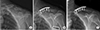
A 56-year-old, right-hand-dominant, male patient was transferred to our hospital for nonunion of mid-shaft clavicular fracture. The patient's first operation was performed at another hospital 5 months prior, where a plate and screws were implanted for repair (Fig. 1). His radiographs upon admission revealed loosening of the implant. On physical examination, he presented with left shoulder discomfort, tenderness along his fracture site of the left clavicle, and limited range of motion of the left shoulder secondary to pain. Neurovascular status was intact. When compared to radiographs obtained after the first operation, radiographs of the clavicle taken at admission demonstrated increased angulation of the fracture site with a visible fracture site gap and distal implant loosening (Fig. 1). He also had avascular necrosis of bilateral femoral heads. We performed a revisionary open reduction and internal fixation of the left clavicle with an autogenous cancellous bone graft from left femoral head after simultaneous left total hip arthroplasty. We recommended right total hip arthroplasty.
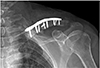
The surgery of the clavicle was performed by a shoulder specialist at our hospital. An incision was made along the clavicular axis on the previous operation scar. The previous plate and screws leaving out a lag screw were removed. Fibrous and granulated tissue of the nonunion site was excised and the ends of each fragment were prepared with curettage. Fracture reduction and fixation were achieved with a longer locking compression plate. The autogenous cancellous bone graft from the left femoral head was packed in the fracture gap after total hip arthroplasty. The wound was closed in a layered fashion after confirmation of satisfactory fracture reduction and implant position with an image intensifier (Fig. 2).
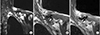
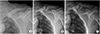
Postoperative neurovascular status was intact, and we found no abnormal findings until postoperative day 3. On postoperative day 4, the patient presented with numbness in the left hand and decreased strength. A grade 1/5 in thumb extension and flexion, wrist extension, and finger extension, flexion, and abduction was found in addition to a grade 3/5 in wrist flexion and elbow flexion and extension. A haematoma formation at the operation site was suspected because of the acute onset of symptoms on postoperative day 4. On postoperative day 5, an ultrasound was performed for evaluation of a possible haematoma, but no definite mass or haematoma was found around the clavicle. On postoperative day 6, diffusion magnetic resonance imaging (MRI) was performed to evaluate for possible acute stroke, but there was no evidence of acute infraction. Additionally, on postoperative day 6, cervical spine MRI was performed to evaluate the cervical spine. Cervical spine MRI demonstrated a fibrotic mass lesion around a small bony fragment inferior to the clavicle. This lesion was compressing the adjacent brachial plexus causing signal changes within the brachial plexus (Fig. 3). Based on the MRI findings, we removed the implant and made a superior angulation to decompress the brachial plexus. There was no haematoma intraoperatively, and we left grafted bone. We put a Velpeau sling on the patient's left shoulder after surgery (Fig. 4).
On postoperative day 1, the patient presented with improved strength of thumb flexion and finger flexion to a grade 3/5 with decreased numbness of the left hand. On postoperative day 3, wrist extension improved to a grade 2/5 and left hand numbness was nearly resolved. Electromyography at 12 days postoperatively demonstrated diffuse brachial plexopathy, especially affecting the upper trunk. The patient underwent physical and exercise therapy until discharge. At the time of discharge, on postoperative day 30, thumb extension and finger extension and abduction were graded as 3/5, and all other left hand strengths were graded as 4/5. A Velpeau sling was not applied after discharge. On radiographs 3 and 6 months postoperatively, callus bridging and consolidation were visible (Fig. 4). On examination, there was no excess motion or tenderness at the fracture site, shoulder abduction and flexion were graded as 3/5 and 4/5, respectively, and all hand and elbow functions were fully recovered.
Discussion
There are several studies and case reports related to brachial plexus injury from a clavicular fracture. Although a rare complication, brachial plexus injury represented about 1% of lesions seen over a period of 20 years.4) A literature review study of 301 cases with complications after treatment of clavicle fracture nonunion showed that 18 cases (6%) had complications related to metal work, 45 cases (15%) had complications related to soft tissues, 7 cases (2%) had complications related to scarring, and 24 cases (8%) had complications related to failure of union. Brachial plexus injury was included in the soft tissue related complications in 2 cases (0.7%).3) Most studies and case reports indicated that the main cause of brachial plexus injury after clavicular fracture was compression or entrapment by aneurysm, callus, scar tissue, or bone fragment. These studies report that symptoms of brachial plexus injury presented with a delayed onset of several weeks or even years.456) Other causes of brachial plexus injury include supraclavicular high energy traction injury and acute direct compression or stretching on the brachial plexus by fracture displacement during acute trauma.47) In these situations, the symptoms of brachial plexus injury occurred acutely.78) In the present case, the patient presented with symptoms 4 days postoperatively. This onset of symptoms led to uncertainty as to the cause of the patient's neurological deficits. The onset appeared delayed when compared to acute brachial plexus injury yet appeared premature for symptoms caused by compression or entrapment.
Open reduction-internal fixation with autogenous bone grafting is an accepted technique for the treatment of clavicle nonunion in cases with an atrophic fracture and/or shortening of the fracture site to regain the necessary clavicle length.9) In our case, we attempted this technique without shortening the nonunion site because we suspected that the correction of the superior angulation of the nonunion site would have a minor effect on length. During the second surgery, after confirming there was no haematoma or space occupying lesion causing direct compression on the brachial plexus, the implant was removed and a superior angulation was made for decompression with a goal of avoiding more violation of soft tissue and grafted bone.
We are unaware of any studies that examine clavicle lengthening during surgery as a cause of brachial plexus injury. Although we recognize that compression or entrapment is the main cause of brachial plexus injury after clavicle fracture, we hypothesize that, as seen in this unique case, a slight lengthening of the clavicle during surgery can cause neurologic deficit. One case report stated that an inferior bony spur resulted in neurologic symptoms of brachial plexus injury after nonunion surgery of the clavicle, and the neurologic complications resolved after excision of that spur.8) Our case also had a small free bone fragment (Fig. 3). This free fragment may have exacerbated compression or stretching of the brachial plexus when the fracture site was straightened during our first surgery.
Our case suggests that brachial plexus injury may be caused by stretching and compression after reduction and straightening of the nonunion site around adhesions or scar tissue. This injury may be exacerbated by a spur or bone fragment under the nonunion site. These differentials should be considered in patients with abnormal neurologic findings in the subacute phase and with a history of a normal immediate neurologic examination after surgery for clavicular fracture nonunion.
 XML Download