

 PDF
PDF Citation
 Print
Print


Abstract
Objectives
We report a case of 3-column fracture caused by low-energy trauma in a patient with Baastrup disease who complained of acute radiating pain and motor weakness in the lower limbs after 3 weeks of conservative treatment. Subsequently, posterior fusion surgery was performed.
Summary of Literature Review
Baastrup disease is characterized by enlargement and close approximation of adjacent spinous processes, and it mostly affects the L4-5 level of the spine. In patients with Baastrup disease affecting multiple levels of the lumbar spine, low-energy trauma can cause an unstable 3-column fracture with neurological compromise. Early recognition and surgical treatment prior to the emergence of a neurological deficit are required.
Materials and Methods
An 84-year-old woman presented with back pain after falling down backward and colliding with the edge of a shelf at ground level. Considering the patient's general condition and age, she was initially treated with close observation and placement of a spinal brace with serial radiographic follow-up.
Results
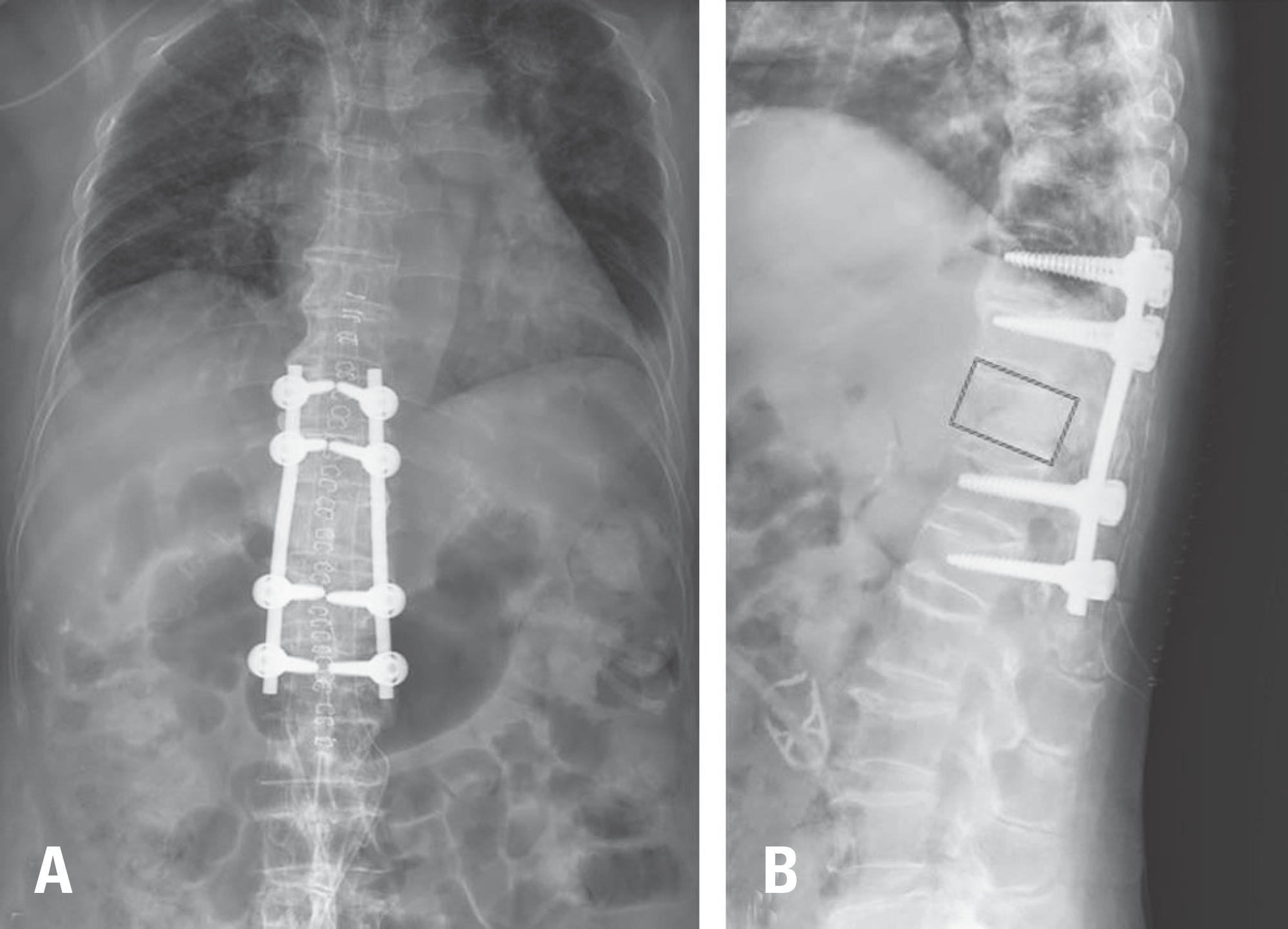
Computed tomography found 3-column fracture at the T11 level, which is quite rare in cases of minor trauma. At a 3-week follow-up, she complained of gradual lower extremity weakness, and her general lower extremity motor function decreased to grade 1–2. The patient underwent posterior fusion 2 levels above and below the affected vertebral body (T9-10-12-L1). Surgery was uneventful and the patient's motor function recovered.
Conclusions
In patients with Baastrup disease affecting multiple levels of the lumbar spine, based on our experience, low-energy trauma can cause an unstable 3-column fracture with neurological compromise. We highly recommend scrutiny of the interspinous space in elderly patients, especially those with a spinal fracture caused by low-energy trauma.
Go to : 
REFERENCES
1. Bywaters EG, Evans S. The lumbar interspinous bursae and Baastrup's syndrome. An autopsy study. Rheumatol Int. 1982; 2(2):87–96.
2. Kwong Y, Rao N, Latief K. MDCT findings in Baastrup disease: disease or normal feature of the aging spine? AJR Am J Roentgenol. 2011 May; 196(5):1156–9. DOI: 10.2214/AJR.10.5719.

3. Maes R, Morrison WB, Parker L, et al. Lumbar interspinous bursitis (Baastrup disease) in a symptomatic population: prevalence on magnetic resonance imaging. Spine (Phila Pa 1976). 2008 Apr 1; 33(7):E211–5. DOI: 10.1097/BRS.0b013e318169614a.
4. Pinto PS, Boutin RD, Resnick D. Spinous process fractures associated with Baastrup disease. Clin Imaging. 2004 May-Jun; 28(3):219–22. DOI: 10.1016/S0899-7071 (03)00156-6.
5. Alonso F, Bryant E, Iwanaga J, et al. Baastrup's Disease: A Comprehensive Review of the Extant Literature. World Neurosurg. 2017 May; 101:331–4. DOI: 10.1016/j.wneu.2017.02.004.
6. Beks JW. Kissing spines: fact or fancy? Acta Neurochir (Wien). 1989; 100(3-4):134–5.
7. Jacobson HG, Tausend ME, Shapiro JH, et al. The sway-back syndrome. Am J Roentgenol Radium Ther Nucl Med. 1958 Apr; 79(4):677–83.
8. Baastrup CI. On the Spinous Processes of the Lumbar Vertebrae and the Soft Tissues Between them, and on Pathological Changes in that Region. Acta Radiologica. 1933; 14(1):52–5. DOI: 10.3109/00016923309132353.
9. Rustagi T, Drazin D, Oner C, et al. Fractures in Spinal Ankylosing Disorders: A Narrative Review of Disease and Injury Types, Treatment Techniques, and Outcomes. J Orthop Trauma. 2017 Sep; 31(Suppl 4):S57–s74. DOI: 10.1097/bot.0000000000000953.
10. Mata S, Fortin PR, Fitzcharles MA, et al. A controlled study of diffuse idiopathic skeletal hyperostosis. Clinical features and functional status. Medicine (Baltimore). 1997 Mar; 76(2):104–17. DOI: 10.1097/00005792-199703000-00003.
Go to :
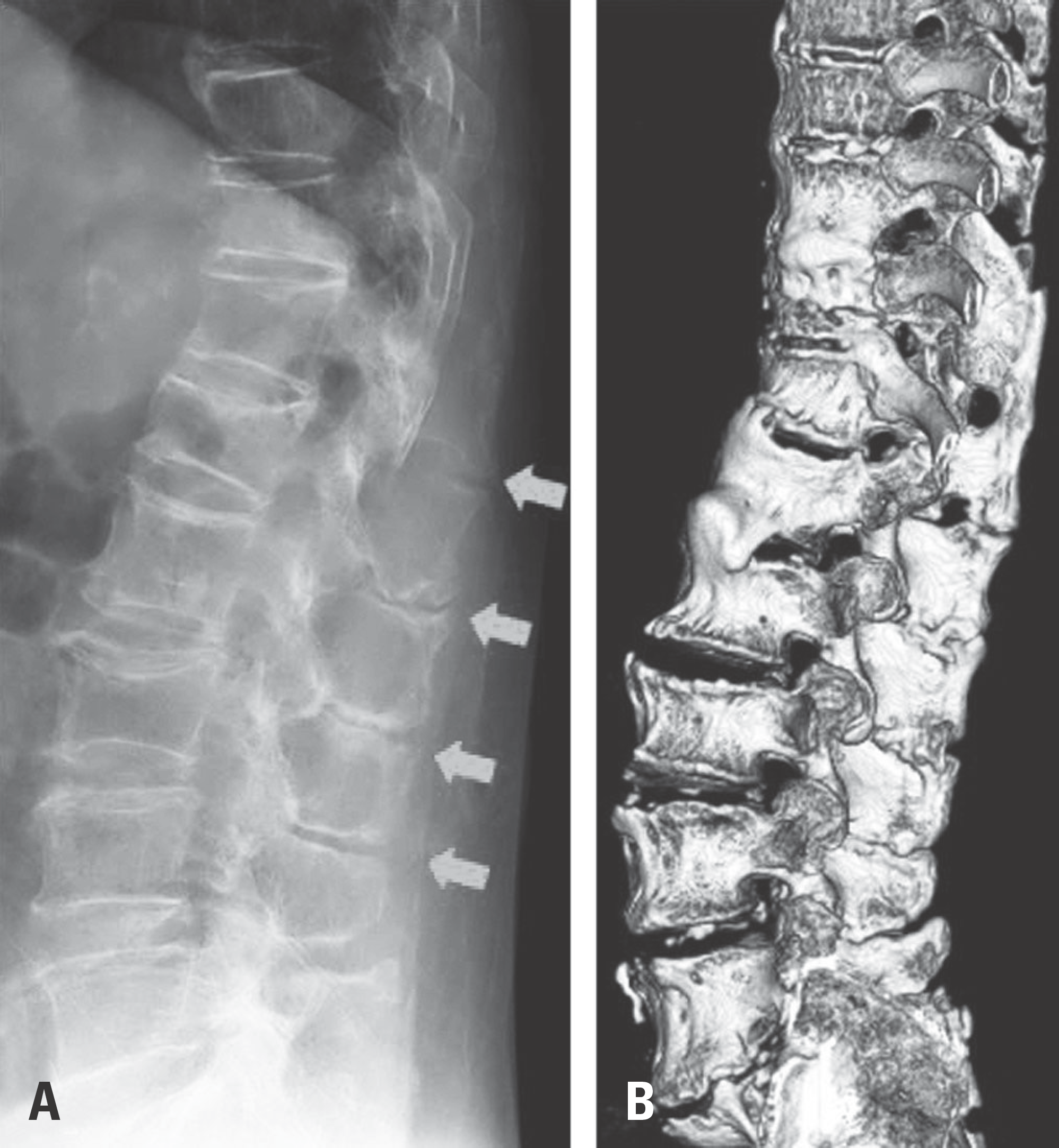 | Fig. 1.
(A) Initial radiographs showing severe narrowing of the interspi-nous process (red arrows). (B) Three-dimensional reconstruction of computed tomography images showing Baastrup disease (kissing spine). |
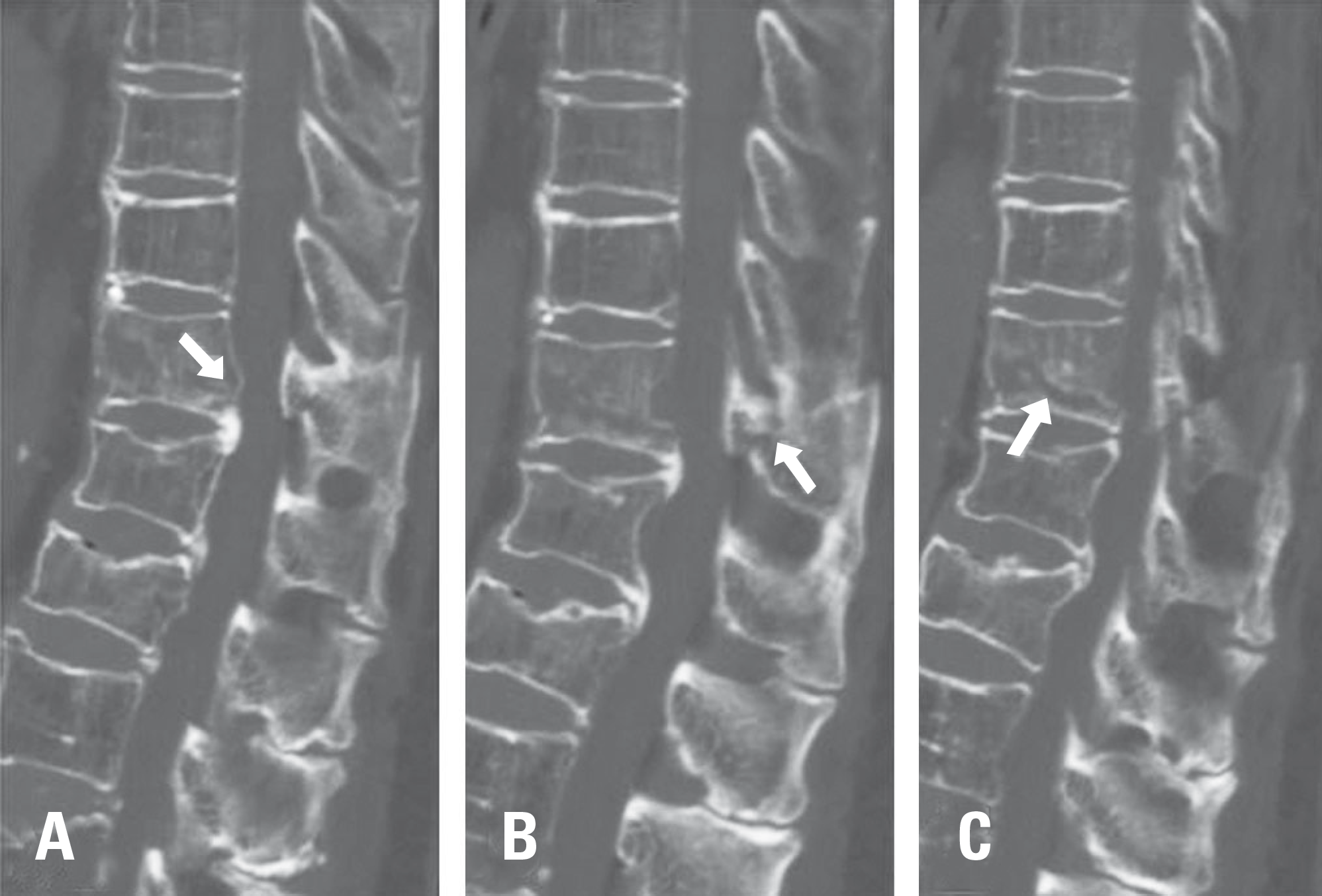 | Fig. 2.Serial sagittal computed tomography images showing the 3-col-umn fracture with (A) posterior vertebral wall bulging (red arrow), (B) a displaced posterior column fracture, and (C) a collapsing vertebral body. |
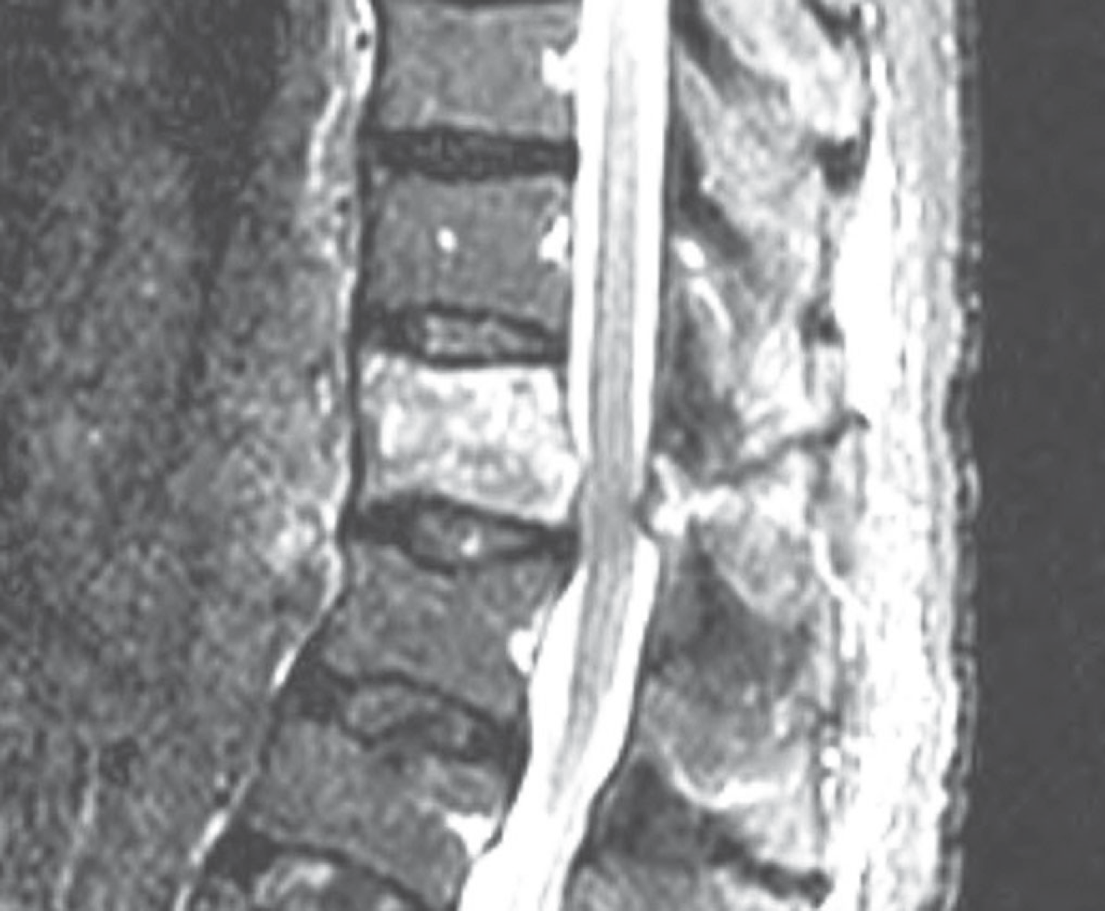 | Fig. 3.Magnetic resonance imaging scan showing a mild cord-compressing lesion with buckling of the ligamentum flavum. |
 XML Download