Abstract
Summary of Literature Review
Osteoid osteoma is a benign osteoblastic tumor that usually arises in the long bones. Osteoid osteoma involving the sacrum is extremely rare.
Materials and Methods
A 29-year-old male patient presented with pain localized in his sacral area for 10 months. His pain was worse at night, relieved by non-steroidal anti-inflammatory drugs, and independent of physical activity. Bone scintigraphy showed increased uptake in the second sacral vertebra (S2). Computed tomography revealed a nidus located in the S2 spinous process. Magnetic resonance imaging showed bone and soft tissue edema around the nidus.
Results
En bloc excision including the nidus revealed a diagnosis of osteoid osteoma and provided immediate relief of the patient's long-lasting sacral pain.
Conclusions
When a young patient presents with localized sacral pain that is worse at night, relieved by non-steroidal anti-inflammatory drugs, independent of physical activity, and lasts longer than expected, proper imaging studies should be performed to rule out osteoid osteoma. Although less invasive treatment modalities have been introduced, classical en bloc excision is currently the gold standard for managing osteoid osteoma.
REFERENCES
2. Schajowicz F. Tumors and Tumorlike Lesions of Bone and Joints. New York: Springer;1981. 36-47.
3. Hadjipavlou AG, Lander PH, Marchesi D, et al. Minimally invasive surgery for ablation of osteoid osteoma of the spine. Spine. 2003 NOV; 28(22):472–7. DOI: 10.1097/01. BRS.0000092386.96824.DB.

4. Chung SS, Seo JS, Moon SH, et al. Osteoid osteoma in the sacrum: a case report. J Korean Soc Spine Surg. 2006 JUN; 13(2):147–51. https://doi.org./10.4184/jkss.2006.13.2.147. DOI: 10.1097/01. BRS.0000092386.96824.DB.
5. OZKOC G. Osteoid osteoma of the sacrum mimicking sacroiliitis: a case report. Turk J Rheumatol. 2013 Jan; 28(1):51–3. DOI: 10.5606/tjr.2013.2863.

6. Ghanem I. The management of osteoid osteoma: update and controversied. Curr Opin Pediatr. 2006 Feb; 18(1):3641. DOI: 10.1097/01.mop.0000193277.47119.15.
7. Etemadifar MR, Hadi A. Clinical findings and results of surgical resection in 19 cases of spinal osteoid osteoma. Asian Spine J. 2015 Jun; 9(3):386–93. DOI: 10.4184/asj.2015.9.3.386.

8. Fukuda S, Susa M, Watanabe I, et al. Computed tomography-guided resection of osteoid osteoma of the sacrum: a case report. Journal of Medical Case Reports. 2014 Jun; 8:206. DOI: 10.1186/1752-1947-8-206.

9. Kneisl JS, Simon MA. Medical management compared with operative treatment for osteoid-osteoma. J Bone Joint Surg Am. 1992 Feb; 74(2):179–85. DOI: 10.2106/00004623-199274020-00004.

10. Feletar M, Hall S. Osteoid osteoma: a case for conservative management. Rheumatology. 2002 May; 41(5):585–6. DOI: 10.1093/rheumatology/41.5.585.

Fig. 1.
Plain radiographs.(A) An anteroposterior plain radiograph is normal. (B) A lateral plain radiograph is normal.
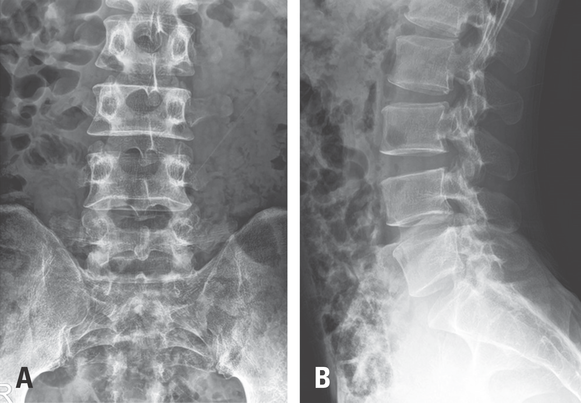
Fig. 2.
Bone scintigraphy shows increased uptake in the middle of the second sacral vertebra (S2) (arrows).
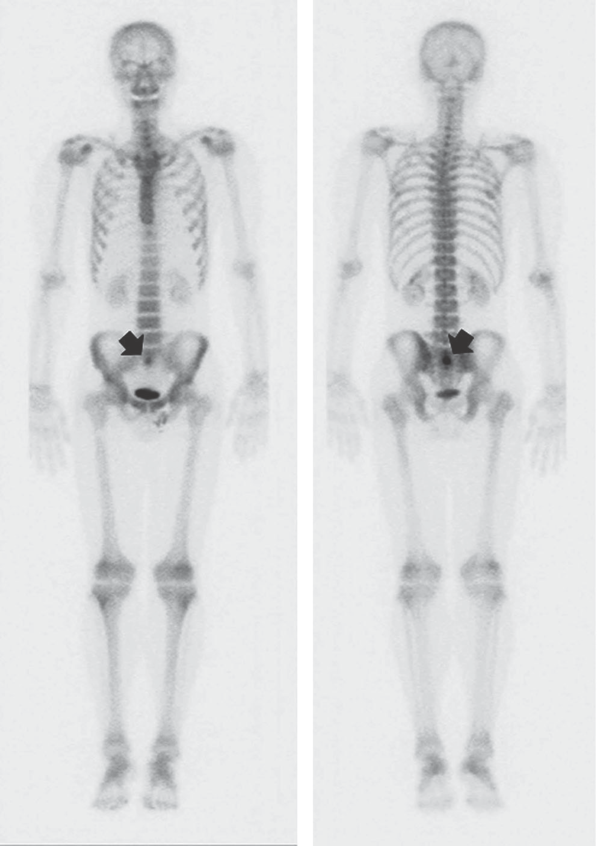
Fig. 3.
Magnetic resonance imaging.(A) A gadolinium-enhanced T1-weighted coronal magnetic resonance image shows a mass with high signal intensity in S2 (arrow).(B) A gadolinium-enhanced T1-weighted axial magnetic resonance image shows a mass with nonspecific surrounding edema in the bone marrow and soft tissue (arrow).
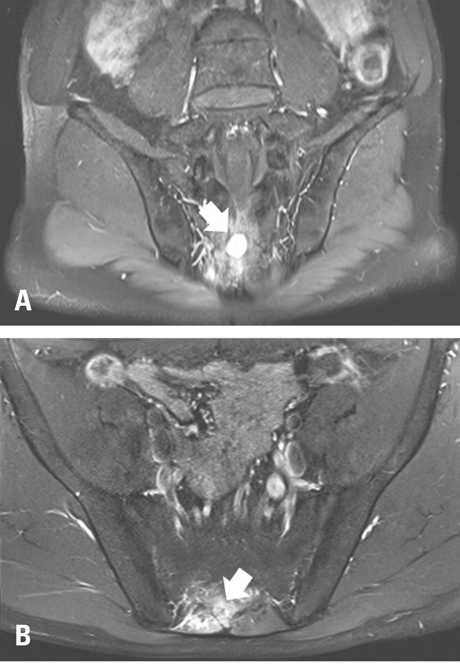
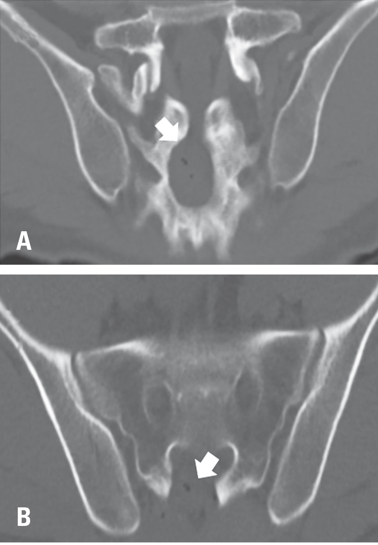
Fig. 4.
Preoperative computed tomography (CT).(A) A coronal CT image shows an oval radiolucent nidus within surrounding sclerotic reactive bone in the spinous process of S2, and a central dot-like calcification within the nidus is also noted (arrow). (B) An axial CT image shows an osteoid osteoma bulging into the S2 spinal canal (ar-row).
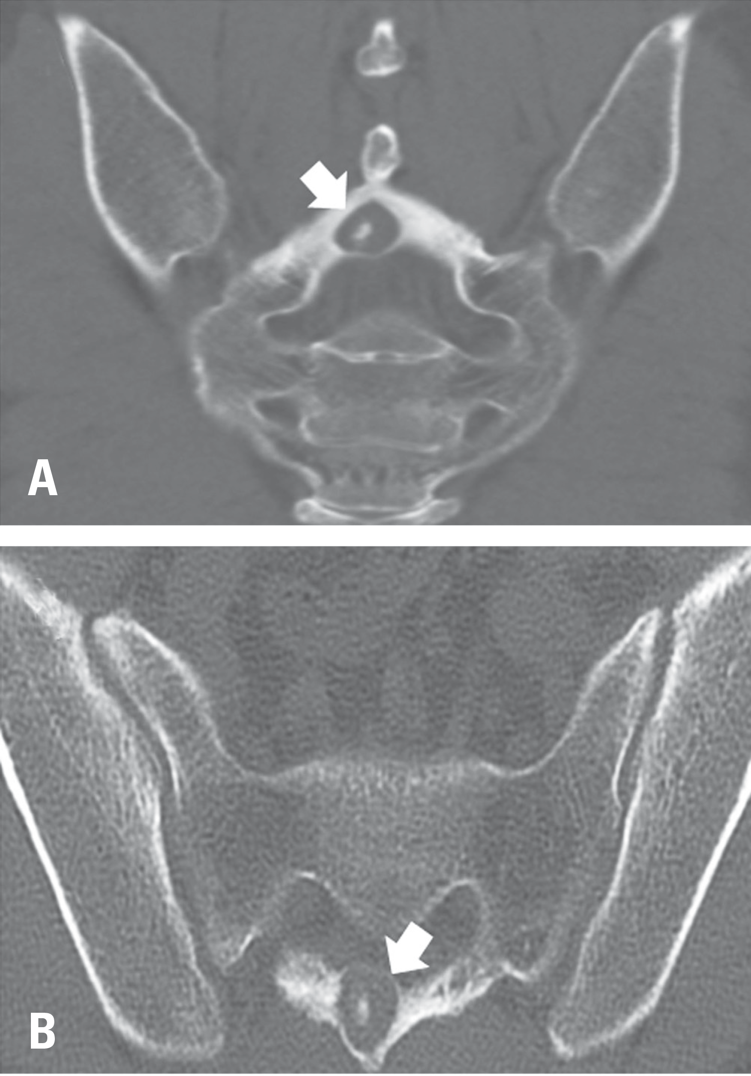
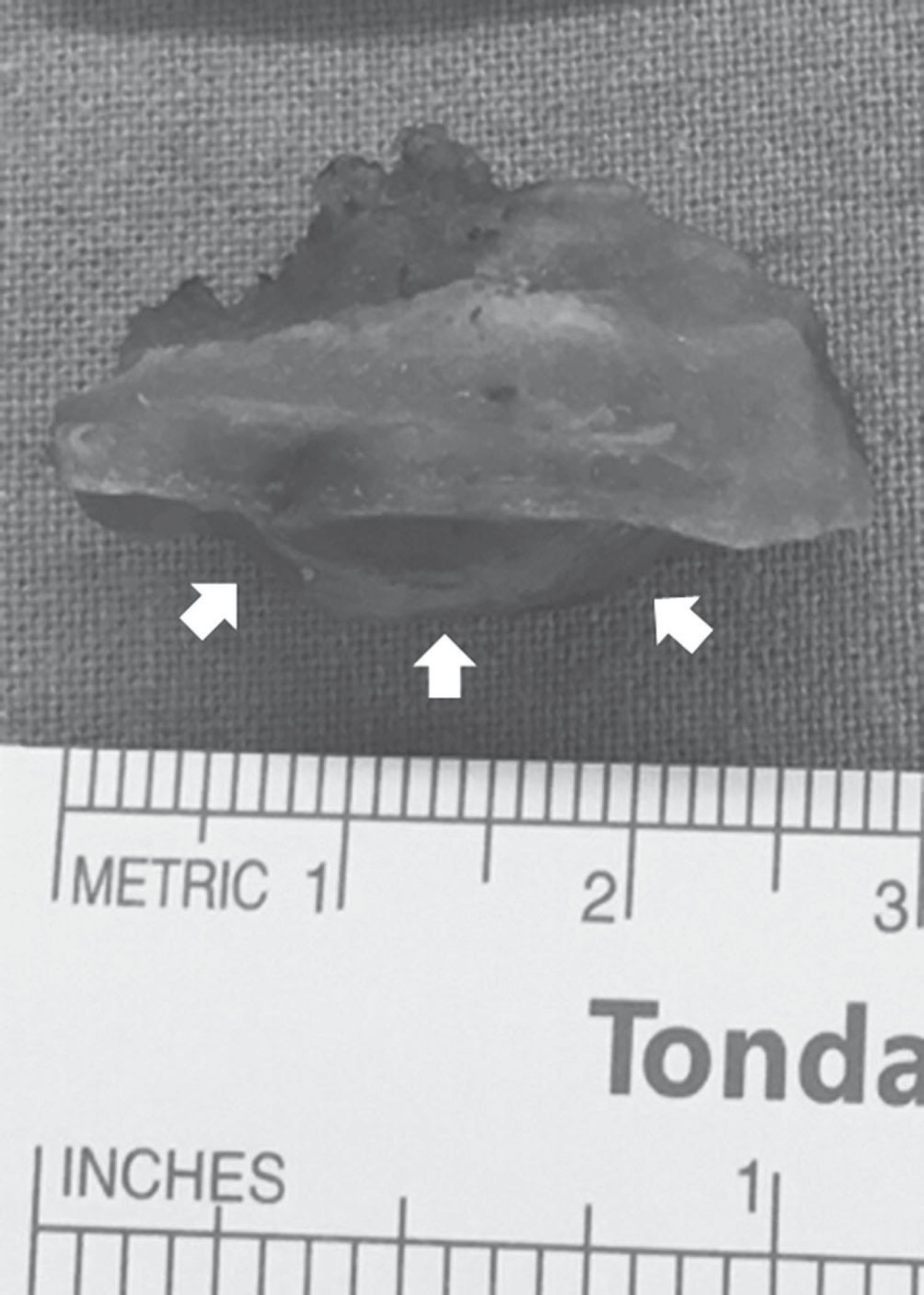
Fig. 5.
The surgically excised specimen shows ventral bulging of the osteoid osteoma (arrows) as seen on the computed tomography image (Fig 4B).


 PDF
PDF Citation
 Print
Print


 XML Download