Abstract
Duodenal varices can develop in patients with portal hypertension secondary to liver cirrhosis. Although upper gastrointestinal bleeding is often severe and fatal, the definite treatment or guideline has not been established. Although endoscopy is the primary therapeutic modality, the use of radiologic interventions, such as transjugular intrahepatic portosystemic shunt, balloon or vascular plug-assisted retrograde transvenous obliteration, and percutaneous transhepatic variceal obliteration, can be considered alternative treatment methods for duodenal varices. Herein, we report a case of duodenal varix in a patient with poor hepatic functional reserve and vascular anatomy, which are contraindications for an occlusion balloon or a vascular plug, successfully treated with coil-assisted retrograde transvenous obliteration.
Go to : 
Index terms
Duodenum, Varix, Embolization, TherapeuticREFERENCES
1. Weishaupt D, Pfammatter T, Hilfiker PR, Wolfensberger U, Marincek B. Detecting bleeding duodenal varices with multislice helical CT. AJR Am J Roentgenol. 2002; 178:399–401.

2. Saad WE, Lippert A, Saad NE, Caldwell S. Ectopic varices: anatomical classification, hemodynamic classification, and hemodynamic-based management. Tech Vasc Interv Radiol. 2013; 16:158–175.

3. Henry Z, Uppal D, Saad W, Caldwell S. Gastric and ectopic varices. Clin Liver Dis. 2014; 18:371–388.

4. Boyer TD, Haskal ZJ. American Association for the Study of Liver Diseases. The role of transjugular intrahepatic portosystemic shunt in the management of portal hypertension. Hepatology. 2005; 41:386–400.
5. Copelan A, Chehab M, Dixit P, Cappell MS. Safety and efficacy of angiographic occlusion of duodenal varices as an alternative to TIPS: review of 32 cases. Ann Hepatol. 2015; 14:369–379.

6. Lee EW, Saab S, Gomes AS, Busuttil R, McWilliams J, Durazo F, et al. Coil-assisted retrograde transvenous obliteration (CARTO) for the treatment of portal hypertensive variceal bleeding: preliminary results.Clin Transl Gastroenterol. 2014; 5:e61.
7. Lee SJ, Jeon GS. Coil-assisted retrograde transvenous obliteration for the treatment of duodenal varix.Di-agn Interv Radiol. 2018; 24:292–294.
8. Kim MJ, Jang BK, Chung WJ, Hwang JS, Kim YH. Duodenal variceal bleeding after balloon-occluded retro-grade transverse obliteration: treatment with transjugular intrahepatic portosystemic shunt. World J Gastroenterol. 2012; 18:2877–2880.

9. Kim DJ, Darcy MD, Mani NB, Park AW, Akinwande O, Ramaswamy RS, et al. Modified balloon-occluded ret-rograde transvenous obliteration (BRTO) techniques for the treatment of gastric varices: vascular plug-as-sisted retrograde transvenous obliteration (PARTO)/coil-assisted retrograde transvenous obliteration (CARTO)/balloon-occluded antegrade transvenous obliteration (BATO). Cardiovasc Intervent Radiol. 2018; 41:835–847.

Go to : 
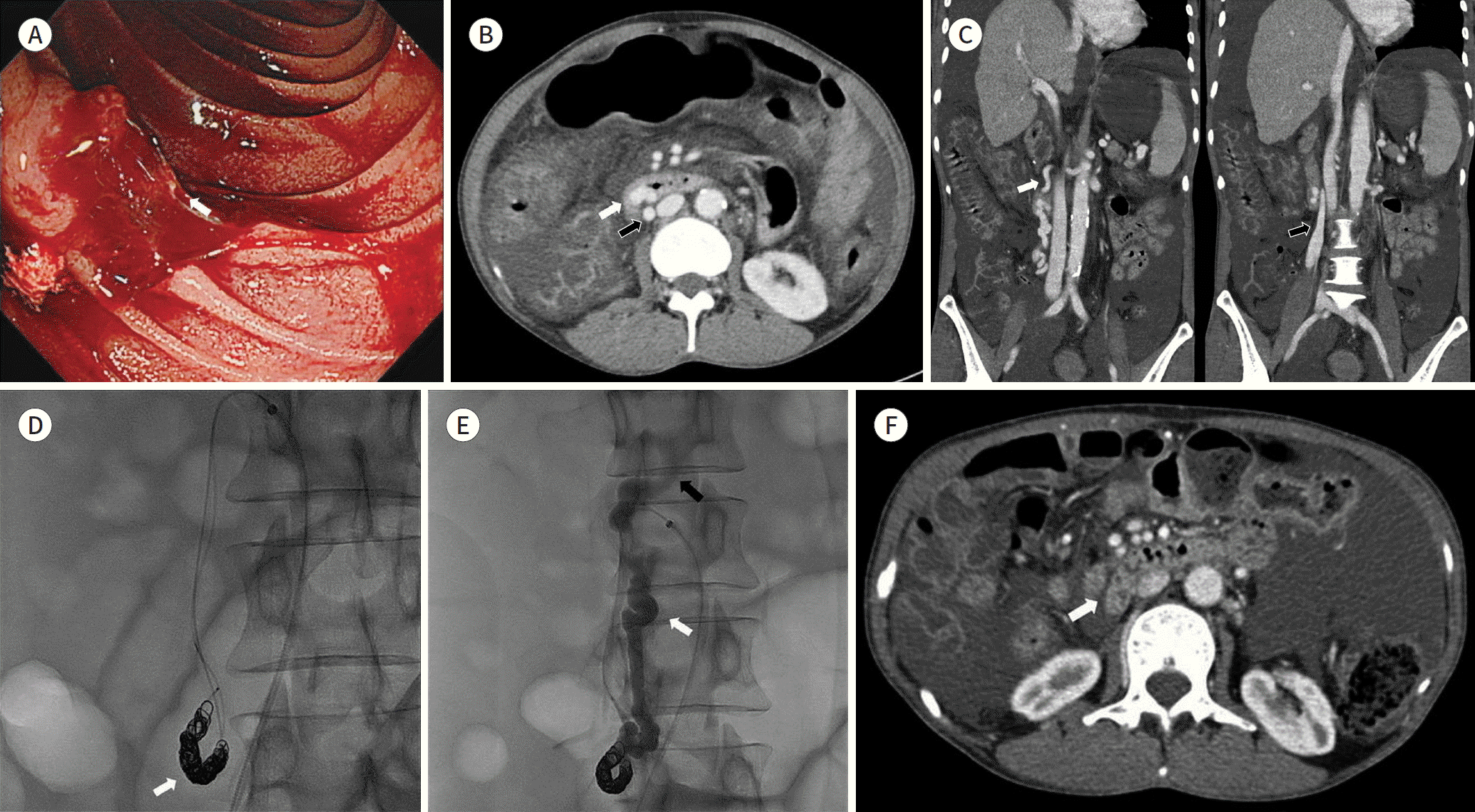
 | Fig. 1.A 50-year-old woman with massive hematemesis. A. Endoscopy shows active bleeding from the duodenal varix (arrow). B. Axial contrast-enhanced computed tomography demonstrates a varix in the 3rd portion of the duodenum (white arrow) supplied by the inferior pancreaticoduodenal vein and draining into the right ovarian vein to form the mesocaval shunt (black arrow). C. Coronal contrast-enhanced computed tomography shows the mesocaval shunt formed by the inferior pancreaticoduodenal (white arrow) and right ovarian (black arrow) veins. D. Coil-assisted retrograde transvenous obliteration was successfully performed using a double microcatheter technique. Detachable coils were deployed in the ovarian vein through a more proximally placed microcatheter (arrow). E. Gelfoam mixed with a contrast agent was injected to embolize the duodenal varices (black arrow) and inferior pancreaticoduodenal vein (white arrow). F. Axial contrast-enhanced computed tomography obtained 6 months after coil-assisted retrograde transvenous obliteration shows complete obliteration of the duodenal varices (arrow). |


 PDF
PDF ePub
ePub Citation
 Print
Print


 XML Download