

 PDF
PDF ePub
ePub Citation
 Print
Print


INTRODUCTION
Primary tracheal tumor is a relatively rare disease. Of all cases of primary tracheal tumor, more than 90% are malignant tumors, such as adenoid cystic carcinoma and squamous cell carcinoma. Tracheal leiomyomas account for approximately 1% of all primary tracheal tumors (123). This tumor usually occurs in the fourth decade of life, although one third of patients are younger than 20 years (4).
While the combination of posteroanterior (PA) and lateral chest radiographs are primary method for evaluation of the trachea and central airways, findings may not always be apparent on chest radiographs. It has been reported that tracheal tumors are found only in 18% of chest radiographs (5). Therefore, further evaluation with cross sectional imaging is usually necessary.
Digital tomosynthesis (DTS) can provide stereoscopic images and remove the shadows of bones and overlying structures, giving superior visibility of airway lesions that are difficult to be recognized on chest radiograph (6). We describe the case in which DTS was useful for the evaluation of tracheal leiomyoma that was overlooked on initial chest radiograph.
CASE REPORT
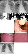
A 58-year-old woman presented with chronic cough and wheezing for 3 months. During physical examination, the clinician could hear a wheezing sound when the patient was breathing. She underwent chest PA and lateral radiographs, but no abnormality was seen on chest radiographs (Fig. 1A). Clinician suspected airway disease and recommended DTS for the evaluation of central airway. Chest DTS were performed using a commercially available unit (Volume RAD; GE Healthcare, Chicago, IL, USA) with a setting of 120 kV, 125 mA, 0.2 copper filter, manual exposure control, source to image distance of 180 cm. Within a time period of approximately 11.4 s, 60 projection images were acquired with a tube angle range of ± 15°, and 53 coronal images of 3 mm thickness were reconstructed without overlap. The estimated mean effective dose was 0.1 mSv. DTS demonstrated a 6 mm sized endotracheal nodule 2.3 cm proximal to carina (Fig. 1A). Tracheal tumor was suspected and computed tomography (CT) was performed to evaluate the lesion. On CT images, a 6 × 4 mm sized homogeneous endotracheal nodule was revealed in the anterior wall of the trachea 2.3 cm above the carina and borderline sized right upper paratracheal lymph nodes were seen (Fig. 1B). There was no evidence of air-trapping, distal atelectasis or obstructive pneumonia. Bronchoscopy revealed a whitish polypoid nodule arising from the mid-trachea (Fig. 1C). Bronchoscopic biopsy was performed to confirm the diagnosis of nodule. Biopsy specimens revealed the tumor was composed of proliferating benign spindle cells, which is consistent with leiomyoma (Fig. 1D). After the pathologic confirmation, the tumor was removed by neodymium-yttrium-aluminum-garnet laser treatment by rigid bronchoscopy under general anesthesia.
The post-procedural course was favorable. Follow-up DTS 2 days after procedure showed no evidence of post-procedural complication (Fig. 1E). She did not have recurrence of the tumor during the follow-up period of 15 months.
DISCUSSION
This case was endotracheal leiomyoma that was not found on initial chest radiograph but detected on DTS. Although chest radiograph is the most basic test for the evaluation of thoracic disease, it often does not provide an useful information for screening of tracheobronchial tree because airway abnormalities may be obscured by mediastinal, apical soft tissue or bony structures. Therefore, chest CT is considered the standard technique for the evaluation of airway abnormalities. However, because of ionizing radiation and high costs, standard dose chest CT has less value as a screening tool. DTS is a type of limited-angle tomography that uses low-dose projections to reconstruct sectional images and can provide more information than chest radiograph with lower radiation doses than CT (67). Although low dose CT plays a sufficient role as a screening test for lung and airway disease, the mean effective dose of low-dose CT is about 10 times that of DTS.
In our case, the size and location of the tumors found on DTS were almost identical to CT. Therefore, DTS may be a clinically useful method for suspected central airway disease while the result of chest radiograph is normal or equivocal.
Diagnosis of tracheal leiomyoma is difficult to establish before pathologic confirmation because of its diverse and non-specific imaging findings. It is typically seen as a well-defined, single nodule or mass. It is identifiable as an endotracheal nodule, or an intratracheal and extratracheal mass on chest radiographs. However, in some cases, it is not seen on chest radiographs. On CT, the lesion is seen as an airway intraluminal nodule or an iceberg tumor. It has an oval or a lobulated contour and homogeneous or heterogeneous enhancement on contrast enhanced CT. Stippled calcification could be found in a large tumor (4).
Although surgical resection with bronchoplastic reconstruction has long been the standard treatment, improvements in the technique have led to therapeutic bronchoscopy replacing surgery for treatment of benign tumors of the tracheobronchial tree (89). Bronchoscopic intervention has the advantage of avoiding complications and morbidity associated with surgery (9). Post-procedural imaging follow-up with CT is usually performed both one week and one month after procedure to evaluate postprocedural complication and tumor recurrence. However, repeated CT examination eventually lead to radiation exposure and cost problems. In our case, follow-up DTS showed no evidence of post-procedural complication or tumor recurrence for 15 months after procedure. The estimated mean effective dose of DTS was 0.1 mSv, which was 30 times less than that of routine dose chest CT and 10 times less than that of low dose chest CT (7). Therefore, DTS can be useful when repeated imaging studies during a short period are needed to evaluate the complications after interventional procedures of airway disease or the recurrence of lesions on follow-up.
In conclusion, we reported a rare case of endotracheal leiomyoma in which DTS was useful for the evaluation of a tracheal tumor that was overlooked on initial chest radiographs. Although DTS could not completely replace CT as a diagnostic tool, it can be used for the initial or additional diagnostic tool in patients with clinically suspected large airway disease.
 XML Download