

 PDF
PDF ePub
ePub Citation
 Print
Print


INTRODUCTION
Extrauterine adenomyomas are defined as circumscribed tumor-like masses composed of smooth muscle tissue, endometrioid glands, and stroma and they are similar to their more common uterine counterparts in most respects (1). They are extremely rare, especially those originating in the liver. To date only 3 cases of hepatic extrauterine adenomyoma have been reported, and those reports are focused on histopathology (2).
Herein, we report for the first time the specific radiologic findings of extrauterine adenomyoma of the liver in a 43-year-old woman, which was initially diagnosed as a hepatic adenoma.
CASE REPORT
A 43-year-old woman presented at the digestive internal medicine department with a 7-day history of epigastric pain. A general physical examination and a systemic examination were unremarkable. She had undergone total laparoscopic hysterectomy for uterine leiomyoma 4 years prior. Otherwise, she had no underlying medical diseases. Initial laboratory evaluations including tumor marker (alpha-fetoprotein) and liver function tests were within normal ranges, and serological tests for hepatitis B surface antigen and anti-hepatitis C virus antibodies were negative.
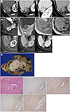
Upper gastrointestinal endoscopy was performed to investigate her epigastric pain, and there were no abnormal findings. Contrast-enhanced CT scanning was performed to further investigate her symptoms. It revealed a 3 × 2 × 2 cm well defined bilobed mass in the subcapsular area of segment VI of the liver. The medial lobe of the mass exhibited low density without enhancement, and the lateral lobe exhibited an enhanced solid portion with peripheral low-density dots. The lateral lobe was shaped like a lotus root. The enhancement pattern was subtle enhancement in the arterial phase and persistent weak enhancement in the portal and delayed phases without washout (Fig. 1A). The CT characteristics of the lesion did not facilitate a definitive diagnosis, so we recommended MRI.
On MRI, the medial lobe and the periphery of the lateral lobe that exhibited low density on CT were depicted as cystic portions that exhibited high signal intensity on T2-weighted images. There was focal high signal intensity with fluid-fluid level within the medial cystic portion on in-phase and out-of-phase T1-weighted images, suggesting a hemorrhagic component. The solid portion of the lateral lobe of the mass exhibited subtle low signal intensity on T1-weighted images and slightly high signal intensity on T2-weighted images. Gadolinium-enhanced fat-suppressed T1-weighted images depicted heterogenous weak arterial enhancement and persistent enhancement (Fig. 1B).
The patient underwent wedge resection of hepatic segment VI. Intraoperatively a mass of approximately 3 × 2 × 2 cm in size was located in the subcapsular region in segment VI of the liver. A gross specimen photograph is shown in Fig. 1C. Wedge resection of the liver revealed a bilobed mass located subcapsularly. The mass was well defined, whitish, and solid with multiple cysts filled with dense brown fluid. Microscopically, the mass was composed of thick muscular tissue lined with benign endometrial gland and stroma that resembled uterine endometrium. The endometrioid glands were positive for estrogen receptor and progesterone receptor, and the endometrioid stromas were positive for CD10. The smooth muscle component was positive for smooth muscle actin (Fig. 1D).
DISCUSSION
Adenomyomas are benign tumors consisting of benign endometrial glands, endometrioid stroma, and smooth muscle tissue, and they typically originate within the uterus (1). Occasionally they are located outside the uterus, and such tumors are termed extrauterine adenomyomas. The majority of such cases originate from the ovary and are located in the pelvis, and extrapelvic locations such as the liver or abdominal wall are extremely rare. The first case of extrauterine adenomyoma was reported by Paul et al. (2) and Cozzutto in 1981 (3)e reported that there had been 37 cases of extrauterine adenomyoma eligible for their review since then. Of these 37 cases, 21 were pelvic, 8 were extrapelvic, and 8 cases involved multiple sites. In their report there were only 3 cases of extrauterine adenomyoma involving the liver, and these reports focused on histopathology (145).
The above-mentioned previously reported 3 cases were only evaluated via abdominal CT, and all of the masses were typically located in the subcapsular region of the posterior right hepatic lobe. All had a cystic portion but there were no definite intralesional hyperattenuating hemorrhages on CT. However, there were hemorrhagic components on gross and pathologic findings. Two exhibited heterogenous arterial enhancement and the remaining 1 was not evaluated via contrast-enhanced CT. In the present case the mass was also located in the subcapsular area of the posterior right hepatic lobe, and it exhibited subtle arterial enhancement and weak persistent enhancement in the portal and delayed phases. It had cystic portions with hemorrhagic foci. We surmise that these locational and morphologic findings are specific imaging features of extrauterine adenomyoma of the liver.
Morphologic imaging finding of uterine adenomyomas and extrauterine adenomyomas including liver and other sites is similar in that they are solid and cystic masses with some internal hemorrhagic foci and show heterogenous enhancement (26).
Because the most common benign hepatic mass with hemorrhage is adenoma, of which inflammatory subtype can exhibit enhancement during the arterial phase that persists in the portal and delayed phases (7) we initially mistook the present case for hepatic adenoma. However, hepatic extrauterine adenomyoma is likely to contain a cystic and hemorrhagic component anywhere within it, and hepatic adenoma is prone to central necrosis and hemorrhage because the vascular supply is limited to the surface of the tumor. Furthermore, hepatic adenoma can have a fat component and pseudocapsule in as many as 30% of cases (78). Otherwise, hepatic endometrioma, abscess, hemangioma, hepatocellular carcinoma (HCC), hemorrhagic metastasis can also show solid and cystic mass with intralesional hemorrhage. However, unlike hepatic adenomyoma having a dominant solid smooth muscle component with scattered endometrial glands which show cystic and hemorrhagic foci, hepatic endometrioma has less or no solid component of smooth muscle and usually shows predominant cystic lesion with intralesional hemorrhage (9). Hepatic abscess typically demonstrates rim enhancement with perilesional edema and hemangioma exhibits centripetal enhancement pattern. HCC also reveals typical enhancement pattern that arterial enhancement and delayed washout. Hemorrhagic metastasis can show one or more liver lesions with primary tumor elsewhere (7).
The mechanism of hepatic extrauterine adenomyoma is still uncertain. However, various theories have been proposed to explain the pathogenesis of extrauterine adenomyoma. In our report, we are discussing 2 of the major theories. The first theory is the implantation theory. All patients of 3 previously reported hepatic adenomyoma cases and our case had undergone laparoscopic surgeries such as myomectomy or hysterectomy for uterine leiomyoma and hysterectomy with salpingectomy for adenomyosis, leiomyoma and endometriosis. During surgery, peritoneal fluid may have viable endometrial cells and smooth muscle cells from cut uterus. And these could experimentally be implanted and grown in a peritoneum or peritoneal cavity. The second theory is the sub-coelomic mesenchyme transformation theory that extrauterine adenomyoma originates from the metaplasia of the layer that lies underneath the mesothelium of the abdominal peritoneum. The cases of hepatic extrauterine adenomyomas including our case were located in the subcapsular region of the liver, which was partially covered by the hepatic peritoneum (1910).
Clinically, patients with hepatic extrauterine adenomyoma can present with abdominal pain or back pain (145). It may be that pain is caused when hemorrhage occurs in the mass.
In summary, a definitive diagnosis of extrauterine adenomyoma is only established postoperatively after confirmation via histopathology. However, extrauterine adenomyoma of the liver exhibits specific radiologic features such as a subcapsular location in the posterior right hepatic lobe, a weak persistent enhancement pattern, and a cystic portion with internal hemorrhage. Therefore when this kind of benign hemorrhagic mass of the liver is encountered, extrauterine adenomyoma should be considered.
 XML Download