

 PDF
PDF ePub
ePub Citation
 Print
Print


Myung Hwan Lee1 , Eun-Kyung Kim1, Eun Ju Lee2, Ha Yan Kim2, Jung Hyun Yoon1, ∗
, Eun-Kyung Kim1, Eun Ju Lee2, Ha Yan Kim2, Jung Hyun Yoon1, ∗
Abstract
Purpose
To evaluate the optimal measurement location, cut-off value, and diagnostic performance of S-Shearwave in differential diagnosis of breast masses seen on ultrasonography (US).
Materials and Methods
During the study period, 225 breast masses in 197 women were includ-ed. S-Shearwave measurements were made by applying a square region-of-interest automati-cally generated by the US machine. Shearwave elasticity was measured three times at four different locations of the mass, and the highest shearwave elasticity was used for calculating the optimal cut-off value. Diagnostic performance was evaluated by using the area under the receiv-ing operator characteristic curve (AUC).
Results
Of the 225 breast masses, 156 (69.3%) were benign and 69 (30.7%) were malignant. Mean S-Shearwave values were significantly higher for malignant masses (108.0 ± 70.0 kPa vs. 43.4 ± 38.3 kPa;p < 0.001). No significant differences were seen among AUC values at different measurement locations. With a cut-off value of 41.9 kPa, S-Shearwave showed 85.7% sensitivity, 63.9% specificity, 70.7% accuracy, and positive and negative predictive values of 51.7% and 90.8%, respectively. The AUCs for US and S-Shearwave did not show significant differences (p = 0.179).
Go to : 
Index terms
Breast, Ultrasonography, Neoplasm, Elasticity Imaging TechniquesREFERENCES
1. American College of Radiology. Breast imaging reporting and data system. 5th ed. Reston, VA: American College of Radiology. 2013.
2. Hong AS, Rosen EL, Soo MS, Baker JA. BI-RADS for sonography: positive and negative predictive values of sonographic features. AJR Am J Roentgenol. 2005; 184:1260–1265.

3. Kim EK, Ko KH, Oh KK, Kwak JY, You JK, Kim MJ, et al. Clinical application of the BI-RADS final assessment to breast sonography in conjunction with mammography. AJR Am J Roentgenol. 2008; 190:1209–1215.
4. Costantini M, Belli P, Lombardi R, Franceschini G, Mulè A, Bonomo L. Characterization of solid breast masses: use of the sonographic breast imaging reporting and data system lexicon. J Ultrasound Med. 2006; 25:649–659. quiz 661.
5. Itoh A, Ueno E, Tohno E, Kamma H, Takahashi H, Shiina T, et al. Breast disease: clinical application of US elastography for diagnosis. Radiology. 2006; 239:341–350.
6. Lee EJ, Jung HK, Ko KH, Lee JT, Yoon JH. Diagnostic performances of shear wave elastography: which pa-rameter to use in differential diagnosis of solid breast masses? Eur Radiol. 2013; 23:1803–1811.
7. Yoon JH, Jung HK, Lee JT, Ko KH. Shearwave elastography in the diagnosis of solid breast masses: what leads to false-negative or false-positive results? Eur Radiol. 2013; 23:2432–2440.
8. Berg WA, Cosgrove DO, Doré CJ, Schäfer FK, Svensson WE, Hooley RJ, et al. Shearwave elastography im-proves the specificity of breast US: the BE1 multinational study of 939 masses. Radiology. 2012; 262:435–449.
9. Burnside ES, Hall TJ, Sommer AM, Hesley GK, Sisney GA, Svensson WE, et al. Differentiating benign from malignant solid breast masses with US strain imaging. Radiology. 2007; 245:401–410.
10. Cho N, Moon WK, Kim HY, Chang JM, Park SH, Lyou CY. Sonoelastographic strain index for differentiation of benign and malignant nonpalpable breast masses. J Ultrasound Med. 2010; 29:1–7.
11. Chang JM, Moon WK, Cho N, Yi A, Koo HR, Han W, et al. Clinical application of shear wave elastography (SWE) in the diagnosis of benign and malignant breast diseases. Breast Cancer Res Treat. 2011; 129:89–97.
12. Choi K, Kong D, Hah Z, Lee HK. A reliability index of shear wave speed measurement for shear wave elastography. Piscataway: IEEE;2015.
13. Deffieux T, Gennisson JL, Bercoff J, Tanter M. On the effects of reflected waves in transient shear wave elastography. IEEE Trans Ultrason Ferroelectr Freq Control. 2011; 58:2032–2035.
14. Tozaki M, Isobe S, Fukuma E. Preliminary study of ultrasonographic tissue quantification of the breast using the acoustic radiation force impulse (ARFI) technology. Eur J Radiol. 2011; 80:e182–187.
15. Wojcinski S, Brandhorst K, Sadigh G, Hillemanns P, Degenhardt F. Acoustic radiation force impulse imaging with Virtual Touch TM tissue quantification: mean shear wave velocity of malignant and benign breast masses. Int J Womens Health. 2013; 5:619–627.
16. Tozaki M, Isobe S, Sakamoto M. Combination of elastography and tissue quantification using the acoustic radiation force impulse (ARFI) technology for differential diagnosis of breast masses. Jpn J Radiol. 2012; 30:659–670.
17. Teke M, Göya C, Teke F, Uslukaya Ö, Hamidi C, Çetinçakmak MG, et al. Combination of virtual touch tissue imaging and virtual touch tissue quantification for differential diagnosis of breast lesions. J Ultrasound Med. 2015; 34:1201–1208.
18. Evans A, Whelehan P, Thomson K, McLean D, Brauer K, Purdie C, et al. Quantitative shear wave ultrasound elastography: initial experience in solid breast masses. Breast Cancer Res. 2010; 12:R104.
Go to :
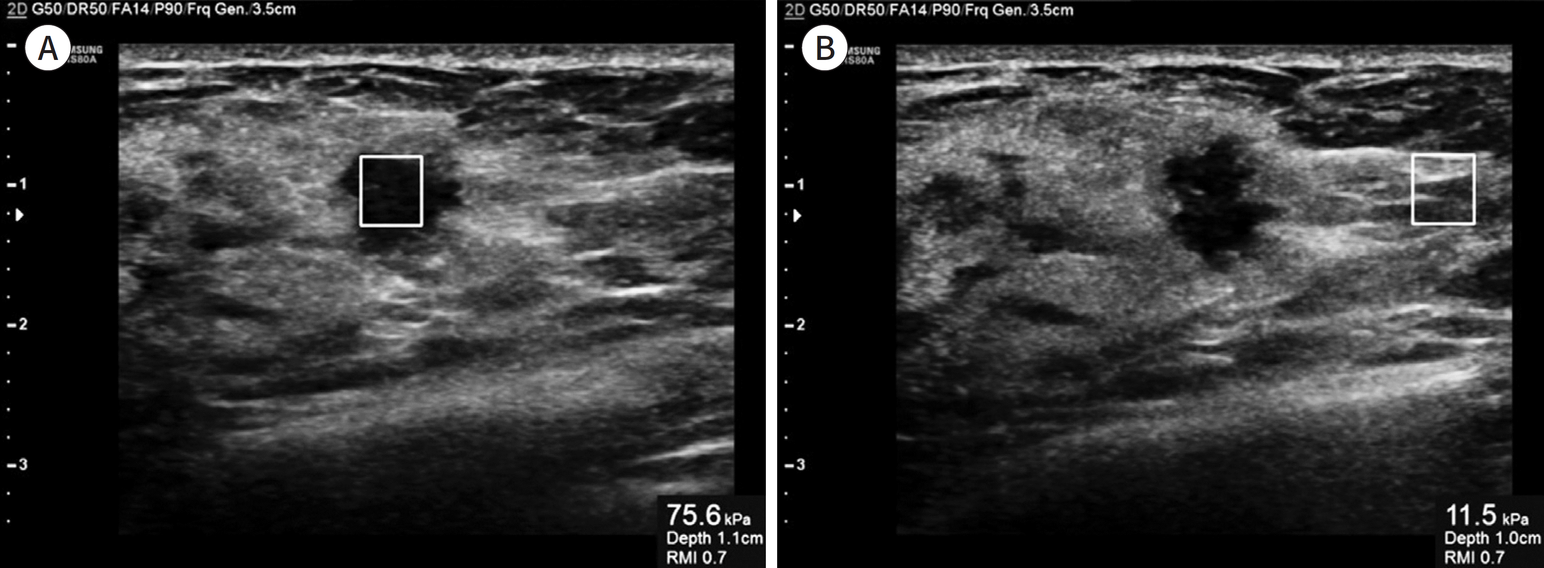 | Fig. 1.Example of application of S-Shearwave to breast ultrasonography examination. A. A square ROI is set at the center of the breast mass for shearwave elasticity measurement. B. An ROI is set at the normal breast parenchyma adjacent to the breast mass, with the ROI set at the same depth as that of the breast mass. |
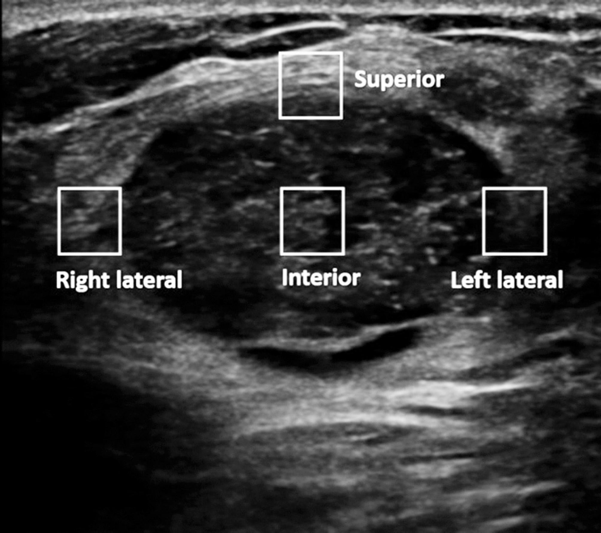 | Fig. 2.After generating the S-Shearwave, shearwave elasticity is measured three times at four different locations of the mass by applying square regions-of-interest – interior (mass center), superior border, right lateral border, and left lateral border of the mass. |
Table 1.
Comparison of Mean S-Shearwave Values between Breast Masses
| Benign (n = 156) | Malignancy (n = 69) | p† | |||||
|---|---|---|---|---|---|---|---|
| Mass | Parenchyma | p∗ | Mass | Parenchyma | p∗ | ||
| Mass center | 29.0 ± 30.0 | 18.3 ± 15.3 | < 0.001 | 72.0 ± 67.4 | 13.1 ± 9.2 | < 0.001 | < 0.001 |
| Superior margin | 33.9 ± 77.8 | 18.3 ± 15.3 | < 0.001 | 77.6 ± 58.1 | 13.1 ± 9.2 | < 0.001 | < 0.001 |
| Right lateral margin | 26.8 ± 26.8 | 18.3 ± 15.3 | < 0.001 | 52.0 ± 47.4 | 13.1 ± 9.2 | < 0.001 | < 0.001 |
| Left lateral margin | 27.2 ± 27.7 | 18.3 ± 15.3 | < 0.001 | 53.4 ± 41.5 | 13.1 ± 9.2 | < 0.001 | < 0.001 |
Table 2.
Diagnostic Performance of S-Shearwave According to Measurement Location
| Mass Center | Superior Margin | Right Lateral Margin | Left Lateral Margin | Overall p | |
|---|---|---|---|---|---|
| Cut-off level∗ (kPa) | > 25.8 | > 32.2 | > 24.1 | > 29.5 | - |
| Sensitivity | 80.0 (70.6–89.4) | 82.9 (74.0–91.7) | 68.6 (57.7–79.5) | 70.0 (59.3–80.7) | 0.024 |
| Specificity | 63.2 (55.6–70.8) | 63.9 (56.3–71.4) | 70.3 (63.1–77.5) | 74.2 (67.3–81.1) | 0.014 |
| PPV | 49.6 (40.3–58.8) | 50.9 (41.7–60.1) | 51.1 (41.0–61.2) | 55.1 (44.7–65.4) | 0.521 |
| NPV | 87.5 (81.4–93.6) | 89.2 (83.4–95.0) | 83.2 (76.8–89.6) | 84.6 (78.5–90.6) | 0.189 |
| Accuracy | 68.4 (62.4–74.5) | 69.8 (68.8–75.8) | 69.8 (63.8–75.8) | 72.9 (67.1–78.7) | 0.586 |
| AUC | 0.716 (0.656–0.777) | 0.734 (0.675–0.792) | ) 0.695 (0.629–0.760) | 0.721 (0.657–0.785) | > 0.999 |
Table 3.
Diagnostic Performance of S-Shearwave and US for the 225 Breast Masses
| S-Shearwave∗ | US Alone | p | |
|---|---|---|---|
| Sensitivity | 85.7 (77.5–93.9) | 98.6 (96,2–100.0) | 0.002 |
| Specificity | 63.9 (56.3–71.4) | 58.3 (51.0–66.5) | 0.332 |
| PPV | 51.7 (42.6–60.8) | 51.1 (43.8–60.7) | 0.888 |
| NPV | 90.8 (85.4–96.2) | 98.9 (96.2–100.0) | 0.002 |
| NPV Accuracy | 90.8 (85.4–96.2)70.7 (64.7–76.6) | 98.9 (96.2–100.0) 70.7 (65.7–77.5) | 0.002 0.821 |
| AUC | 0.748 (0.692–0.804) | 0.794 (0.755–0.832) | 0.179 |
 XML Download