

 PDF
PDF ePub
ePub Citation
 Print
Print


INTRODUCTION
The aim of voiding cystourethrography (VCUG) is to demonstrate the anatomy and function of the bladder and the urethra. It is a dynamic procedure that reflects the function and coordination of the lower urinary system. The most important indication for performing VCUG is to evaluate the presence or absence of vesicoureteral reflux (VUR) (123). The associated conditions of VUR are congenital anomalies of the urinary tract (anorectal malformation, myelodysplasia, or prune-belly syndrome), febrile urinary tract infection (particularly, if recurrent), hydronephrosis or hydroureter, hematuria, voiding abnormalities (dysuria, dysfunctional voiding such as neurogenic bladder, or incontinence), neonatal ascites, trauma, and postoperative evaluation of urinary tract (Table 1) (14).
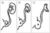
The VUR is defined as the retrograde passage of urine from the urinary bladder into the ureter and often to the calyces. The normal ureterovesical junction (UVJ) is closed by contracture during voiding to protect the upper urinary tract from reflux, which is called the antireflux action of the UVJ (Fig. 1) (125). Slanted entry and submucosal tunneling of sufficient length in the ureter are the key components involved in the antireflux action (125). These two components prevent VUR by constriction and compression of intramural ureter when bladder contracts (125). The causes of VUR are divided into primary and secondary. The primary VUR is caused due to defect in antireflux action, which is a consequence of anomaly of UVJ or immaturity of UVJ in young infants (26). The secondary VUR is caused due to malfunction within the urinary system such as bladder-outlet obstruction or neurogenic bladder, and infection (237). In active urinary tract infection, reversible VUR can occur because infection itself can affect UVJ (2).
To achieve the ultimate medical goal, reviewing previous imaging studies and medical records, and communicating with the referred physicians improve complete performance of VCUG (1). Likewise, in all other X-ray imaging studies, the principle of As Low As Reasonably Achievable (ALARA) should be followed by using pulsed fluoroscopy with last image hold technique (89). In addition, understanding and skilled application of protocols and techniques of VCUG is important.
PREPARATION
PSYCHOLOGICAL PREPARATION
Apart from physical preparation, psychological preparation is necessary for both, patients and parents as they may be anxious due to fear of pain and unknown, urethral catheterization, and radiation use (Fig. 2) (1). To help both patients and parents relieve the anxiety from the VCUG, providing a translated explanatory pamphlet provided by The Image Gently Campaign of The Alliance for Radiation Safety in Pediatric Imaging (https://www.imagegently.org) can be used as a solution (110). In addition, introducing the procedure to the patients and the parents is also recommended just before the examination (1). Mild sedation can be considered in case of uncooperative patients; however, alert state is preferred for physiologic voiding (1112).
SUPPLIES
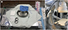
Before the examination, the supplies should be prepared (Fig. 3A). The contrast for VCUG at our institute is prepared by mixing 30 mL of iodine-containing contrast (300 or 350 mg I/mL, Iohexol; BONOREX, Central Medical Service, Seoul, Korea) with 100 mL saline. A tip for contrast is to keep concentration under 30% to avoid chemical irritation (12). Five-French feeding tubes for infants, 8-French feeding tubes for children, or a catheter larger than 8-French feeding tube for adolescents should be used (4). Also, few chlorhexidine balls for skin sterilization, sterile lubricant gel for coating catheter, a plastic extension tubing, a syringe for regurgitating urine bladder, one inch of tape for anchoring catheter to the skin, and sterile surgical drape having a hole are used. A warmer for maintaining body temperature of infant and light for better visibility during catheter insertion are also helpful (Fig. 3B).
IMAGING
CATHETERIZATION WITH GENTLE HANDLING
Parents are recommended to stand beside their children for comforting them during the procedure. If the patient's mother is pregnant, she is advised to be with the patient only during catheter insertion. For girls, sterilization of perineal skin and interlabial area with chlorhexidine balls is the first step. Next, a surgical drape having a hole should be secured and a catheter coated with lubricant gel should be inserted gently. Anchoring a catheter with tape on the inner thigh is recommended in girls. Often, the labial adhesion is encountered. Unless the urethral opening is exposed, VCUG can be performed after treating labial adhesion and reassessing the need for VCUG (1).
For boys, sterilization of perineal and penile skin including penile tip after retracting foreskin with chlorhexidine balls is the first step. The next step is to secure a surgical drape having a hole, followed by insertion of a catheter coated with lubricant gel, and anchoring catheter with tape to lower abdominal skin and penis. The foreskin is common in an infant, which makes it difficult to insert a catheter. Therefore, retracting the foreskin and exposing the urethral opening is a tip for successful insertion of catheter in boys (1). After finishing catheterization, the foreskin should be reduced. If foreskin is retracted unnecessarily for a longer time, it gets trapped behind the glans, causing edema of the glans (4). Insert the catheter as much as you need by winding it, once inside the urinary bladder. Excessive catheter insertion can trigger knotting within the urinary bladder and may need longer removal time (4). Then, drain naturally or regurgitate urine with a syringe to empty the urinary bladder (12).
INSTILLING CONTRAST INTO EMPTIED BLADDER TILL FILLED TO NEAR CAPACITY
Contrast and saline mixture is hanged about 1 m above the table to naturally instill it into the emptied bladder. Do not squeeze plastic bag containing contrast and saline mixture to instill faster because manual pressure can produce artificial VUR. Instill contrast and saline mixture to near full capacity. The estimate of the near full capacity of the urinary bladder is different in children of different ages. Approximate volume of the urinary bladder can be calculated by the formulas (1):
(Kg × 7) mL in an infant
[(age + 2) × 30] mL in children
For example, approximate volume of the urinary bladder is 35 mL in 5 kg infants, 70 mL in 10 kg infant, and 100 mL in 1.5-year-old children. When the bladder is full, the infants begin crying and flexing their toes.
IMAGE OPTIMIZATION
Under the ‘ALARA’ principle, performers should store focused images of critical areas and conditions. Pulsed fluoroscopy with last image hold technique and adjustment of collimators are key strategies to avoid unnecessary exposure to radiation (9). Continuous fluoroscopy should be avoided during filling of urinary bladder; brief and intermittent fluoroscopy is sufficient. Increasing the distance between the child and the X-ray source and removing the antiscatter grid for small patients are also recommended (13). Antiscatter grid itself is source of scattered radiation and is less useful in small patients, because smaller patient leads to less scattered radiation. Moreover, reducing patient dose is closely related to staff protection by reducing scattered radiation from a patient which is proportional to staff radiation.
STORING FOCUSED IMAGING OF CRITICAL AREAS AND CONDITIONS
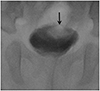
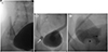
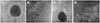
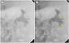
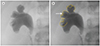
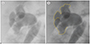
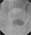
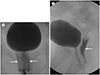
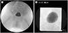
A standardized evaluation of critical areas and conditions should be ensured and are categorized into anteroposterior (AP) projection of urinary bladder during early filling phase (Fig. 4A), full filling phase of urinary bladder (Fig. 4B), the oblique projection of urinary bladder focusing both UVJs (Fig. 4C, D), an AP projection of urinary bladder during voiding (Fig. 4E), renal collecting system with or without presence of reflux (Fig. 4F), an AP projection of urinary bladder post voiding (Fig. 4G), and lateral projection for urethra during voiding in boys. A scout image is not a significant image in the management decision although its dose is low (14). Therefore, obtaining a routine scout image is not recommended. In the early filling phase, the AP image of urinary bladder demonstrates ureteroceles (Fig. 5). If any abnormality is observed, oblique projections of the urinary bladder to evaluate further distal ureters should be used (Fig. 6A). The presence of ureteroceles with or without eversion, or diverticulum is demonstrated on oblique images clearly (Fig. 6B, C). To grade VUR using VCUG, focused images targeting renal collecting system are essential. In the absence of VUR, capturing images at renal collecting system is recommended for the evidence of negative VUR. Images for the urethra, especially in boys, are easy to be missed, however, it is important. To evaluate urethra, lateral projection during voiding is standardly used. The fluoroscopic time and dose-area product are documented on the last image of study (Fig. 4G). Dimming of room lights, being patient and calm, erect positioning, the sound of flowing water, instilling water on the toes or perineum, and encouragement can be helpful when older girls or adolescents hesitate to void (1). Occasionally, infants void after removal of catheter (11).
The cyclic VCUG is refilling the bladder following voiding in multiple times within the catheter in place and is valuable in infant to avoid missing significant reflux and potential ectopic ureter (Fig. 7) (151617). Refluxed contrast medium is diluted by unopacified urine in the first session. But refluxed contrast medium is better demonstrated on the second session for less dilution. In our institution, two sessions of cyclic VCUG are performed in infants to maximize detection of VUR.
INTERPRETATION
GRADING VUR
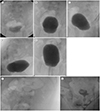
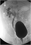
The grading for severity of VUR is according to the International Reflux Study in Children and is based on VCUG (Table 2) (12). The higher the grade and the prolonged the morbidity, worse is the prognosis. Although, mild VUR (grade I and II) resolves spontaneously with age in the first decade of life, timely detection and immediate intervention of severe VUR (grade Ⅲ, IV, and V) is critical to prevent sequels such as renal scarring, hypertension, and end-stage renal diseases (236). To achieve the ultimate goal of VCUG, acquaintance with VUR grading system and appropriate reporting of VUR is essential. Although, the degree of ureteral dilatation depicted in the grading system, criteria for mild, moderate and severe dilatation are not clear (12). The reflux nephropathy (renal scarring) is characterized by atrophy, tubular destruction, and interstitial fibrosis, and almost always accompanies blunting or distortion of the calyx (318). Therefore, it is important to examine calyceal blunting clearly.
In key features, grade I reflux does not reach the renal pelvis and has a questionable clinical impact (Fig. 8) (23). Grade II reflux reaches the renal pelvis but does not dilate fornices (Fig. 9). Grade III reflux does not or minimally deform fornices (Fig. 10). It is borderline grade between grade II and grade IV reflux. Grade IV reflux presents blunt fornices, but the impression of papillae is not visible in majority of papillae (Fig. 11). Grade V reflux is characterized by loss of papillary impression (Fig. 12). In reporting, the maximal degree of VUR is described (12).
BLADDER AND URETHRA
On VCUG, the inner wall of urinary bladder on AP projection is smooth and its contour is round. The interureteric ridge which is the elevated border of trigone in cephalad, is visible on the lateral projection at about distal one-quarter of the posteroinferior urinary bladder (Fig. 13). Any other space occupying lesions, fistulous or diverticular structures suggesting mass, urachal remnant, or diverticulum observed during VCUG should be reported.
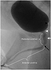
Urethral evaluation is critical in boys (Fig. 13). Therefore, it is better to prepare the field of view and lateral position to demonstrate optimal view of urethra (4). Posterior urethral valve is the result of remaining and fused anterior mucosal folds which should be absorbed during normal development. Posterior urethral valve may lead to decompensation of the urinary bladder, distal ureteral obstruction, and end-stage renal disease (1). Oligohydramnios, bladder distention, bilateral hydroureteronephrosis, and occasionally fetal ascites are the manifestation of the severe obstruction of posterior urethral valve (12). Normal fold at posterior urethra is often seen, therefore, along with flap-like filling defect, disproportional dilatation of posterior urethra during voiding is also observed in posterior urethral valve (1). Removal of the catheter during voiding is recommended for proper evaluation of posterior urethral valve (1).
MISCELLANEOUS CONDITIONS
CONTRAST REFLUX OR INADVERTENT CATHETER INSERTION INTO THE VAGINA
It is important to insert the catheter via urethral opening and check that the catheter is properly inserted before infusing the contrast. If the catheter enters the vagina, remove it immediately. During voiding, contrast reflux into the vagina is common in girls (Fig. 15). There are various causes of vaginal reflux which are not clear; however, adhesions of the labia minora, ectopic ureteral insertion, and dysfunctional pelvic floor musculature are discussed (221).
INTRA-RENAL REFLUX
Intra-renal reflux refers to reflux of contrast into renal papilla via ducts of Bellini (1). It is not included in the international criteria for grading VUR; however, it should be described separately because it is an important factor for renal scarring in ascending infection (1). The prevailing sites for intrarenal reflux are upper pole and lower pole of the kidney, respectively, due to presence of compound papillae (Fig. 16) (2).
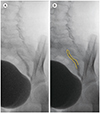
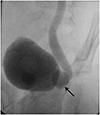
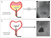
PARAURETERAL (HUTCH) DIVERTICULUM
Diverticulum at UVJ is called paraureteral or Hutch diverticulum (Fig. 17). It is associated with VUR by negating normal arrangement of UVJ (Fig. 18) (2). Therefore, VUR from Hutch diverticulum does not resolve without surgical correction and leaves renal scar (2). In this regard, radiologists are recommended to capture images of the urinary bladder in oblique projection focusing UVJ and should report the presence of Hutch diverticulum.
ERRONEOUS PERFORMANCE
Obtaining images without collimation (Fig. 19A) and excessive number of images are common errors made by beginners (Fig. 19B). Radiologists need to remember the required adjustments to achieve ALARA principle in practice before performing VCUG.
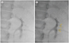
Although, contrast mixture was strictly prepared according to the standards, insufficient opacity of contrast may be due to its dilution with remaining urine in the bladder (Fig. 20A). Therefore, natural drainage or regurgitation with the syringe after anchoring catheter on the skin is necessary (Fig. 20B).
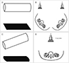
The presence or absence of calyceal blunting is an important factor in determining VUR grading. The orthographic projection is a method in which an object is imaged using projection parallel to the principle axes (Fig. 21A, B) (22). The actual contour of calyx can be altered by oblique projection (Fig. 21C, D). Therefore, an accurate evaluation of the orthogonal plane is required. The orthogonal plane for calyceal evaluation is tangential to the renal hilar axis which is directed anteromedially (Fig. 21B). It is recommended to prepare the patient's body for the tangential projection of the expected renal hilum.
CONCLUSION
VCUG is the most common and standard test demonstrating VUR. Radiologists need to know the clinical issues of VCUG in advance, prepare the proper supplies, and take appropriate action and judgment accordingly. They should be familiar with the essential elements of grading, calyceal blunting, papillary impressions, and various conditions that may be associated with it.


 XML Download