

 PDF
PDF ePub
ePub Citation
 Print
Print


INTRODUCTION
Currently, the World Health Organization (WHO) classifies uterine smooth muscle neoplasms that cannot be clearly diagnosed as benign or malignant based on generally applied histopathologic parameters as “smooth muscle tumors of uncertain malignant potential (STUMP)” (1). Extrauterine STUMPs are very rare; only one metastasizing case, in the retroperitoneum, has been reported (2). Herein, we report on the first case of a STUMP that originated from the superior mesenteric vein (SMV), with emphasis on the radiologic findings.
CASE REPORT
A 59-year-old woman was referred to our hospital for a pancreatic mass incidentally found on abdominal ultrasonography, which had been performed as part of her general health screening. She had an unremarkable medical history and no symptoms except mild, intermittent epigastric pain; her laboratory findings were also not remarkable. As for a tumor marker, the result of a carbohydrate antigen 19-9 test was also within the normal range.
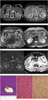
The patient underwent computed tomography (CT) and magnetic resonance imaging (MRI) for further evaluation. The contrast-enhanced CT images showed a 4 × 3 × 3 cm sized smooth-margined mass compressing the pancreas head, with a sharp beak-shaped interface (Fig. 1A). The mass was encasing the entire circumference of the SMV (Fig. 1A). Progressive homogeneous enhancement was seen on the arterial and portal venous phases (Fig. 1A). Similarly, gadoxetic acid-enhanced MR images revealed progressive homogeneous enhancement, and the lesion on MRI showed mild hypointensity on a T1-weighted image (Fig. 1B) and mild hyperintensity on a T2-weighted image when compared with the skeletal muscle (Fig. 1B). The lesion also showed hyperintensity on a diffusion-weighted image (DWI) obtained with a b value of 600 sec/mm2 and hypointensity on an apparent diffusion coefficient (ADC) map (Fig. 1C). The main pancreatic duct was not dilated. Based on these imaging findings, the mass was first considered to be a SMV-origin tumor, such as leiomyoma. The differential diagnosis was a nonepithelial tumor of the pancreas with SMV invasion.
During an exploratory laparotomy a round encapsulated tumor firmly attached to the wall of the SMV was identified in the pancreas head. En bloc resection, with an involved part of the vein, was performed and the tumor was easily dissected from the pancreas. Grossly, the excised mass was 4.4 × 4.3 × 2.5 cm in size, and the cut surface revealed a firm, circumscribed grayish white mass encircling the SMV (Fig. 1D). Histologically, the tumor was characterized by interlacing bundles of compact uniform spindled cells with little collagen. The tumor showed diffuse moderate cytologic atypia, four mitoses events per 50 high-power fields, and no tumor cell necrosis (Fig. 1D). Immunohistochemical staining for smooth muscle actin (SMA), S-100, CD117, and Ki-67 was performed, and the results were positive for SMA (Fig. 1D). Additionally, 30% of tumor cells were positive for Ki-67. The final pathological diagnosis was a STUMP.
DISCUSSION
There is an ambiguous group of uterine smooth muscle neoplasms, with overlapping features between leiomyoma and leiomyosarcoma, that can be challenging for even the most experienced pathologist to diagnose. Therefore, the current WHO guidelines indicate that uterine smooth muscle neoplasms, that do not fit the definitions for any other categories, should be classified as STUMPs (1).
A number of investigators have studied parameters for assessing the diagnosis and prognosis of uterine smooth muscle neoplasms. One of the most widely used classification systems was proposed by Bell et al. (3). The histologic distinctions between the five subgroups are based on assessing a combination of features including cytologic atypia, mitotic index, and coagulative tumor cell necrosis. STUMPs are defined as tumors with the following features: 1) no, or no more than, mild cytologic atypia; no tumor cell necrosis; and a mitotic index of 5 ≤ mitoses < 20 per 10 high-power fields; 2) diffuse moderate to severe cytologic atypia, no tumor cell necrosis, and a mitotic index of < 10 mitoses per 10 high-power fields; 3) focal or multifocal moderate to severe cytologic atypia, no tumor cell necrosis, and a mitotic index of 1 < mitoses < 20 per 10 high-power fields; 4) no, to mild, cytologic atypia; tumor cell necrosis; and a mitotic index < 10 mitoses per 10 high-power fields.
In this case, the tumor showed diffuse moderate cytologic atypia, four mitoses events per 50 high-power fields, and no tumor cell necrosis; the histologic appearance of the smooth muscle neoplasm in our case fulfilled the Bell's criteria for atypical leiomyoma with a low risk of recurrence. For this reason, our final diagnosis was a STUMP, even though STUMP is typically a term used only with uterine neoplasms. We found only one case report of an extrauterine STUMP that arose from the retroperitoneum with lung metastasis after a hysterectomy (4). To the best of our knowledge, this is the first case report of a STUMP that originated from the SMV, without a primary uterine STUMP.
Regardless of their anatomic locations, classic leiomyomas have signal intensities similar to those of skeletal and smooth muscles on T1- and T2-weighted MR images. Upon histologic examination, non-degenerated leiomyomas are composed of whorls of uniform smooth muscle cells with varying amounts of intervening collagen. Cellular leiomyoma is a specific subtype of leiomyoma composed of compact smooth muscles cells with little or no collagen that can have relatively increased T2-weighted signal intensity and homogeneous enhancement on contrast-enhanced images. On the other hand, high signal intensity on T2-weighted images can also be caused by myxoid or cystic degeneration (5). However, mild hyperintense tumors on T2-weighted images were more likely to be highly cellular tumors, whereas markedly hyperintense tumors with heterogeneous architecture tended to be degenerated tumors (6). Because our case showed mild hyperintensity on T2-weighted images compared with the skeletal muscle and homogeneous enhancement on contrast-enhanced T1-weighted images, the cause of the increased signal intensity, unlike with a usual leiomyoma, was likely high cellularity rather than myxoid or cystic degeneration. Pathologic findings also revealed a uniform cellular mass composed of compact smooth muscle cells with little collagen. Only one study described the MR characteristics of three uterine STUMPs (7); one of the three STUMPs resembled the MR findings of ordinary leiomyoma, but, in the other two cases, more than half of the mass showed hyperintensity on T2-weighted images.
In this case, the mass showed high signal intensity on a DWI and low signal intensity on an ADC map. Because previous investigations have shown that ADC values of the tissues are dependent on the cellularity and amount of tissue fibrosis (8), we presumed that high cellularity was responsible for the low ADC value in this case.
Prognostic significance is not well-known because of insufficient cases of extrauterine STUMPs. A number of studies have shown that uterine STUMPs are usually clinically benign, but that they can occasionally recur or metastasize to distant sites (9). Therefore, they should be considered tumors with low malignant potential, but patients who are diagnosed with STUMPs require close, long-term follow-up. Additionally, radiologic clues for suspected STUMPs might be important.
In conclusion, we report on the first case report of an extrauterine STUMP, that originated from the SMV, without a primary uterine STUMP. Because STUMPs can occasionally recur or metastasize, radiologic clues about the suspect STUMP might be important. Imaging findings of STUMPs are not well established, but a STUMP can show slightly high signal intensity on T2-weighted imaging with homogeneous or heterogeneous contrast-enhancement. Additionally, diffusion restriction could be seen in the STUMP due to its high cellularity.
 XML Download