Abstract
Digital breast tomosynthesis (DBT) has been shown to be a promising imaging technique for breast cancer detection. DBT involves acquisition of a series of images in different planes over a limited angular range, and their subsequent reconstruction into a quasi-three-dimensional breast volume. This reduces the effects of tissue overlap. This review aims to describe the key features of DBT, including technique, results from recent retrospective and prospective clinical studies, and issues with DBT as a screening tool.
References
1. Duffy SW, Tabár L, Smith RA. The mammographic screening trials: commentary on the recent work by Olsen and G⊘tzsche. CA Cancer J Clin. 2002; 52:68–71.

2. Independent UK Panel on Breast Cancer Screening. The benefits and harms of breast cancer screening: an independent review. Lancet. 2012; 380:778–1786.
3. Tabar L, Fagerberg G, Chen HH, Duffy SW, Smart CR, Gad A, et al. Efficacy of breast cancer screening by age. New results from the Swedish two-county trial.Cancer. 1995; 75:2507–2517.
4. Nyström L, Andersson I, Bjurstam N, Frisell J, Nordenskjöld B, Rutqvist LE. Long-term effects of mammography screening: updated overview of the Swedish randomised trials.Lancet. 2002; 359:909–919.
5. Berry DA, Cronin KA, Plevritis SK, Fryback DG, Clarke L, Zelen M, et al. Effect of screening and adjuvant therapy on mortality from breast cancer.N Engl J Med. 2005; 353:1784–1792.
6. Roubidoux MA, Bailey JE, Wray LA, Helvie MA. Invasive cancers detected after breast cancer screening yielded a negative result: relationship of mammographic density to tumor prognostic factors.Radiology. 2004; 230:42–48.
7. Kolb TM, Lichy J, Newhouse JH. Comparison of the performance of screening mammography, physical examination, and breast US and evaluation of factors that influence them: an analysis of 27,825 patient evaluations. Radiology. 2002; 225:165–175.

8. Mandelson MT, Oestreicher N, Porter PL, White D, Finder CA, Taplin SH, et al. Breast density as a predictor of mammographic detection: comparison of interval- and screen-detected cancers.J Natl Cancer Inst. 2000; 92:1081–1087.
9. Kerlikowske K, Grady D, Barclay J, Sickles EA, Ernster V. Effect of age, breast density, and family history on the sensitivity of first screening mammography. JAMA. 1996; 276:33–38.

10. Haas JS, Kaplan CP. The divide between breast density notification laws and evidencebased guidelines for breast cancer screening: legislating practice. JAMA Intern Med. 2015; 175:1439–1440.
11. Lee CI, Bassett LW, Lehman CD. Breast density legislation and opportunities for patient-centered outcomes research. Radiology. 2012; 264:632–636.

12. Park JM, Franken EA Jr, Garg M, Fajardo LL, Niklason LT. Breast tomosynthesis: present considerations and future applications. Radiographics. 2007; 27(Suppl 1):S231–240.

13. Helvie MA. Digital mammography imaging: breast tomosynthesis and advanced applications.Radiol Clin North Am. 2010; 48:917–929.
14. Niklason LT, Christian BT, Niklason LE, Kopans DB, Castleberry DE, Opsahl-Ong BH, et al. Digital tomosynthesis in breast imaging. Radiology. 1997; 205:399–406.

15. Gur D, Zuley ML, Anello MI, Rathfon GY, Chough DM, Ganott MA, et al. Dose reduction in digital breast tomosynthesis (DBT) screening using synthetically reconstructed projection images: an observer performance study.Acad Radiol. 2012; 19:166–171.
16. Rafferty EA, Park JM, Philpotts LE, Poplack SP, Sumkin JH, Halpern EF, et al. Diagnostic accuracy and recall rates for digital mammography and digital mammography combined with one-view and two-view tomosynthesis: results of an enriched reader study. AJR Am J Roentgenol. 2014; 202:273–281.

17. Friedewald SM, Rafferty EA, Rose SL, Durand MA, Plecha DM, Greenberg JS, et al. Breast cancer screening using tomosynthesis in combination with digital mammography. JAMA. 2014; 311:2499–2507.

18. Gilbert FJ, Tucker L, Gillan MG, Willsher P, Cooke J, Duncan KA, et al. The TOMMY trial: a comparison of TOMosynthesis with digital MammographY in the UK NHS breast screening programme–a multicentre retrospective reading study comparing the diagnostic performance of digital breast tomosynthesis and digital mammography with digital mammography alone.Health Technol Assess. 2015; 19:i–xxv. 1–136.
19. Poplack SP, Tosteson TD, Kogel CA, Nagy HM. Digital breast tomosynthesis: initial experience in 98 women with abnormal digital screening mammography. AJR Am J Roentgenol. 2007; 189:616–623.

20. Gilbert FJ, Tucker L, Gillan MG, Willsher P, Cooke J, Duncan KA, et al. Accuracy of digital breast tomosynthesis for depicting breast cancer subgroups in a UK retrospective reading study (TOMMY Trial).Radiology. 2015; 277:697–706.
21. Good WF, Abrams GS, Catullo VJ, Chough DM, Ganott MA, Hakim CM, et al. Digital breast tomosynthesis: a pilot observer study. AJR Am J Roentgenol. 2008; 190:865–869.

22. Andersson I, Ikeda DM, Zackrisson S, Ruschin M, Svahn T, Timberg P, et al. Breast tomosynthesis and digital mammography: a comparison of breast cancer visibility and BIRADS classification in a population of cancers with subtle mammographic findings.Eur Rad/iiol. 2008; 18:2817–2825.
23. Shin SU, Chang JM, Bae MS, Lee SH, Cho N, Seo M, et al. Comparative evaluation of average glandular dose and breast cancer detection between single-view digital breast tomosynthesis (DBT) plus single-view digital mammography (DM) and two-view DM: correlation with breast thickness and density. Eur Radiol. 2015; 25:1–8.

24. Vedantham S, Karellas A, Vijayaraghavan GR, Kopans DB. Digital breast tomosynthesis: state of the art. Radiology. 2015; 277:663–684.

25. Wallis MG, Moa E, Zanca F, Leifland K, Danielsson M. Two-view and single-view tomosynthesis versus full-field digital mammography: high-resolution X-ray imaging observer study. Radiology. 2012; 262:788–796.

26. Förnvik D, Kataoka M, Iima M, Ohashi A, Kanao S, Toi M, et al. The role of breast tomosynthesis in a predominantly dense breast population at a tertiary breast centre: breast density assessment and diagnostic performance in comparison with MRI. Eur Rad/iiol. 2018; 28:3194–3203.

27. Skaane P, Bandos AI, Gullien R, Eben EB, Ekseth U, Haakenaasen U, et al. Comparison of digital mammography alone and digital mammography plus tomosynthesis in a population-based screening program.Radiology. 2013; 267:47–56.
28. Ciatto S, Houssami N, Bernardi D, Caumo F, Pellegrini M, Brunelli S, et al. Integration of 3D digital mammography with tomosynthesis for population breast-cancer screening (STORM): a prospective comparison study. Lancet Oncol. 2013; 14:583–589.

29. Zackrisson S, Lång K, Rosso A, Johnson K, Dustler M, Förnvik D, et al. One-view breast tomosynthesis versus two-view mammography in the Malmö breast tomosynthesis screening trial (MBTST): a prospective, population-based, diagnostic accuracy study. Lancet Oncol. 2018; 19:1493–1503.

30. Kim JY, Kang HJ, Shin JK, Lee NK, Song YS, Nam KJ, et al. Biologic profiles of invasive breast cancers detected only with digital breast tomosynthesis. AJR Am J Roentgenol. 2017; 209:1411–1418.

31. Lee SH, Chang JM, Shin SU, Chu AJ, Yi A, Cho N, et al. Imaging features of breast cancers on digital breast tomosynthesis according to molecular subtype: association with breast cancer detection.Br J Radiol. 2017; 90:20170470.
32. Bouwman RW, van Engen RE, Young KC, den Heeten GJ, Broeders MJ, Schopphoven S, et al. Average glandular dose in digital mammography and digital breast tomosynthesis: comparison of phantom and patient data. Phys Med B/iiol. 2015; 60:7893–7907.

33. Feng SS, Sechopoulos I. Clinical digital breast tomosynthesis system: dosimetric characterization.Radiology. 2012; 263:35–42.
34. Skaane P, Bandos AI, Eben EB, Jebsen IN, Krager M, Haakenaasen U, et al. Two-view digital breast tomosynthesis screening with synthetically reconstructed projection images: comparison with digital breast tomosynthesis with full-field digital mammographic images. Radiology. 2014; 271:655–663.

35. Zuley ML, Guo B, Catullo VJ, Chough DM, Kelly AE, Lu AH, et al. Comparison of two-dimensional synthesized mammograms versus original digital mammograms alone and in combination with tomosynthesis images. Radiology. 2014; 271:664–671.

36. Zuckerman SP, Conant EF, Keller BM, Maidment AD, Barufaldi B, Weinstein SP, et al. Implementation of synthesized two-dimensional mammography in a population-based digital breast tomosynthesis screening program. Radiology. 2016; 281:730–736.
37. Aujero MP, Gavenonis SC, Benjamin R, Zhang Z, Holt JS. Clinical performance of synthesized two-dimensional mammography combined with tomosynthesis in a large screening population. Rad/iiology. 2017; 283:70–76.
38. Gur D, Abrams GS, Chough DM, Ganott MA, Hakim CM, Perrin RL, et al. Digital breast tomosynthesis: observer performance study. AJR Am J Roentgenol. 2009; 193:586–591.

39. Dang PA, Freer PE, Humphrey KL, Halpern EF, Rafferty EA. Addition of tomosynthesis to conventional digital mammography: effect on image interpretation time of screening examinations. Radiology. 2014; 270:49–56.

40. Kopans D, Gavenonis S, Halpern E, Moore R. Calcifications in the breast and digital breast tomosynthesis. Breast J. 2011; 17:638–644.

41. Michell MJ, Iqbal A, Wasan RK, Evans DR, Peacock C, Lawinski CP, et al. A comparison of the accuracy of film-screen mammography, full-field digital mammography, and digital breast tomosynthesis.Clin Radiol. 2012; 67:976–981.
42. Spangler ML, Zuley ML, Sumkin JH, Abrams G, Ganott MA, Hakim C, et al. Detection and classification of calcifications on digital breast tomosynthesis and 2D digital mammography: a comparison.AJR Am J Roentgenol. 2011; 196:320–324.
43. Gilbert FJ, Tucker L, Young KC. Digital breast tomosynthesis (DBT): a review of the evidence for use as a screening tool. Clin Radiol. 2016; 71:141–150.

44. Chu AJ, Cho N, Chang JM, Kim WH, Lee SH, Song SE, et al. 3D computer-aided detection for digital breast tomosynthesis: comparison with 2D computer-aided detection for digital mammography in the detection of calcifications. J Korean Soc Radiol. 2017; 77:105–112.

45. Lourenco AP, Barry-Brooks M, Baird GL, Tuttle A, Mainiero MB. Changes in recall type and patient treatment following implementation of screening digital breast tomosynthesis. Radiology. 2015; 274:337–342.

46. Lång K, Nergården M, Andersson I, Rosso A, Zackrisson S. False positives in breast cancer screening with one-view breast tomosynthesis: an analysis of findings leading to recall, workup and biopsy rates in the Malmö breast tomosynthesis screening trial. Eur Radiol. 2016; 26:3899–3907.

47. Alshafeiy TI, Nguyen JV, Rochman CM, Nicholson BT, Patrie JT, Harvey JA. Outcome of architectural distortion detected only at breast tomosynthesis versus 2D mammography. Radiology. 2018; 288:38–46.
Fig. 1.
Digital breast tomosynthesis images with multiple low-dose X-ray exposures are obtained as the X-ray tube moves over a limited angular range above the breast. These images are reconstructed into a quasi-three-dimensional breast volume.
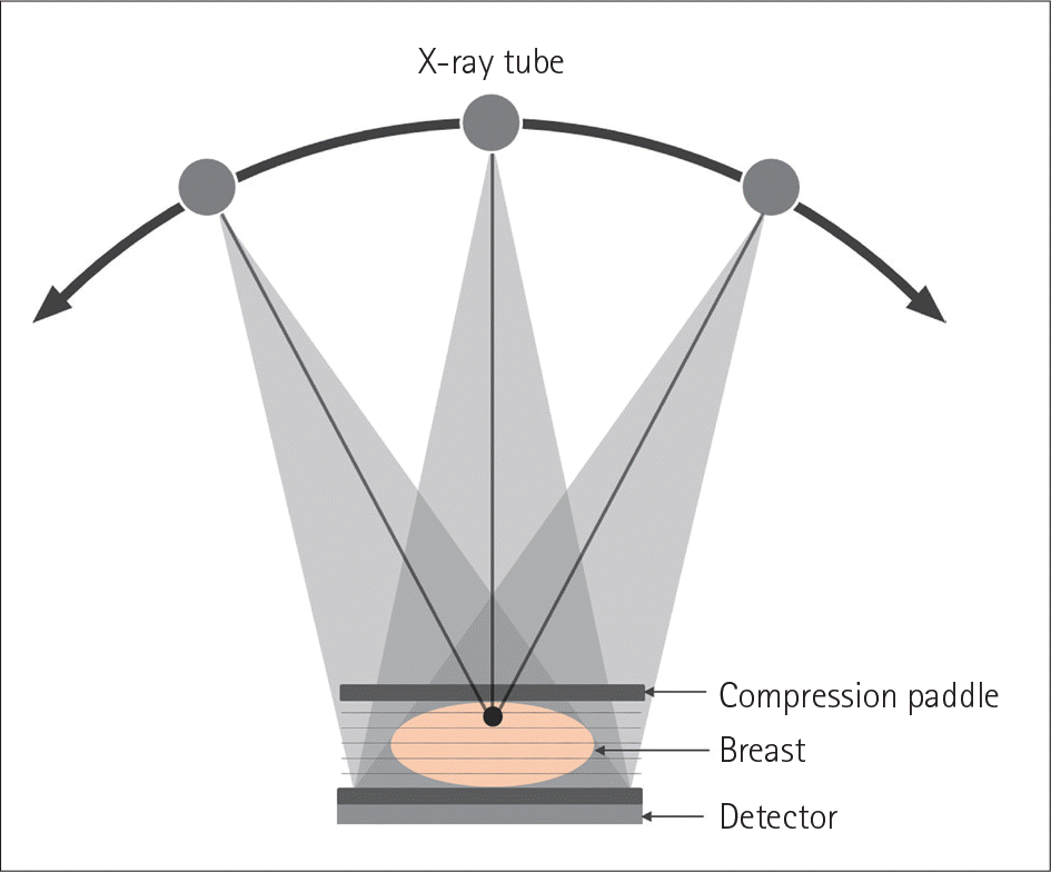
Fig. 2.
A 55-year-old woman who underwent DBT and FFDM for screening. A. CC view of the right breast on FFDM shows heterogeneously dense composition without definite focal abnormality. B. CC view on DBT shows an irregular mass (arrow) in the outer portion of the right breast. C. Ultrasonography shows a 4-mm hypoechoic mass in the right breast. Invasive ductal carcinoma was diagnosed on ultrasound-guided biopsy. CC = craniocaudal, DBT = digital breast tomosynthesis, FFDM = full-field digital mammography
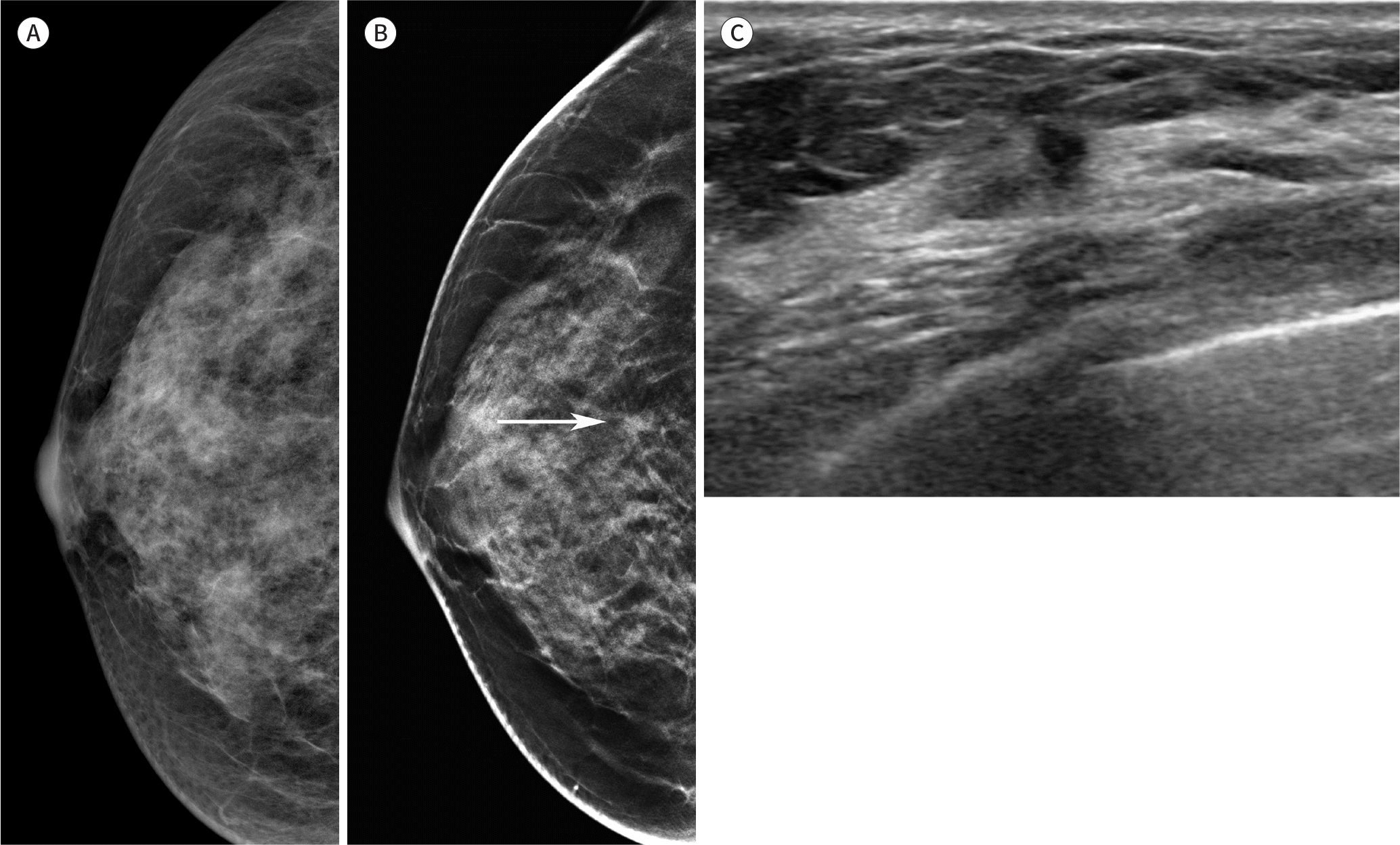
Fig. 3.
Example of superimposed tissue that mimics an asymmetry on FFDM. A. CC view on FFDM shows an asymmetry (arrow) in the inner portion of the left breast that needs to be recalled without DBT images. B, C. CC view on DBT (B) and CC view on synthetic mammogram (C) reveals fibroglandular tissues (arrows) without associated mass. The patient can return for the annual screening. CC = craniocaudal, DBT = digital breast tomosynthesis, FFDM = full-field digital mammography
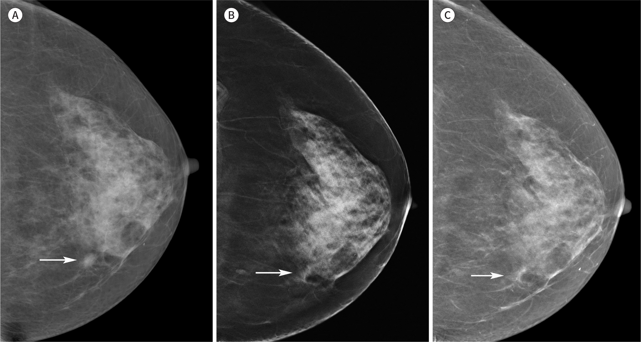
Fig. 4.
A 45-year-old woman with invasive ductal carcinoma. A. CC view on full-field digital mammography shows a subtle architectural distortion (arrow) in the right breast. B. CC view on digital breast tomosynthesis shows a more conspicuous mass with spiculated margins, which is highly suggestive of malignancy (arrow). CC = craniocaudal
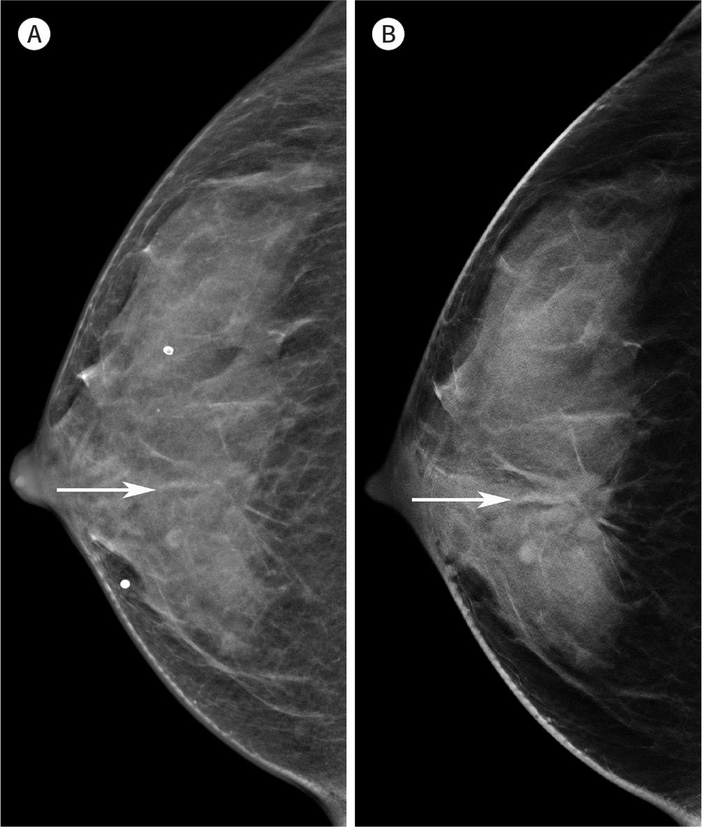
Fig. 5.
A 57-year-old woman who underwent DBT and FFDM for screening. A. The MLO view on FFDM shows an irregular-shaped, isodense mass with indistinct margins (arrows) in the upper portion of the left breast. B. MLO view on DBT demonstrates an oval-shaped, hyperdense mass with circumscribed margins, likely a benign finding. C. Ultrasonography shows a 17-mm, oval-shaped, isoechoic mass in the left breast. It was suspected to be a benign mass and was unchanged at the 5-year follow-up. DBT = digital breast tomosynthesis, FFDM = full-field digital mammography, MLO = mediolateral oblique
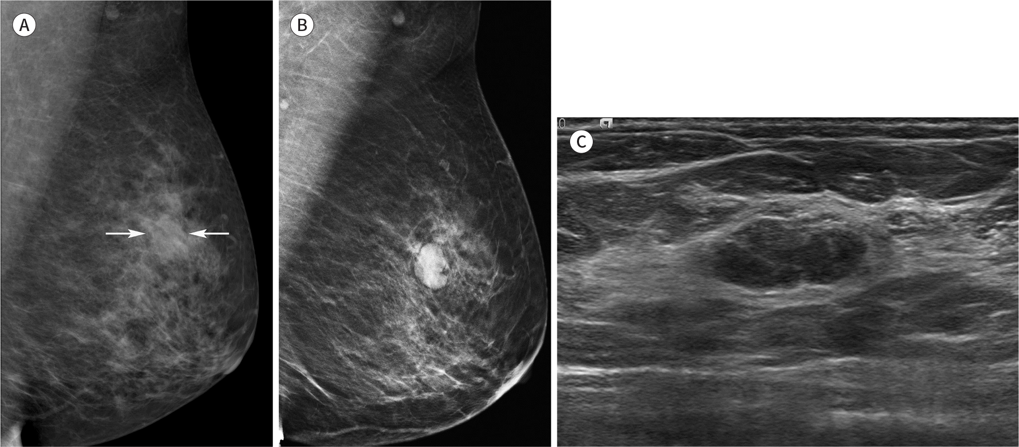
Fig. 6.
A 46-year-old woman with invasive ductal carcinoma. A. Craniocaudal view on FFDM shows an oval-shaped, isodense mass (arrows) in the central portion of the right breast. B. An irregular mass with spiculated margins (arrows) is clearly revealed on digital breast tomosynthesis. An oval isodense mass (arrowheads) is also noted in the inner portion of the right breast, which was not clearly visible on FFDM. It was suspected to be a benign mass on ultrasonography (not shown). FFDM = full-field digital mammography
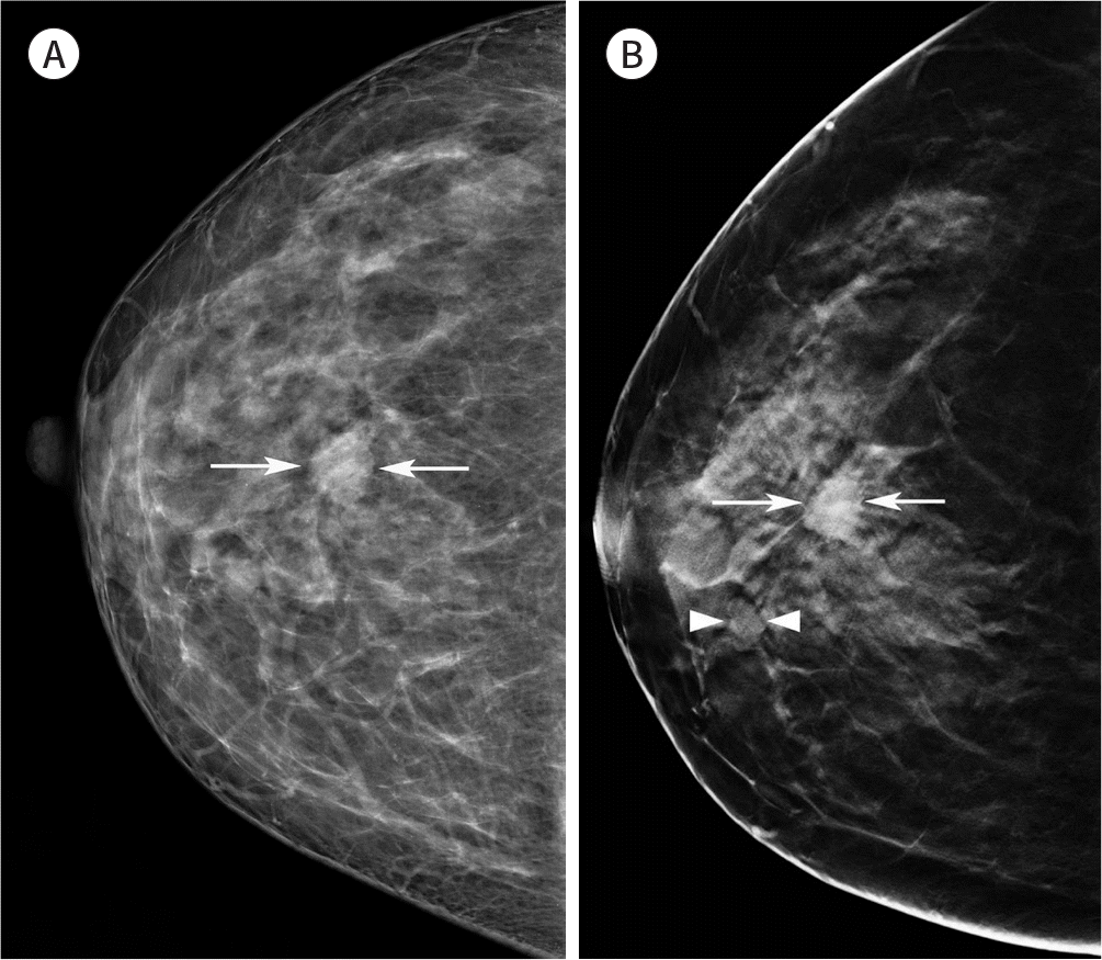
Fig. 7.
A 52-year-old woman with ductal carcinoma in situ of the left breast. A, B. Fine pleomorphic calcifications (squares) in the left upper breast are equally visible on full-field digital mammography (A) and syntheticmam- mography (B).
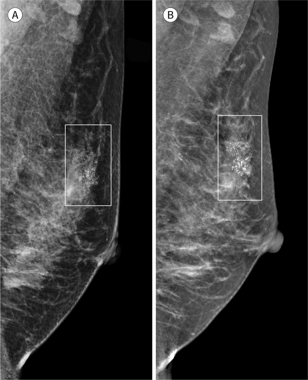
Table 1.
Comparison of Clinical Digital Breast Tomosynthesis Symtems


 PDF
PDF ePub
ePub Citation
 Print
Print


 XML Download