

 PDF
PDF ePub
ePub Citation
 Print
Print


Abstract
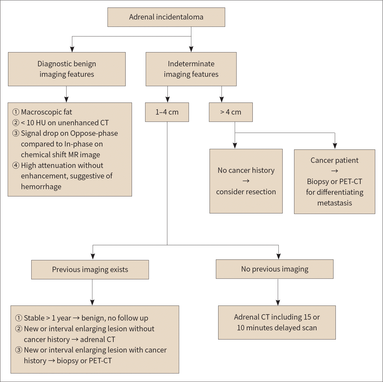
Adrenal incidentaloma refers to adrenal masses that are accidentally found on imaging performed for other reasons, without clinical symptoms of adrenal disease. Generally, adrenal masses measuring less than 1 cm are not considered adrenal incidentalomas. The purpose of radiologic examination in evaluating non-functioning adrenal incidentalomas is to distinguish between benign and malignant masses to establish the treatment plan. In this review, based on previously published research and recommendations, we describe the radiologic approach for adrenal incidentaloma and describe the imaging findings of representative diseases.
Go to : 
References
1. Young WF Jr. Clinical practice. The incidentally discovered adrenal mass.N Engl J Med. 2007; 356:601–610.
2. Blake MA, Cronin CG, Boland GW. Adrenal imaging. AJR Am J Roentgenol. 2010; 194:1450–1460.

3. Willatt JM, Francis IR. Radiologic evaluation of incidentally discovered adrenal masses. Am Fam Physician. 2010; 81:1361–1366.
4. Mansmann G, Lau J, Balk E, Rothberg M, Miyachi Y, Bornstein SR. The clinically inapparent adrenal mass: update in diagnosis and management. Endocr Rev. 2004; 25:309–340.
5. Glazer DI, Mayo-Smith WW. Management of incidental adrenal masses: an update. Abdom Radiol (NY) 2019 Jul 29 [Epub]. Available at.https://doi.org/10.1007/s00261-019–02149–2.
6. Lenert JT, Barnett CC Jr, Kudelka AP, Sellin RV, Gagel RF, Prieto VG, et al. Evaluation and surgical resection of adrenal masses in patients with a history of extra-adrenal malignancy.Surgery. 2001; 130:1060–1067.
7. Frilling A, Tecklenborg K, Weber F, Kühl H, Müller S, Stamatis G, et al. Importance of adrenal incidentaloma in patients with a history of malignancy.Surgery. 2004; 136:1289–1296.
8. Mayo-Smith WW, Song JH, Boland GL, Francis IR, Israel GM, Mazzaglia PJ, et al. Management of incidental adrenal masses: a white paper of the ACR Incidental Findings Committee.J Am Coll Radiol. 2017; 14:1038–1044.
9. Park JJ, Park BK, Kim CK. Adrenal imaging for adenoma characterization: imaging features, diagnostic accuracies and differential diagnoses. Br J Radiol. 2016; 89:20151018.
10. Paulsen SD, Nghiem HV, Korobkin M, Caoili EM, Higgins EJ. Changing role of imaging-guided percutaneous biopsy of adrenal masses: evaluation of 50 adrenal biopsies.AJR Am J Roentgenol. 2004; 182:1033–1037.
11. Korobkin M, Giordano TJ, Brodeur FJ, Francis IR, Siegelman ES, Quint LE, et al. Adrenal adenomas: relationship between histologic lipid and CT and MR findings. Radiology. 1996; 200:743–747.
12. Boland GW, Lee MJ, Gazelle GS, Halpern EF, McNicholas MM, Mueller PR. Characterization of adrenal masses using unenhanced CT: an analysis of the CT literature. AJR Am J Roentgenol. 1998; 171:201–204.
13. Elsayes KM, Emad-Eldin S, Morani AC, Jensen CT. Practical approach to adrenal imaging.Radiol Clin North Am. 2017; 55:279–301.
14. Katabathina VS, Flaherty E, Kaza R, Ojili V, Chintapalli KN, Prasad SR. Adrenal collision tumors and their mimics: multimodality imaging findings.Cancer Imag/iing. 2013; 13:602–610.
15. Untch BR, Shia J, Downey RJ, Carrasquillo JA, Panicek DM, Strong VE. Imaging and management of a small cell lung cancer metastasis/adrenal adenoma collision tumor: a case report and review of the literature. World J Surg Oncol. 2014; 12:45.
16. Mitchell DG, Crovello M, Matteucci T, Petersen RO, Miettinen MM. Benign adrenocortical masses: diagnosis with chemical shift MR imaging.Radiology. 1992; 185:345–351.
17. Korobkin M, Lombardi TJ, Aisen AM, Francis IR, Quint LE, Dunnick NR, et al. Characterization of adrenal masses with chemical shift and gadolinium-enhanced MR imaging. Radiology. 1995; 197:411–418.
18. Mayo-Smith WW, Lee MJ, McNicholas MM, Hahn PF, Boland GW, Saini S. Characterization of adrenal masses (< 5 cm) by use of chemical shift MR imaging: observer performance versus quantitative measures.AJR Am J Roentgenol. 1995; 165:91–95.
19. Israel GM, Korobkin M, Wang C, Hecht EN, Krinsky GA. Comparison of unenhanced CT and chemical shift MRI in evaluating lipid-rich adrenal adenomas. AJR Am J Roentgenol. 2004; 183:215–219.
20. Seo JM, Park BK, Park SY, Kim CK. Characterization of lipid-poor adrenal adenoma: chemical-shift MRI and washout CT.AJR Am J Roentgenol. 2014; 202:1043–1050.
21. Haider MA, Ghai S, Jhaveri K, Lockwood G. Chemical shift MR imaging of hyperattenuating (> 10 HU) adrenal masses: does it still have a role? Radiology. 2004; 231:711–716.
22. Korobkin M, Brodeur FJ, Francis IR, Quint LE, Dunnick NR, Londy F. CT time-attenuation washout curves of adrenal adenomas and nonadenomas.AJR Am J Roentgenol. 1998; 170:747–752.
23. Terzolo M, Stigliano A, Chiodini I, Loli P, Furlani L, Arnaldi G, et al. AME position statement on adrenal incidentaloma. Eur J Endocr/iinol. 2011; 164:851–870.
24. Park BK, Kim CK, Kim B. Adrenal incidentaloma detected on triphasic helical CT: evaluation with modified relative percentage of enhancement washout values.Br J Radiol. 2008; 81:526–530.
25. Liu T, Sun H, Zhang H, Duan J, Hu Y, Xie S. Distinguishing adrenal adenomas from non-adenomas with multidetector CT: evaluation of percentage washout values at a short time delay triphasic enhanced CT. Br J Radiol. 2019; 92:20180429.
26. Northcutt BG, Raman SP, Long C, Oshmyansky AR, Siegelman SS, Fishman EK, et al. MDCT of adrenal masses: can dual-phase enhancement patterns be used to differentiate adenoma and pheochromocytoma? AJR Am J Roentgenol. 2013; 201:834–839.
27. Barzon L, Sonino N, Fallo F, Palu G, Boscaro M. Prevalence and natural history of adrenal incidentalomas. Eur J Endocrinol. 2003; 149:273–285.
28. Choi YA, Kim CK, Park BK, Kim B. Evaluation of adrenal metastases from renal cell carcinoma and hepatocellular carcinoma: use of delayed contrast-enhanced CT. Radiology. 2013; 266:514–520.
29. Park SY, Park BK, Kim CK. The value of adding (18)F-FDG PET/CT to adrenal protocol CT for characterizing adrenal metastasis (≥10 mm) in oncologic patients. AJR Am J Roentgenol. 2014; 202:W153–W160.
30. Ho LM. Adrenal imaging: a three-category approach to managing incidentalomas. Appl Radiol. 2018; 47:8–13.
31. Allen BC, Francis IR. Adrenal imaging and intervention. Radiol Clin North Am. 2015; 53:1021–1035.
32. Rossi A, Incensati R. Bone tissue in adrenal myelolipoma: a case report. Tumori. 1998; 84:90–93.
33. Song JH, Chaudhry FS, Mayo-Smith WW. The incidental adrenal mass on CT: prevalence of adrenal disease in 1,049 consecutive adrenal masses in patients with no known malignancy. AJR Am J Roentgenol. 2008; 190:1163–1168.
34. Daneshmand S, Quek ML. Adrenal myelolipoma: diagnosis and management. Urol J. 2006; 3:71–74.
35. Palmer WE, Gerard-McFarland EL, Chew FS. Adrenal myelolipoma. AJR Am J Roentgenol. 1991; 156:724.
36. Taffel M, Haji-Momenian S, Nikolaidis P, Miller FH. Adrenal imaging: a comprehensive review. Radiol Clin North Am. 2012; 50:219–243.
37. Zhang HM, Perrier ND, Grubbs EG, Sircar K, Ye ZX, Lee JE, et al. CT features and quantification of the characteristics of adrenocortical carcinomas on unenhanced and contrast-enhanced studies. Clin Radiol. 2012; 67:38–46.
38. Kenney PJ, Wagner BJ, Rao P, Heffess CS. Myelolipoma: CT and pathologic features. Radiology. 1998; 208:87–95.
39. Otal P, Escourrou G, Mazerolles C, Janne d'Othee B, Mezghani S, Musso S, et al. Imaging features of uncommon adrenal masses with histopathologic correlation. Radiographics. 1999; 19:569–581.
40. Rozenblit A, Morehouse HT, Amis ES Jr. Cystic adrenal lesions: CT features. Radiology. 1996; 201:541–548.
41. Guo YK, Yang ZG, Li Y, Deng YP, Ma ES, Min PQ, et al. Uncommon adrenal masses: CT and MRI features with histopathologic correlation. Eur J Radiol. 2007; 62:359–370.
42. Udelsman R, Fishman EK. Radiology of the adrenal. Endocrinol Metab Clin North Am. 2000; 29:27–42.
43. Kawashima A, Sandler CM, Ernst RD, Takahashi N, Roubidoux MA, Goldman SM, et al. Imaging of nontraumatic hemorrhage of the adrenal gland. Radiographics. 1999; 19:949–963.
44. Lee JM, Kim MK, Ko SH, Koh JM, Kim BY, Kim SW, et al. Guidelines for the management of adrenal incidentaloma: the Korean Endocrine Society, Committee of Clinical Practice Guidelines. Korean J Med. 2017; 92:4–16.
45. Francis IR. Distinguishing benign from malignant adrenal masses. Cancer Imaging. 2003; 3:102–110.
46. Casola G, Nicolet V, VanSonnenberg E, Withers C, Bretagnolle M, Saba RM, et al. Unsuspected pheochromocytoma: risk of blood-pressure alterations during percutaneous adrenal biopsy. Radiology. 1986; 159:733–735.
47. Hagspiel KD. Manifestation of Hodgkin's lymphoma in an adrenal myelolipoma. Eur Radiol. 2005; 15:1757–1759.
48. Glazer HS, Lee JK, Balfe DM, Mauro MA, Griffith R, Sagel SS. Non-Hodgkin lymphoma: computed tomographic demonstration of unusual extranodal involvement. Radiology. 1983; 149:211–217.
49. Rha SE, Byun JY, Jung SE, Chun HJ, Lee HG, Lee JM. Neurogenic tumors in the abdomen: tumor types and imaging characteristics. Radiographics. 2003; 23:29–43.
50. Szolar DH, Korobkin M, Reittner P, Berghold A, Bauernhofer T, Trummer H, et al. Adrenocortical carcinomas and adrenal pheochromocytomas: mass and enhancement loss evaluation at delayed contrast-enhanced CT. Radiology. 2005; 234:479–485.
51. Park BK, Kim B, Ko K, Jeong SY, Kwon GY. Adrenal masses falsely diagnosed as adenomas on unenhanced and delayed contrast-enhanced computed tomography: pathological correlation. Eur Radiol. 2006; 16:642–647.
52. Yoon JK, Remer EM, Herts BR. Incidental pheochromocytoma mimicking adrenal adenoma because of rapid contrast enhancement loss.AJR Am J Roentgenol. 2006; 187:1309–1311.
53. Park SY, Park BK.Adrenal gland and retroperitoneum. In Korean Society of Urogenital Radiology, eds.Urogenital Radiology-Urogenital Imaging. 2nd ed. Seoul: Ilchokak;2006. p. 250–269.
54. Wajchenberg BL, Albergaria Pereira MA, Medonca BB, Latronico AC, Campos Carneiro P, Alves VA, et al. Adrenocortical carcinoma: clinical and laboratory observations. Cancer. 2000; 88:711–736.
56. Bharwani N, Rockall AG, Sahdev A, Gueorguiev M, Drake W, Grossman AB, et al. Adrenocortical carcinoma: the range of appearances on CT and MRI.AJR Am J Roentgenol. 2011; 196:W706–W714.
57. Korivi BR, Elsayes KM, De Castro SF, Garg N, Qayyum A. An update of practical CT adrenal imaging: what physicians need to know. Curr Radiol Rep. 2015; 3:12.
Go to :
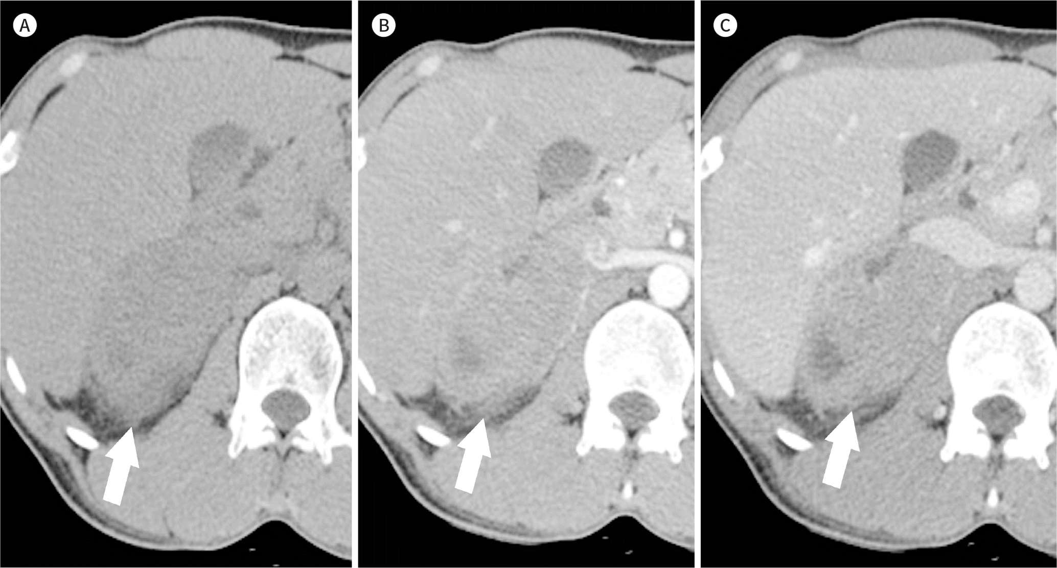
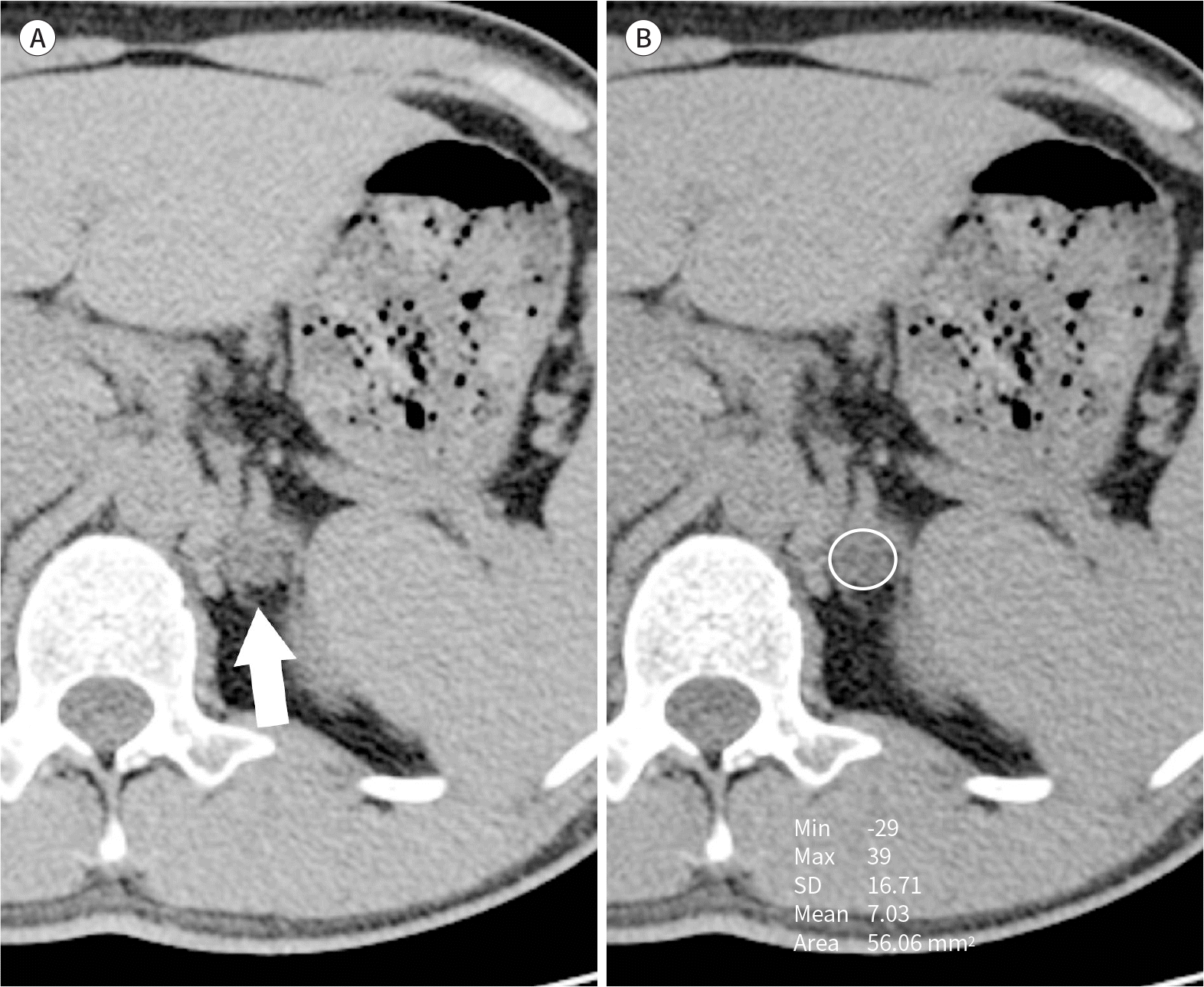 | Fig. 1.Lipid-rich adenoma in the left adrenal gland of a 31-year-old man. A, B. Unenhanced CT shows a 1.3-cm well-defined nodule in the left adrenal gland (A, arrow), with an attenuation value of 7 HU (under 10 HU) (B). HU = Hounsfield unit, SD = standard deviation |
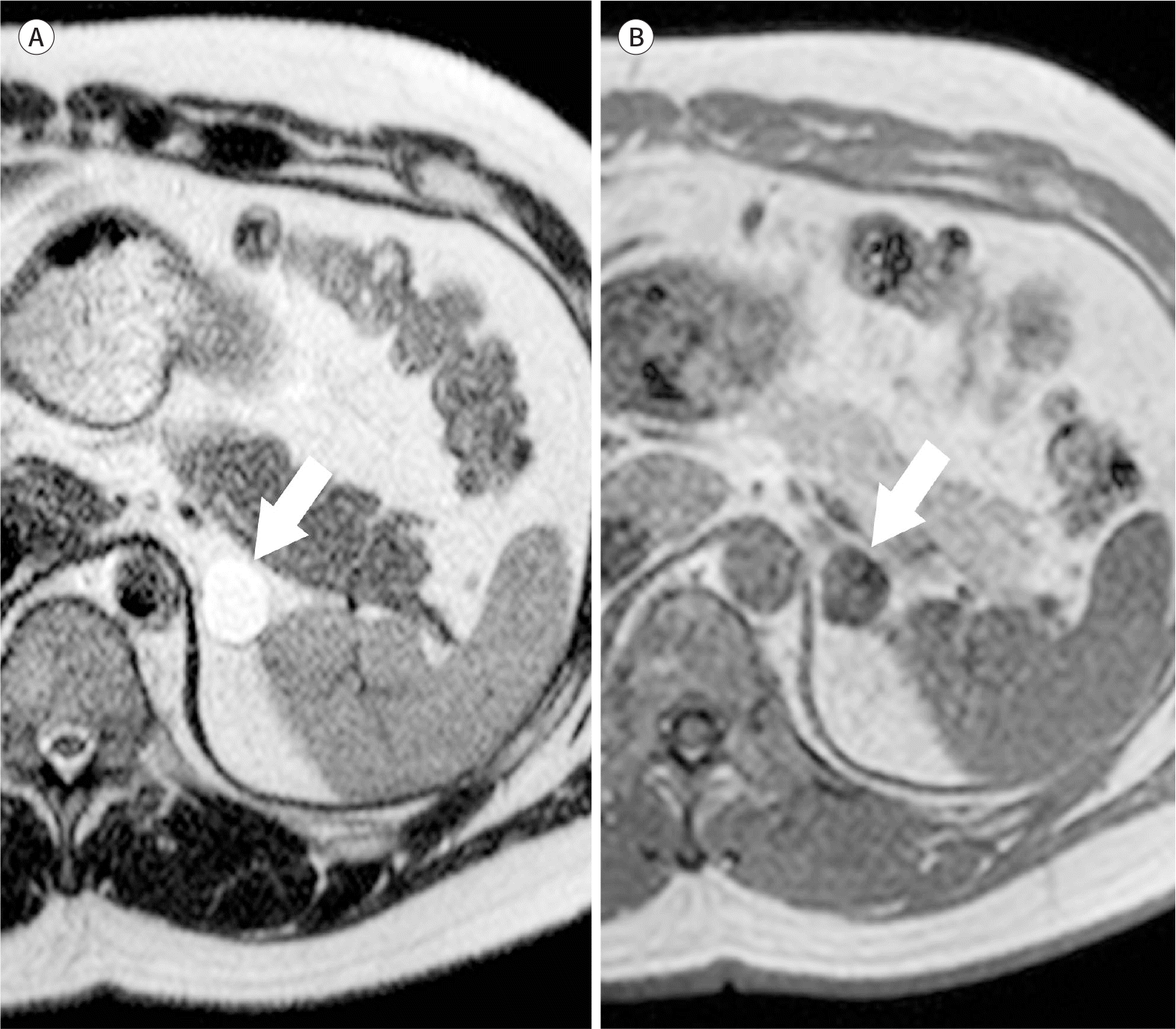
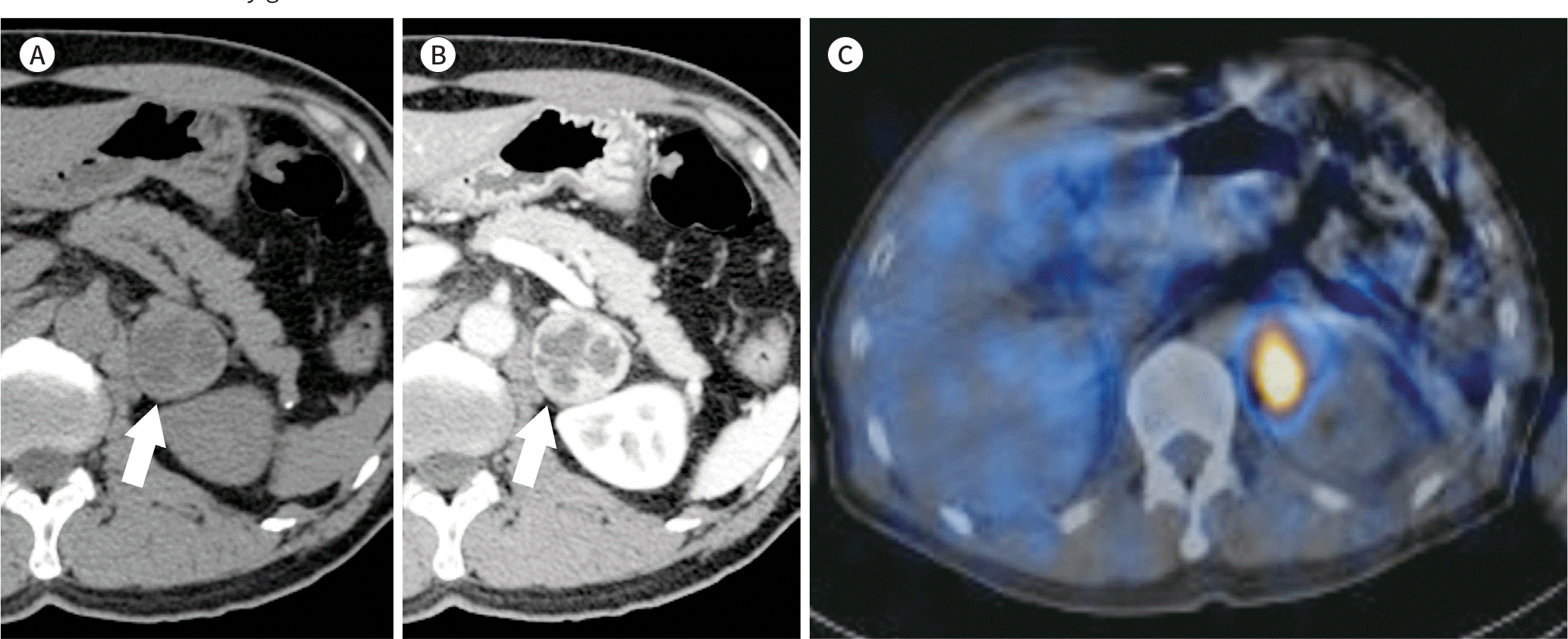
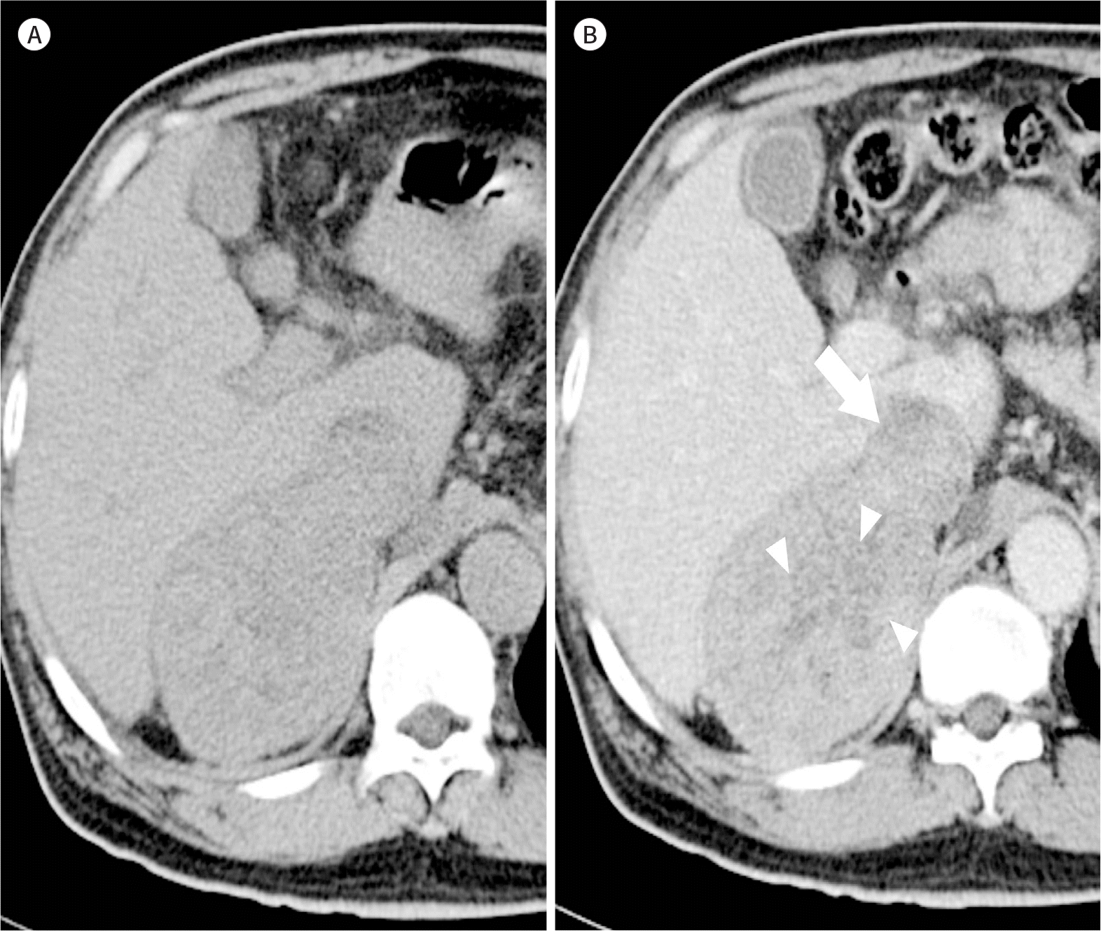
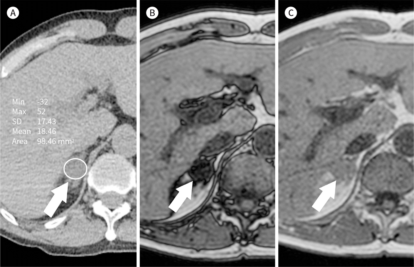 | Fig. 2.Chemical shift imaging. A-C. On unenhanced CT (A) of a 64-year-old man with gastric cancer, a solid mass (arrows) measuring 2.2 cm is observed in the right adrenal gland. The attenuation value is 18.5 HU, which makes it difficult to differentiate between adrenal adenoma and adrenal metastasis. The signal intensity of the right adrenal mass is visually reduced in the opposed-phase image (B) compared to the in-phase image (C). In the quantitative analysis, the adrenal-to-spleen ratio was 0.20, and the signal intensity index was 81.0%. Therefore, the rightadrenal nodule could be diagnosed as adrenal adenoma rather than adrenal metastasis. HU = Hounsfield unit, SD = standard deviation |
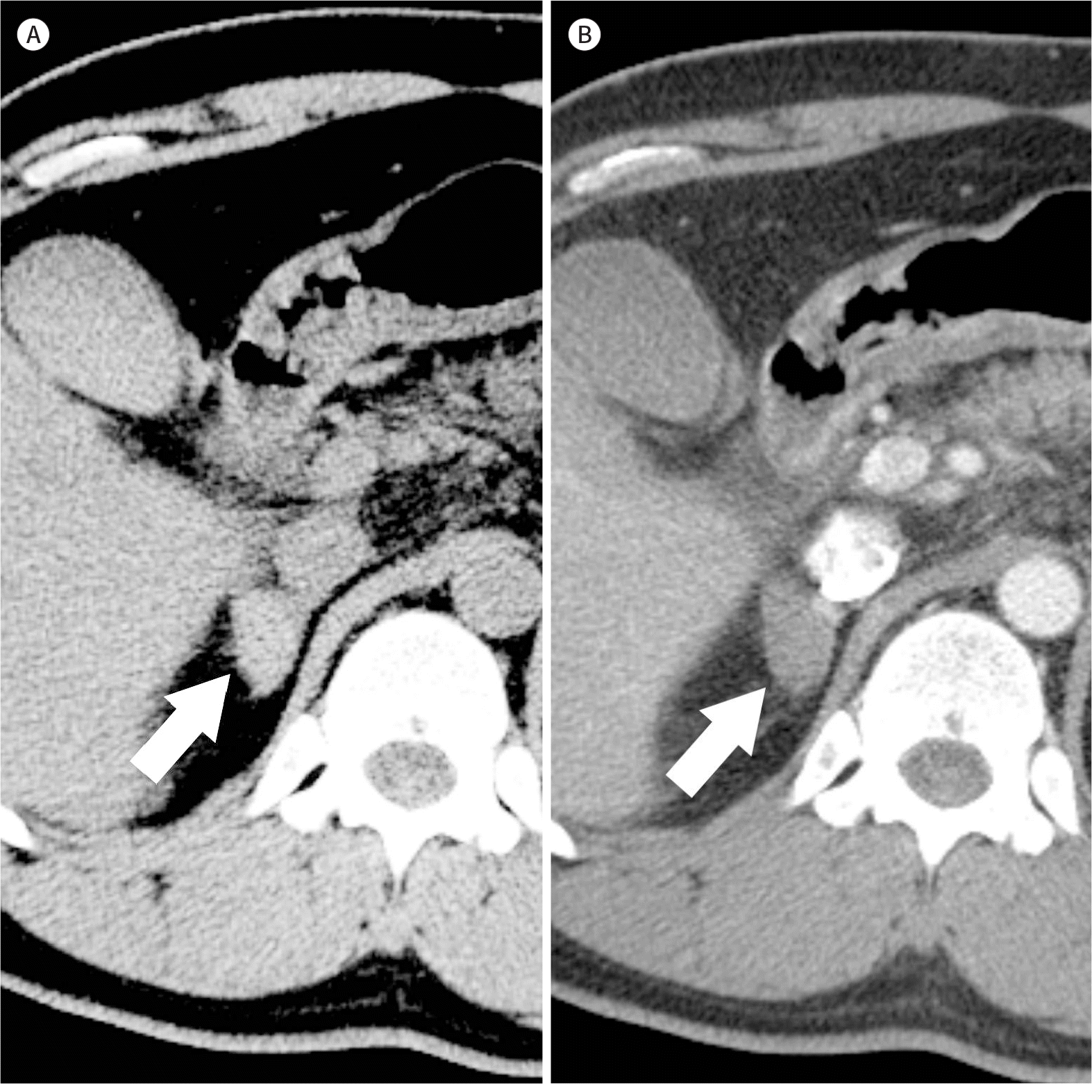
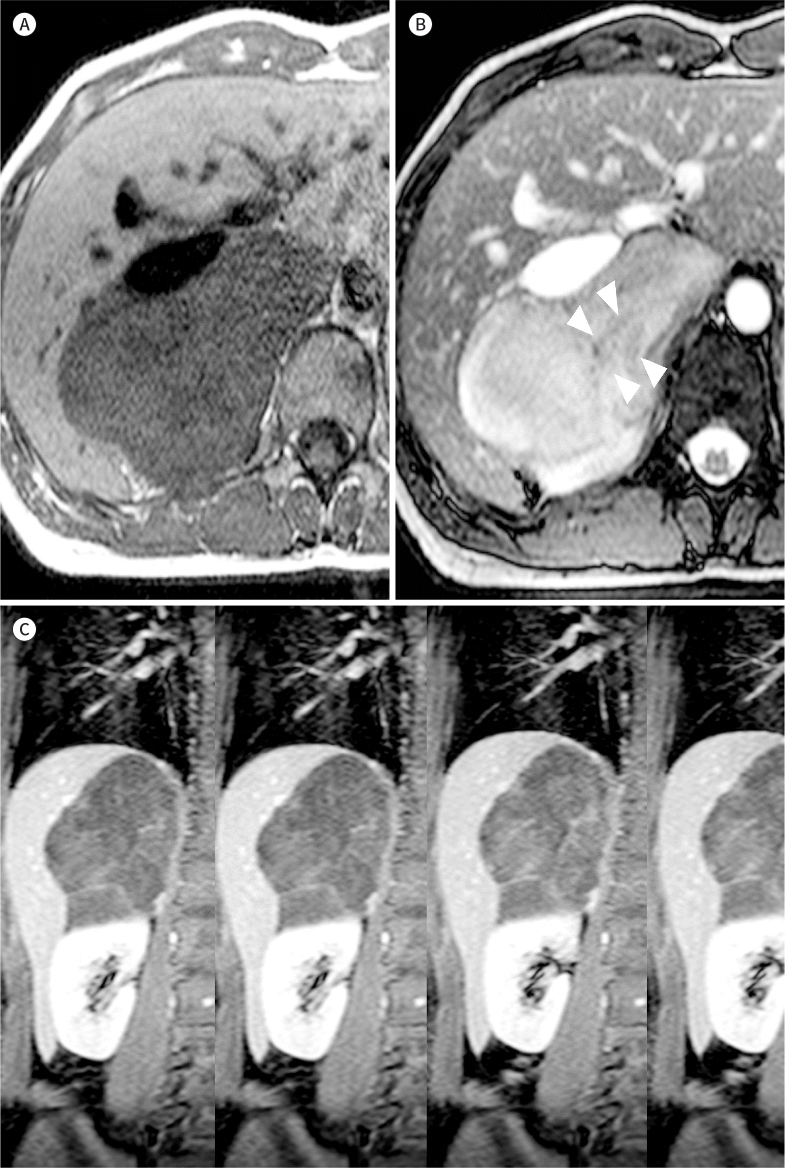
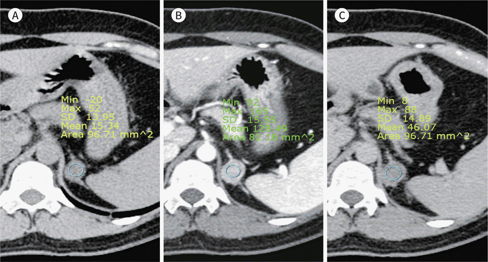 | Fig. 3.Calculation of the APW and RPW using adrenal CT of a 29-year-old woman with Cushing's syndrome. A-C. A 2.2-cm well-defined ovoid mass is seen in the left adrenal gland, which was pathologically confirmed as adrenal adenoma. The attenuation values are 15.3, 128.5, and 46.1 in the unenhanced (A), 1-min delayed (B), and 10-min delayed (C) phases, respectively. APW and RPW, which are 72.8% and 64.1%, respectively, are with the reference values (50% and 40%, respectively) for diagnosing adrenal adenoma. APW = absolute percentage washout, RPW = relative percentage washout |
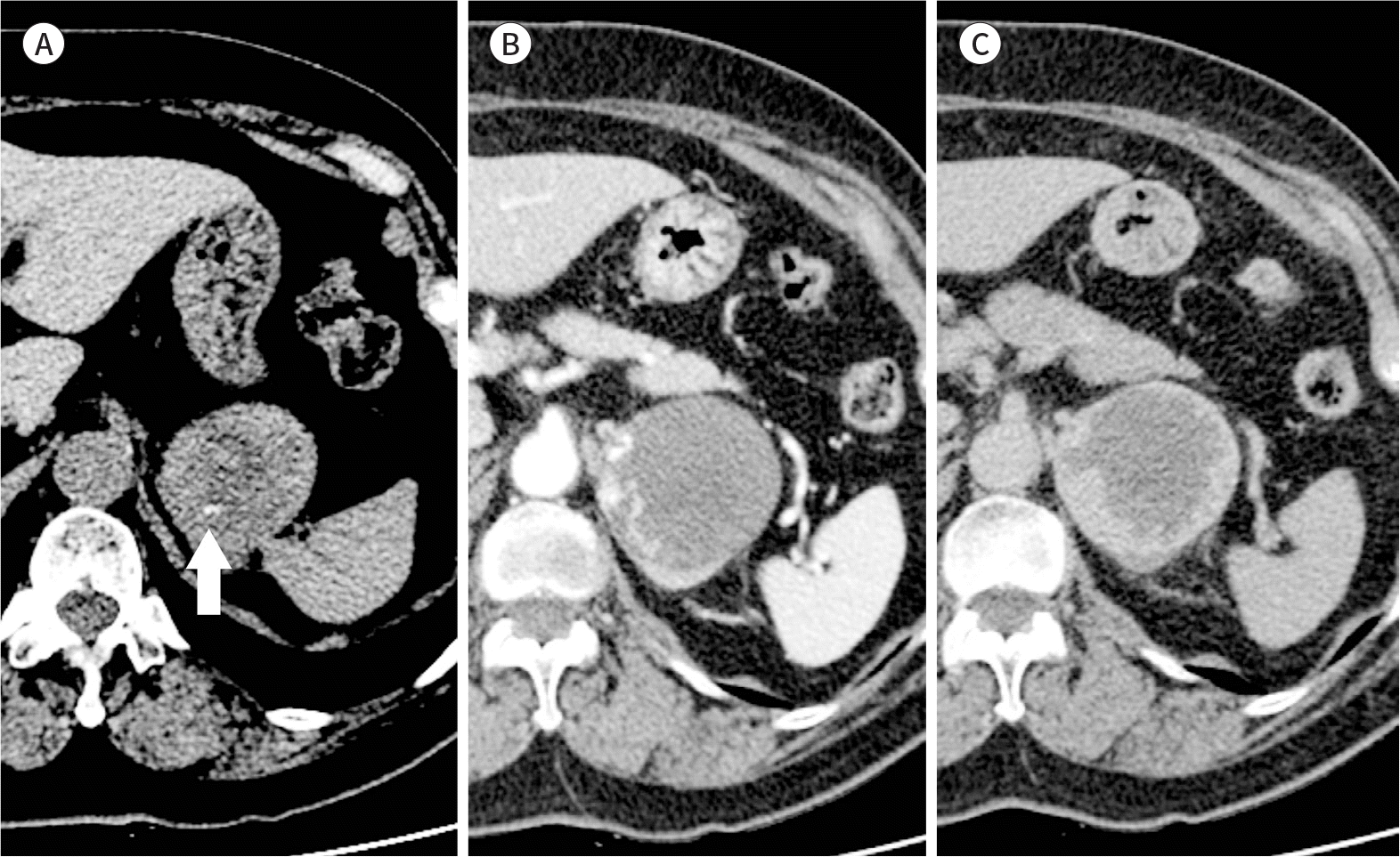
 | Fig. 4.Adrenal myelolipoma in the left adrenal gland of a 39-year-old man. Unenhanced CT shows a 7.5-cm well-demarcated round mass, mainly composed of macroscopic fat in the left adrenal gland. The mass had a high-attenuation focus in the anterior portion (41 HU, arrow), suggestive of hemorrhage. The left adrenalectomy specimen revealed thatthe mass was filled with fat and some portion of hemorrhagic necrosis, consistent with myelolipoma. |
 | Fig. 5.Adrenal cyst in the left adrenal gland of a 40-year-old man. A, B. On MRI, the masses (arrows) show high signal intensity on the T2-weighted image (A) and low signal intensity on the T1-weighted image (B) without internal soft tissue components or internal enhancement. The presumptive diagnosis was adrenal cyst. |
 | Fig. 6.Adrenal hematoma in the right adrenal gland of a 42-year-old man after a traffic accident. A, B. Unenhanced CT (A) shows a 2.5-cm ovoid mass in the right adrenal gland (arrows). On the post-contrast scan (B), the mass shows high attenuation (58 HU) and no enhancement. The final diagnosis was adrenal hemorrhage, considering the history of trauma. |
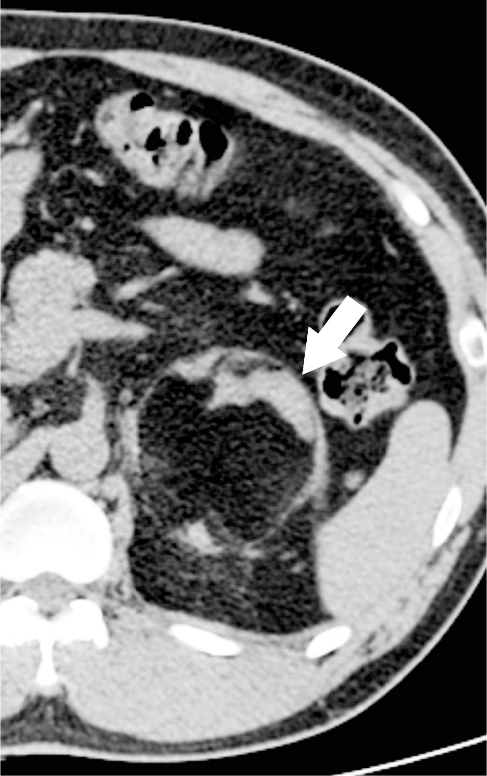
 | Fig. 7.Adrenal hemangioma in the left adrenal gland of a 69-year-old woman. A-C. Unenhanced CT (A) shows a 5.7-cm homogeneous low-attenuation mass in the left adrenal gland. Curvilinear calcification is noted in the mass (arrow). The mass shows peripheral nodular enhancement pattern in the 1-min delayed phase (B) and delayed centripetal enhancement in the 10-min delayed phase (C). The left adrenalectomy specimen revealed that the mass had dilatated vascular networks and dystrophic calcifications, suggestive of hemangioma. |
 | Fig. 9.Diffuse large B cell lymphoma in the right adrenal gland of a 42-year-old man. A-C. Unenhanced CT (A) shows an ovoid, homogeneous mass in the right adrenal gland (arrows). Arterial (B) and portal (C) phase scans shows poor homogeneous enhancement and a few cystic foci in the mass. The adrenalectomy specimen was pathologically confirmed as diffuse large B cell lymphoma. |
 | Fig. 10.Ganglioneuroma in the right adrenal gland of a 28-year-old woman. A, B. The well-defined ovoid mass in the right adrenal gland shows low signal intensity on the T1-weighted image (A) and heterogeneous high signal intensity on the T2-weighted image (B). Curvilinear low signal intensity foci (arrowheads) are seen in the mass on the T2-weighted image, with whorled appearance. C. Dynamic contrast-enhanced T1-weighted images show slow gradual enhancement because of abundant internal myxoid matrices. Right adrenalectomy was performed, and the mass was pathologically confirmed as ganglioneuroma. |
 | Fig. 11.Pheochromocytoma in the left adrenal gland of a 59-year-old man. A-C. Adrenal CT (A, B) shows a well-circumscribed, markedly enhancing round mass (arrows) with areas of necrosis and cystic change in the left adrenal gland. In the MIBG scan (C), the left adrenal mass is very MIBG-avid. The adrenalectomy specimen was pathologically confirmed as pheochromocytoma. MIBG = metaiodobenzylguanidine |
 | Fig. 12.Adrenal cortical carcinoma in the right adrenal gland of a 61-year-old man. A, B. A large (9.2 cm) mass is seen in the right adrenal gland. The mass shows high attenuation on the unenhanced image (A) and heterogeneous enhancement after the contrast media injection (B). There are ill-defined central low-attenuation areas, (arrowheads) suggestive of necrosis, and direct invasion with tumor thrombus formation to the adjacent inferior vena cava (arrow). |
Table 1.
Diagnostic Features of Adrenal Adenoma on Various Imaging Modalities
 XML Download