

 PDF
PDF ePub
ePub Citation
 Print
Print


INTRODUCTION
Epilepsy is characterized by a dynamic imbalance between the excitatory and inhibitory impulses in the cortex.1 Such imbalances are targeted by different antiepileptic drugs (AEDs) via different mechanisms. A failure to achieve sustained seizure freedom occurs after adequate trials of two tolerated and appropriately chosen and used AED schedules either as monotherapies or in combination is regarded as drug-resistant epilepsy (DRE).2 It is important to treat DRE since these patients have an increased risk of mortality as well as other disabilities affecting the quality of life. The likelihood of remission is very low for subsequent therapies with alternative AEDs in these patients. Apart from pharmacotherapy, the alternative modalities currently offered for treating DRE include epilepsy surgery, vagus nerve stimulation, trigeminal nerve stimulation, transcranial magnetic stimulation (TMS), and deep brain stimulation.3
TMS was initially used as a diagnostic tool, but it is now being applied as a reliable treatment modality for several psychiatric and neurological disorders. Repetitive TMS (rTMS) is a noninvasive method based on Faraday's law of electromagnetic induction4 in which small intracranial electrical currents are generated by a rapidly changing extracranial magnetic field, therefore inducing focal electrical brain stimulation.5 This results in antiparallel currents in cortical neurons that modulate them so as to produce desirable neurobiological effects.6 The application of repetitive trains of low-frequency TMS modulates the cortical excitability and produces its relatively long-lasting suppression.7 It also acts on neurotransmitter release, signaling pathways, and gene expression.8
Our literature review revealed that the results regarding the efficacy of rTMS in epilepsy in previous randomized controlled trials (RCTs) are inconsistent and contradictory. A few trials showed that rTMS significantly reduced the seizure frequency, whereas others did not show any significant differences.91011121314151617181920 The meta-analysis of Hsu et al.21 showed a beneficial effect of rTMS in epilepsy, but as in all TMS studies regardless of the presence of a placebo/sham/control arm, the placebo estimate effect could not have been established in their study. rTMS is a long-duration procedure involving a loud and bulky device along with verbal and tactile contact made by the operator with the subject, which may induce a considerable placebo effect. Hence, the present meta-analysis was planned with an aim to generate evidence for the absolute effect of rTMS in DRE in reducing seizure frequency and epileptiform discharges. The null hypothesis of no difference between the effects of rTMS when compared to placebo/sham control was considered initially. The Cochrane systematic review of Chen et al.22 admitted that it is not possible to perform a meta-analysis due to the high variability of rTMS protocols and time points reported for individual studies. However, this problem was addressed in the present study by performing a meta-regression with all possible confounders.
METHODS
Protocol development and registration
We developed and followed a standard meta-analysis protocol in accordance with Preferred Reporting Items for Systematic Reviews and Meta-Analysis (PRISMA)-P 2015 guidelines,23 and registered the protocol in the International Prospective Register of Ongoing Systematic Reviews (systematic review registration-PROSPERO: CRD42018088544). This meta-analysis was conducted and reported in conformance with the PRISMA statement.24
The protocol of the meta-analysis was exempted from the full review and approved by the Institutional Ethics Committee, All India Institute of Medical Sciences (AIIMS), Bhubaneswar as per ICMR 2017 guideline on 21st April 2018.
Search strategy
To collect data from all relevant studies, we searched MEDLINE and Cochrane databases for RCTs on rTMS in patients with DRE published up to December 2018. Search terms were constructed using the following key search elements in the PICO method: “P” (Drug-Resistant Epilepsies / Epilepsies, Drug-Resistant / Resistant Epilepsies, Drug / Resistant Epilepsy, Drug / Epilepsy, Drug-Resistant / Medication Resistant Epilepsy / Epilepsies, Medication Resistant / Epilepsy, Medication Resistant / Medication Resistant Epilepsies / Resistant Epilepsies, Medication / Resistant Epilepsy, Medication / Intractable Epilepsy / Epilepsies, Intractable / Intractable Epilepsies / Epilepsy, Drug Refractory / Epilepsy, Intractable / Refractory Epilepsy / Epilepsies, Refractory / Epilepsy, Refractory / Refractory Epilepsies / Drug Refractory Epilepsy / Drug Refractory Epilepsies / Epilepsies, Drug Refractory / Refractory Epilepsies, Drug Refractory Epilepsy), “I” (rTMS / Transcranial Magnetic Stimulation / Magnetic Stimulation, Transcranial Magnetic Stimulations, Transcranial / Stimulation, Transcranial Magnetic/ Stimulations, Transcranial Magnetic / Transcranial Magnetic Stimulations / Transcranial Magnetic Stimulation, Single Pulse / Transcranial Magnetic Stimulation, Paired Pulse / Transcranial Magnetic Stimulation, Repetitive), “C” (Sham / Placebo), and “O” (seizure frequency / seizure rate). The reference lists of published studies were also searched, and unpublished data were searched for by checking the International Clinical Trials Registry Platform, which is a central database containing trial registration data sets provided by different international trial registries including ClinicalTrials.gov.
Study selection criteria
RCTs on rTMS in patients with DRE published in English-language peer-reviewed journals were included. All of the studies included in this meta-analysis had seizure frequency as an outcome measure. The included studies were not restricted by date of publication, tool used, number of stimulations/sessions, stimulation site, type of coil used for stimulation, or method of localization of the site stimulation. Letters to the editor, case series, and case reports were excluded.
Types of participants
We included studies examining adult human subjects of both sexes irrespective of age with a diagnosis of DRE including unclassified types of epilepsy and postsurgical epilepsy patients who did not achieve freedom from seizures despite trials of two AEDs either as monotherapies or in combination. Exclusion criteria applied to all of the included studies were the presence of pacemakers or other electronic implants, the presence of metal or magnetic objects in the brain, or the use of any other method for cortical stimulation.
Type of intervention
rTMS at any frequency using either a round or figure-of-eight coil for any duration and at any intensity added to current therapy or used as a single therapy was the intervention of interest. More than 50 designs of stimulation coils are used in rTMS, but the two most common types are figure-of-eight and round coils. Figure-of-eight coils, which induce a more-focal current, are used at the coordinates representing the ictal focus in the EEG. Round coils, which induce more homogeneous and widespread currents, are mostly used at the vertex. Sham/placebo stimulations were performed using specially designed coils that looked like rTMS coils and produced cutaneous skin sensations similar to those induced by the rTMS coils.
Outcome measures
Seizure frequency: seizure frequency was estimated based on seizure calendars maintained either by the patients themselves or their relatives, and changes in seizure frequency were assessed.
Interictal epileptiform discharges: All patients underwent 18-channel EEG recordings, and the total duration of artifact-free discharges was analyzed for epileptiform discharges.
Study selection and data collection
Relevant studies were selected in a stepwise manner. All articles were first screened based on their title and abstract, and then the full texts of all articles that passed the selection process were retrieved and read. Inclusion criteria were determined prior to performing the literature search, and those studies that met the inclusion criteria were included in the meta-analyses.
Data extraction and management
Data were abstracted and their quality was assessed independently by three investigators (R.M., B.R.M., and A.M.) using guidelines published by the Cochrane Collaboration. Any disagreement was resolved by discussion between these three authors in consultation with the clinical pharmacologist cum statistical advisor (A.S.). The extracted data included information on the study design, participants, intervention type, stimulation site, outcome measure, and intervention protocol (stimulation frequency, number of trains, intertrain interval, and motor threshold). The data were in the form of plots in two studies,1416 which were interpreted using a plot digitizer since the authors did not respond to our request for numerical data. The data in one of the studies14 were converted from range to SD to ensure uniformity in entered data.
Data analysis
The meta-analysis was conducted using Cochrane Program Review Manager software (version 5.3; Cochrane, Copenhagen, Denmark),25 while the meta-regression was performed using the “Metapackage” function of R software (version 3.4; R Foundation for Statistical Computing, Vienna, Austria).26
Assessment of risk of bias in included studies
The risk of bias in individual studies was assessed using the standardized risk-of-bias critical appraisal instrument of the Cochrane Collaboration. This tool rates the bias of a clinical trial in three categories (low, unclear, and high) in the following domains: random sequence generation (selection bias), allocation concealment (selection bias), blinding of participants and personnel (performance bias), blinding of outcome assessment (detection bias), incomplete outcome data (attrition bias), selective reporting (reporting bias), and other types of bias (if any). Three reviewers (R.M., B.R.M., and A.M.) independently evaluated and recorded their judgments and justifications in each domain for each included study
Measures of the treatment effect
The primary outcome measure of interest in this meta-analysis was the seizure frequency, which can be estimated from the seizure calendar maintained by the relatives of the patient. Interictal epileptiform discharges in the EEG recordings were also accounted for as an outcome measure, which were available for only three studies.161719 The mean difference was calculated to estimate the effect size in order to assess the differences in seizure frequency and interictal epileptiform discharges between active and sham/placebo stimulations.
Unit-of-analysis issue
This meta-analysis considered “stud”y as a unit of design. In studies in which two different protocols were used to assess the reduction in seizure frequency, the two protocols were considered as separate units of analysis.
Assessment of heterogeneity
Given that statistical heterogeneity is inevitable due to the clinical and methodological diversity in clinical studies, it is important to consider the extent of the inconsistency or to quantify the inconsistency across the included studies. The chi-square test has commonly been used to assess whether observed differences in results are compatible with chance alone. A low probability value (or a large chi-square statistic relative to its degrees of freedom) provides evidence of heterogeneity of intervention effects (i.e., a variation in the effect estimates beyond chance). I2 statistics describe the percentage of the variability in an effect for an estimate that is due to heterogeneity and were used to quantify inconsistency.
Meta-regression
Since different study characteristics such as the type of coil, duration of active intervention, posttreatment follow-up period, and rTMS frequency can potentially modify the effect size of the intervention, we performed a meta-regression across the studies to estimate how the outcome variable (the intervention effect) changes with a unit increase in the explanatory variable (the potential effect modifier), which can be described as a regression coefficient. The statistical significance of the regression coefficient can indicate whether there is a linear relationship between the intervention effect and the explanatory variable.
Sensitivity analysis
Sensitivity analysis was used to test the robustness of the results obtained in the present meta-analysis. In the case of high heterogeneity, forest plots were constructed again after excluding individual studies one at a time, and observing the effect of excluding a particular study on individual parameters.
RESULTS
Description of included studies
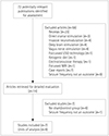
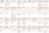
The database search yielded 72 results, and after excluding articles based on their title and abstract, 14 potentially eligible articles remained. The full-text review conducted by the 3 reviewers concluded that 8 of the 14 articles reported on RCTs, but the study of Joo et al.11 compared the focal vs. nonfocal stimulation for different numbers of stimuli, and so was excluded. Seven RCTs that compared rTMS with sham or placebo controls were therefore finally included in this meta-analysis (Table 1).14151617181920
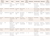
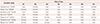
The entire selection process is illustrated by the PRISMA flow diagram in Fig. 1. Three of the seven included studies were placebo-controlled,141517 one study compared between rTMS at different intensities,19 one study compared rTMS at different intensities with placebo,14 one study compared using a figure-of-eight coil, round coil, and sham control,20 and one study compared rTMS with AED treatment.18 Six studies were excluded since they had only a single arm with no comparator (Table 2). The included studies used either standard figure-of-eight or round coils to deliver rTMS, while the sham methods differed. The risk of bias was assessed by appraising the following six domains for each trial: allocation concealment, randomization method, blinding, completeness of data, selective outcome reporting, and other types of bias. Most of the studies were deemed to have an unclear risk of selection bias since the allocation concealment method was not reported. In another study the outcome assessors were not blinded, and so a high risk of detection bias was assumed.18 The study of Tergau et al.14 performed an interim analysis, and was considered to be at a high risk of attrition bias due to incomplete outcome data; moreover, since the blinding of participants and personnel was not described, the risk of bias was unclear (Table 3).
Effect of intervention on seizure frequency
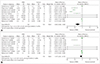
To evaluate the effect of low-frequency rTMS on the seizure frequency in DRE, the mean difference and the SD of included studies were entered into Cochrane Program Review Manager (version 5.3) using a random-effects model. The test for heterogeneity was significant (chi square=314.07, df=8, p<0.00001, I2=97%). In the forest plot, the CI for the results of individual studies (depicted graphically using horizontal lines) showed less overlap and hence significant heterogeneity. The very high variance observed in the study of Tergau et al.14 was contributed by one patient who had frequent seizures (up to 50 per day). The random-effects model analysis revealed a pooled effect size of −5.96 (95% CI=−8.98 to −2.94), indicating an overall effect size significantly favoring the rTMS group (Z=3.87, p=0.0001) over the control group with regard to seizure frequency (Fig. 2A). The very high heterogeneity prompted a sensitivity analysis, which indicated that a considerable degree of heterogeneity was contributed by the study of Seynaeve et al.20 Excluding that study reduced the heterogeneity (chi square=30.70, df=6, I2=80%) but did not change the overall effect on seizure frequency. There was an overall effect size of −1.47 (95% CI=−2.81 to −0.13, Z=2.15, p=0.03) favoring the rTMS group over the control group (Fig. 2B). Therefore, irrespective of the inclusion or exclusion of the study of Seynaeve et al.,20 the effect of rTMS in reducing seizure frequency remained statistically significant in DRE.
Effect of intervention on interictal epileptiform discharges
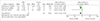
Information on the effect of rTMS on interictal epileptiform discharges was available for three of the seven studies. The test for heterogeneity was not significant (chi square=1.46, df=2, p=0.48, I2=0%). The overall effect size for interictal epileptiform discharges significantly favored the rTMS group, at −9.36 (95% CI=−13.24 to −5.47, Z=4.72, p<0.00001) (Fig. 3).
Effect of independent variables: meta-regression
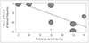
In the meta-regression, when adjusted for other potential variables such as the type of coil used, stimulation frequency, and the total duration of the active intervention, the seizure frequency worsened by 2.00±0.98 (mean±SD, p=0.042) for each week of lengthening of the posttreatment follow-up period. This suggests that rTMS exerts only a short-term effect (Fig. 4). The type of coil used for rTMS did not significantly affect the effect size or heterogeneity of the outcome (p=0.75). Similarly, the duration of the active intervention (p=0.22) and the stimulation frequency (p=0.46) did not significantly affect the effect size.
DISCUSSION
TMS has previously been used previously as an effective mapping tool for the presurgical localization of epileptogenic foci and for evaluating pathophysiological mechanisms noninvasively, as well as for studying the mechanism of action of AEDs.27 It is already evident that repetitive pulses of such stimulation can modulate the functionality of eloquent cortical neurons.7 However, the RCTs performed around the world have not provided consistent results, and thus to obtain conclusive evidence we performed this meta-analysis given that alternative approaches are desperately need for drug-resistant cases of epilepsy.
Two of the included studies showed statistically significant reductions in the seizure rate from baseline.1619 Three randomized, blinded trials failed to show any statistically significant difference in seizure frequency following rTMS treatment compared with controls.151820 Though the reduction in seizure frequency in one of the included studies was not significant, power analysis of the study data suggested that the smallness of the sample meant that a reduction in seizure frequency of less than 70% would not have been significant, resulting in the possibility of a type 2 error.15 Tergau et al.14 compared stimulation frequencies of 0.33 Hz and 1 Hz against placebo, and found a significant reduction in seizure frequency compared to baseline only for the 0.33-Hz stimulation, but the difference relative to the placebo could not be established. Seynaeve et al.20 concluded that rTMS was not an effective intervention, but the analysis performed in that study was both incomplete and misleading, since the authors did not analyze pooled data and did not perform comparisons in a pairwise manner. We analyzed the published data for individual patients and compared sham vs. a round coil and sham vs. a figure-of-eight coil, and found that rTMS was effective in reducing the seizure frequency.
All the participants recruited across the studies had DRE, making the results applicable to the overall population of DRE patients. However, the results must be interpreted while keeping in mind the small number of studies and the smallness of the samples, as well as methodological and design dissimilarities. However, we addressed possible variability by performing a meta-regression for potential effect modifiers. rTMS had a significant effect on reducing the seizure frequency and interictal epileptiform discharges in patients with DRE. Increasing the number of days of rTMS treatment was linearly related to the reduction of the seizure frequency. However, the intervention only produced a short-term effect, with the seizure propensity increasing with the time since applying rTMS. Different studies have used different types of coils and different numbers of frequencies, but our analysis suggests that these parameters did not significantly affect the outcomes. Three of the seven studies included in our meta-analysis evaluated the secondary end point of the mean change in interictal epileptiform discharges. Fregni et al.16 and Sun et al.19 demonstrated statistically significant reductions in epileptiform discharges, whereas Cantello et al.17 found no significant difference in the mean reduction in the number of epileptiform discharges. However, overall rTMS induced significant reductions in interictal epileptiform discharges. Since the reduction in seizure frequency and interictal epileptiform discharges were correlated, the results of this meta-analysis can be reliably interpreted.
The results of the present study agree with those found in the systematic review of 11 studies performed by Hsu et al.21 That study found a small but significant effect of low-frequency rTMS on medically intractable epilepsy. In contrast to that systematic review additionally including single-arm nonrandomized studies in which all patients received the intervention, the present review included only RCTs comparing intervention and control participants, which should have addressed the placebo effect. Bae et al.28 estimated that there was a significantly lower reduction in seizure frequency for sham procedures.
It has been seen that modulation of cortical networks by rTMS depend on its frequency, intensity, and duration and on the intertrain intervals and the rTMS protocols used in different studies in our meta-analyses are not essentially similar.29 The effect of rTMS on individual neurons might also depend on their orientation relative to the induced electrical and magnetic fields, and also any underlying anatomical lesions in patients. A variability in the reduction in seizure frequency can also result from neurophysiological differences between ethnic groups. This study considered DRE subjects and the various analyzed studies included AEDs in different numbers and combinations, which might have resulted in different affects of rTMS on synaptic alterations. Age, sex, and genetic factors can also contribute to differences in responses.30 Any or all these factors could account for the different responses induced by apparently similar rTMS protocols.
The main limitation of this meta-analysis is the small number of available RCTs and the eligible studies not applying a consistent intervention protocol. The data for interictal epileptiform discharges were available for only three of the eligible studies. In two of these studies the data had to be converted into numerical format using a plot digitizer, and in one study the SD was calculated from the range by applying relevant formulae.
In conclusion, the overall beneficial effects of low-frequency rTMS in reducing the seizure frequency and interictal epileptiform discharges indicate that this technique may be recommended as a therapeutic intervention for DRE. However, the variability in rTMS procedures warrants further investigations before drawing definitive conclusions. No major adverse event was found to be associated with rTMS, thereby demonstrating its safety.31 However, increasing the posttreatment follow-up duration was associated with a worsening of the seizure frequency, indicating the presence of only a short-term effect of rTMS. A standard protocol for its application needs to be established in order to reduce heterogeneity, and further RCTs involving adequate sample sizes and durations should be performed to validate the efficacy and safety of the procedure.
 XML Download