

 PDF
PDF ePub
ePub Citation
 Print
Print


INTRODUCTION
The aging of the world's population has resulted in a tremendous increase in the incidence of aging-related brain diseases. Cognitive impairment is one such disease that has become very common among the elderly population. All kinds of aging-related brain diseases adversely impact the health and quality of life of elderly people, and they are expensive to diagnose, treat, and manage, hence representing a huge economic burden on the affected families as well as society as a whole.1 Previous studies have investigated different types of aging populations comprising people older than 70 years, and have found that 14% of this population has mild cognitive impairment (MCI), which involves a cognitive dysfunction severity between normal aging and dementia.23 When this condition worsens, the patient will suffer from Alzheimer's disease (AD).1 According to Alzheimer's Association International, 44 million people currently suffer from dementia worldwide, and this number will increase to 76 million in 2030 and 135 million in 2050.4 Such growth will inevitably lead to enormous increases in the economic and health burdens on societies worldwide.
Cognitive function can be quantified using several psychological questionnaires into different levels based on the cognitive decline5 and mental disability.6 In clinical situations, several imaging protocols such as CT and MRI are sometimes used to detect structural changes in the brain or cerebral vasculature. Such imaging modalities are valuable diagnosing the severity of cognitive impairment,78910 but they are also expensive and can only be performed in large hospitals. Due to the similarity between the retinal and cerebral vasculatures, imaging biomarkers on high-resolution retinal images are promising features comparable with low-resolution brain images obtained by CT or MRI. Moreover, retinal imaging is less expensive and can be applied in community hospitals, thus providing the potential for the large-scale screening of aging populations.
The advances of computer programs developed for medical imaging have facilitated several computerized measurements on captured retinal images for discovering the relationship between retinal changes and clinical diseases.1112 There are increasing data on the retinal changes and some diseases, including those affecting the brain. Several studies have revealed a relationship between retinal vascular changes and the incidence of dementia.13 Several clinical investigations have attempted to determine whether the retinal vasculature could be an imaging biomarker for cognitive impairment, and the results suggest that there is an association between retinal changes and cognitive impairment. However, some reported findings have been found to be inconsistent, and so in this study we performed a comprehensive meta-analysis to determine the relationship between retinal vascular geometric changes and cognitive impairment.
METHODS
Search strategy
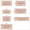
We performed a comprehensive search of the following electronic databases up to July 2018: China National Knowledge Infrastructure (CNKI), Chinese VIP Information, Wanfang, PubMed, and Embase. Our main aim was to include all published studies that investigated the relationship between retinal vascular geometric changes and cognitive impairment. We used the following search terms to identify these published studies: retina*, retinopathy, fundus, vascular, microvascular, blood vessel, microvascula*, microvasculature, fractal dimension (FD), Df, FD, tortuosity, curvature, width, caliber, central retinal arteriolar equivalents (CRAE), CRAE, central retinal venular equivalents (CRVE), CRVE, branching angle, geometric changes, cogniti*, Alzheimer's disease, AD, MCI, dementia, aphrenia, loss of memory, dysfunction, impairment, abnormalities, and decline. The above terms were combined in various ways to form a complete retrieval system. The reference lists of identified studies were also examined rigorously to determine whether they met our inclusion criteria. We also performed a manual search of the gray literature, including theses, dissertations, and conference proceedings. We obtained raw data by contacting the original authors by email.
The study protocol was conducted in accordance with the ethical guidelines of the 1995 Declaration of Helsinki and was approved by Ethics Committee of Affiliated Hospital Nantong University (IRB No. 2018-L084).
Selection criteria and exclusion criteria
The inclusion criteria for the clinical trials were as follows:
1) Involving patients with different levels of cognitive impairment, from memory loss to MCI and AD. These patients were diagnosed using various instruments, including the National Institute of Neurological and Communicative Disorders and Stroke and the Alzheimer's Disease and Related Disorders Association criteria (NINDS-ADRDA criteria)14 and the Mini Mental State Examination.15
2) Reporting on measures and quantitative data on retinal microvascular changes, including CRAE, CRVE, FD, and tortuosity.
3) A case–control study design.
4) Published in the medical literature written in English or Chinese.
-
5) Published between December 2012 and August 2018.
The exclusion criteria for the clinical trials were as follows:
Quality assessment and data extraction
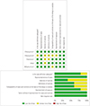
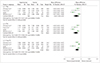
We appraised the quality of all included studies using the Newcastle-Ottawa Scale16 and RevMan (version 5.3, The Nordic Cochrane Centre, The Cochrane Collaboration, Copenhagen, Denmark). A high-quality case–control study had the following characteristics: adequate definition and representativeness of the case, clear definition and selection of controls, reasonable comparability of cases and controls based on the study design or analysis, ascertainment of exposure, using the same method to as certain cases and controls, and a low no-response rate. Two reviewers (W.C. & C.C.) evaluated and cross-checked the quality of the studies, and if there were any discrepancies, another author (W.H.) was consulted to reach a consensus. Fig. 1 illustrates the risk of bias among the five included studies.
Two reviewers (W.C. & C.C.) independently extracted data using a data-extraction form. To identify all of the published literature studies, they considered the name of the first author and the year of publication. The following data were extracted for the reported studies: year of publication, inclusion and exclusion criteria, sample size, age, racial group, sex, the number or percentage of diabetes mellitus patients, pre-existing eye diseases, ophthalmic camera and imaging conditions, retinal microvascular abnormalities, cognitive measures and outcomes, and other confounding risk factors such as the history of smoking and hypertension. All duplicated studies were excluded in this systematic review.
Statistical analysis
The meta-analysis was performed using RevMan software (version 5.3). In the statistical analysis we calculated the mean difference (MD) and its 95% confidence interval (CI) as effect factors. Heterogeneity was determined with the chi-square test, and quantified by calculating the inconsistency index I2. We determined the impact of the combined effects by synthesizing all studies using different statistical models according to the heterogeneity. The results were considered to be statistically significant when the two-sided p value was less than 0.05. In addition, funnel plots were constructed to detect the presence of publication bias and other types of bias. Finally, sensitivity analyses were performed to improve the accuracy of the tests.
RESULTS
Summary characteristics of included studies
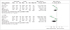
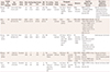
While the 5 included studies involved 2,343 subjects, only 671 of them had cognitive impairment or AD. Frost et al.,17 Cheung et al.,18 and Williams et al.19 all used NINDS-ADRDA criteria to diagnose AD. Most of the participants included in the present meta-analysis were aged between 60 to 80 years. Both males and females were included in the case and control groups. The participants in the included studies were Asian or Caucasian. Each study had a cross-sectional design. Most of the included participants in both the case and control groups had hypertension. Nearly one-third of the participants in the study of Jiang3 were smokers. All of the studies except for that of Frost et al.17 captured retinal images after performing pupil dilation. In three studies,171820 a digital ophthalmic camera was positioned at 45° to acquire images. In all of these studies, retinal microvascular abnormalities were determined by measuring the FD, tortuosity, and caliber. The retinal vascular caliber was measured using the Singapore I Vessel Assessment method,21 and caliber data were summarized into CRAE and CRVE by using Knudtson et al.22 revised formulas. The vascular tortuosity of retinal arterioles and venules was calculated as a ratio of the integral of the curvature squared along the path of the vessel relative to the total path length.23 FD was calculated using the box-counting method to represent the complexity of a skeletonized retinal arteriole and venule network, which represents a ‘global’ measure summarizing the entire branching pattern of the retinal vascular tree.24 The risk factors (additional adjustments) were vascular diseases, reflecting that some participants have vascular problems of different intensities. Table 1 provides a detailed description of all of the features included in these studies.
Quality of included studies
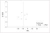
The methodological quality of the included studies is graphically demonstrated in Fig. 2. The representativeness of cases as well as the method of ascertainment for cases and controls were quality controlled in the included studies. The definition and selection of the controls were only vaguely described for some of the included studies, and the ascertainment of exposure for cases and controls might have been opaque in some of the studies. In general, the overall quality of the included studies indicated a low risk of bias, although for some the risk of bias was unclear.
Association between retinal vascular caliber and cognitive impairment
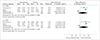
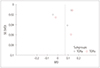
The forest plot in Fig. 3 depicts the association between cognitive impairment and CRAE or CRVE. In four studies3171819 there was a considerable heterogeneity between the controls and the patients with cognitive impairment (p<0.0001, I2=93%), with MD total-effect values of −4.24 (95% CI: −11.33, 2.84) and −8.82 (95% CI: −18.74, 1.09), respectively. The Z values for CRAE and CRVE were 1.17 (p=0.24) and 1.74 (p=0.08), respectively. These data indicate that there was no significant association between CRAE or CRVE and cognitive impairment. The sensitivity analysis showed that the heterogeneity decreased slightly when excluding one study19 while analyzing the results for CRAE and CRVE. A particularly interesting finding was that the previous reported association between CRAE and cognitive impairment remained unchanged, while a decreased CRVE was found to be related to dementia.
Association between retinal vascular tortuosity and cognitive impairment
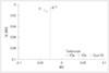
Fig. 4 shows that there was severe heterogeneity (p<0.00001, I2=96% or 94%) across three of the included studies.31819 The MD total-effect estimates were 0.06 (95% CI: −0.07, 0.19) and 0.08 (95% CI: −0.04, 0.20), respectively. The Z values for arteriolar tortuosity (TORa) and venular tortuosity (TORv) were 0.91 (p=0.36) and 1.31 (p=0.19), respectively. These results indicated that retinal vascular tortuosity was not significantly correlated with cognitive impairment. The sensitivity analysis showed that when the study of Williams et al.19 was excluded from the meta-analysis, the heterogeneity of TORa and TORv decreased from 96% and 94%, respectively, to 0%. Furthermore, it was found that the tortuosity was greater in the patients with cognitive impairment.
Association between retinal vascular FD and cognitive impairment
The forest plot in Fig. 5 depictsthe association between retinal FD and cognitive impairment. In four of the original studies,3171819 the authors measured the arteriolar FD (FDa) and the venular FD (FDv),for which the MD total-effect values were −0.03 (95% CI: −0.05, −0.01) and −0.03 (95% CI: −0.05, −0.02), respectively, and the Z values were 3.08 (p=0.002) and 3.72 (p<0.0001), respectively. In these four selected studies,3171819 heterogeneity was also observed in the total FD (TFD) group (p=0.0009, I2=82%). The effect value of MD was −0.02 (95% CI: −0.04, −0.01). The test for the overall TFD effect produced a Z value of 3.72 (p=0.0002). These results suggest that retinal FD is related to cognitive impairment. The sensitivity analysis showed that when one study3 was eliminated, the heterogeneity in the FDa and TFD groups decreased to 27% and 35%, respectively, from 82%, while in the FDv group it decreased from 84% to 35%.
DISCUSSION
This study performed a meta-analysis to determine the relationship between retinal vascular geometric changes and cognitive impairment or dementia. Our investigation of 2,343 individuals indicated that retinal vascular FD rather than caliber and tortuosity was associated with cognitive dysfunction. The findings of our meta-analysis were similar to those of previous qualitative systematic reviews performed by Heringa et al.25 and Ding et al.26 However, some of our findings are inconsistent with previously reported investigations. The Los Angeles Latino Eye Study27 found that variables related to the retinal microvasculature caliber such as CRAE and CRVE can provide surprising insights into cognitive impairment in Latino populations. However, their reported data were qualitative rather than quantitative, and so could not been synthesized in our meta-analysis. A similar situation was found in the Circulatory Risk in Communities Study study,28 whose authors reported that arteriolar narrowing and overall retinal abnormalities may be used as markers to identify persons who are more likely to be affected by dementia. In contrast, our meta-analysis did not confirm the presence of such a relationship between arteriolar narrowing and cognitive impairment. Our study mainly disclosed that patients with cognitive impairment might have a smaller vascular FD or a lower complexity of fundus microvascular networks. This is consistent with the finding of Jiang et al.29 that AD patients had sparser retinal microvascular networks than controls, indicating the presence of retinal microvascular dysfunction in AD individuals. Ong et al.30 also indicated that decreased FDa and FDv values were associated with cognitive impairment, which is in complete agreement with the results of our meta-analysis. It is also interesting that Mroczkowska et al.31 found that mild-AD patients with relatively minor cognitive impairment exhibited signs of microvascular dysfunction even in the absence of a history of significant systemic vascular disease. This suggests that retinal changes are potentially useful as an early indicator of cognitive impairment.
The exact biological mechanism underling the association between retinal FD changes and cognitive impairment remains unclear. We consider that there are several possible reasons. The branching complexity is less when FD is smaller, and so rarefaction of retinal vessels is associated with decreased FD, representing corresponding geometric changes in the cerebral microvasculature, further indicating hypoperfusion that may switch on hypoxia-induced pathways, which would increase the amyloid-beta load1932 or tau pathology.33 All of these undesirable events would lead to the development of AD in patients. Moreover, amyloid plaques in those AD patients may result in vascular damage that undermines the microvasculature. The risk of cerebrovascular disease increases in patients with impaired microcirculation and decreased blood flow.3435 which may further lead to decreased retinal FD since the retinal vasculature is an observable microcirculation tissue.
Our study was subject to several limitations. At the study and outcome levels, publication bias and the risk of bias may interfere with the detected outcomes. At the review level, our findings were also influenced by eligible research studies that produced incomplete retrievable results. The reliability of the results is impaired by the huge heterogeneity of data present in the selected reports. We therefore performed a sensitivity analysis in addition to applying a random-effects model to tackle the considerable heterogeneity. We speculated that sex and the smoking ratio did impact the results of the retinal FD sensitive analysis. The cerebrovascular perfusion is better in females than in males,36 and so the vascular density of males may be smaller than that of females. Smoking is a risk factor that damages vascular health and increases vascular stiffness,37 and thus might change the branching pattern of the retinal vasculature. For the sensitive analysis of TORa and TORv, we believed that the disparity could be attributed to the participants of this study being Caucasians, while participants of other studies were Asians; in other words, that race can influence retinal measurements. The small number of included studies made it difficult to perform subgroup analysis and a meta-regression analysis. Moreover, the conclusions cannot be generalized since only small populations were analyzed in the included studies. The correlation between the severity of cognitive impairments and the extent of retina microvascular abnormalities therefore needs further investigations to confirm the significance of a retinal imaging biomarker as a predictor for cognitive impairments.
In conclusion, a smaller retinal microvascular FD might be associated with cognitive impairment. Such a relationship could be used to establish a novel retinal image biomarker that might allow the early detection of cognitive impairment in an affordable and convenient way. Further large-sample and well-controlled original studies are required to confirm the present findings.
 XML Download