

 PDF
PDF ePub
ePub Citation
 Print
Print


Dear Editor,
Periventricular leukomalacia (PVLM) refers to hypoxic-ischemic damage to the brain at a gestational age of 24–34 weeks.1 The prevalence of PVLM can reportedly reach 30% in preterm neonates.1 PVLM is one of the most important causes of visual impairments in children.2 Here we report the oculomotor and vestibular findings in a patient with PVLM.
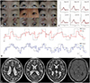
A 28-year-old woman presented with long-standing strabismus that had been first noticed at the age of 1 years, but without diplopia. She was born at a gestational age of 32 weeks due to placenta previa, and had no family history of visual or neurologic disorders. She had developed problems in learning and wayfinding but did not have cerebral palsy. Her visual acuity was 20/30 in the right eye and 20/20 in the left eye. Fundoscopy revealed large optic cups in both eyes, but the intraocular pressures were normal. The pupils were equal in size and normally reactive to light and accommodative stimuli. There was no ptosis. She showed hypotropia, as well as limitation of supraduction and adduction of the right eye (Fig. 1A). Bell's phenomenon was found in both eyes. When evaluated with a bright target, she showed fixation difficulties but no spontaneous nystagmus. Smooth pursuit was impaired bilaterally in response to a target moving sinusoidally at a peak velocity of 10°/s. Saccades could not be elicited by a visual target jumping with an amplitude of 15° (Fig. 1B). The findings of optokinetic tests were normal. Horizontal head shaking or positional maneuvers did not evoke nystagmus. In addition, she exhibited poor concentration, uncoordinated fine movements, and unstable tandem gait. She also showed inaccurate visually guided reaching performance during finger-to-nose tests. Video head impulse tests showed normal vestibulo-ocular reflex gains for the horizontal and vertical canals (Fig. 1C). Bithermal caloric tests produced normal results. MRI revealed reduced volumes of the middle and posterior parts of the periventricular white matter along with ventricular enlargement (Fig. 1D). All tests followed the tenets of the Declaration of Helsinki and informed consent was obtained from the patient.
Hypoxic injury of the posterior visual pathway is a common cause of visual impairments in prematurely born children.1 Children with PVLM may show impaired visual functions such as visual field defects and defects in visual cognition.2 Large optic cups, as were evident in our patient, are presumed to be a consequence of axonal damage to the optic radiation in the developing brain.1 Neuroimaging indeed shows a thinning of the white matter and secondary enlargement of the lateral ventricles in PVLM.3 In addition to afferent visual dysfunction, PVLM may affect ocular motility.4 Our patient showed a monocular limitation of upward gaze, suggesting the presence of double-elevator palsy. Although the pathogenesis of double-elevator palsy remains uncertain, positivity for Bell's phenomenon indicates the presence of a supranuclear lesion.5 The enlarged third ventricle adjacent to the rostral midbrain may have caused retrograde trans-synaptic degeneration in the immature brain during a sensitive period.
Our patient could not generate saccades in response to visual targets. She also showed poor performance in smooth pursuit, which involves a cerebral network that includes the medial temporal (MT) and middle superior temporal (MST) areas. Thus, the prenatal white-matter lesions adjacent to the trigonal area may have injured the arcuate fiber bundles connecting the striate cortex to the MT/MST areas. Otherwise, afferent visual impairments that are often found in PVLM may have led to a deficit in motion perception. This study found dissociated impairments in the vestibulo-ocular reflex and visually guided eye movements in a patient with PVLM.
The failure to generate visually guided saccades in association with preservation of the vestibulo-ocular reflex may be attributed to dysfunction of the saccadic system at the cerebral cortical/subcortical level or to cerebral visual impairments of motion perception.
 XML Download