

 PDF
PDF ePub
ePub Citation
 Print
Print


Dear Editor,
Cortical deafness is a rare condition that is usually caused by bilateral damage to the temporal lobes. We report a case of cortical deafness and tinnitus following bilateral putaminal hemorrhage.
A 44-year-old Chinese woman with hypertension was referred to our neurology department for the evaluation of hearing loss and tinnitus on June 29, 2019. She had developed right hemiplegia and dysarthria suddenly on May 1, 2017, and head CT performed at that time showed a left putaminal hemorrhage. Conservative treatment produced an almost full recovery. However, she developed sudden-onset left hemiplegia and unconsciousness on November 15, 2017, at which time head CT disclosed another intracerebral hemorrhage centered in the right putamen and extending to the right temporal lobe. She reported being unable to hear anything and had mild symmetric tinnitus after she regained consciousness by conservative treatment. Her movement function gradually returned to normal, whereas the hearing loss remained unchanged and her tinnitus worsened, which she described as the persistent roar of airplane engines.
On examination she did not respond to any auditory stimuli. Her speech, writing, and reading abilities were intact. She could communicate verbally with people by lip reading. There were no other remarkable findings except for bilateral brisk deep-tendon reflexes and extensor plantar responses.
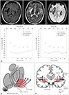
Brain MRI showed encephalomalacia in T1- and T2-weighted imaging (Fig. 1A, B) and hemosiderin deposits in susceptibility-weighted imaging (Fig. 1C) that were mainly bilaterally in the putamina and in the right temporal lobe. Pure-tone audiometry showed severe bilateral sensorineural hearing loss with the absence of bone conduction (Fig. 1D). She exhibited normal distortion-product otoacoustic emissions, which suggested normal functioning of the peripheral hearing system. Brainstem auditory-evoked potentials showed normal waveforms and latencies of waves I to V, indicating functional integrity of the auditory pathways within the brainstem (Supplementary Fig. 1 in the online-only Data Supplement). She was finally diagnosed with cortical deafness and referred to rehabilitation centers.
Cortical deafness is a rare condition that is usually caused by bilateral damage to the temporal lobes, and subcortical lesions have also been reported to lead to this condition.1 The underlying etiologies include stroke, mitochondrial encephalomyopathy, lactic acidosis, and stroke-like episodes (MELAS) syndrome,2 multiple sclerosis.3 To the best of our knowledge, only six cases of cortical deafness following bilateral putaminal hemorrhage have been reported456789 (Supplementary Table 1 in the online-only Data Supplement). The auditory radiations travel densely from the medial geniculate body (MGB) up to the sublenticular region and radiate to auditory cortices, partly by coursing through the white matter immediately ventral to the posterior half of the putamen, and partly by penetrating the ventral and lateral portions of the posterior half of the putamen.9 Thus, bilateral putaminal hematomas can interrupt all of the projection fibers from the MGB bodies to the cortices to result in severe hearing loss9 (Fig. 1E, F). The resulting deafness could improve gradually and be replaced by auditory agnosia in some of these patients,56 while other patients remain permanently deaf,489 as in the present case.
Tinnitus after bilateral putaminal hemorrhage has not been reported previously. Tinnitus is often triggered by peripheral mechanisms, but central mechanisms are also crucial. We speculated that our patient developed tinnitus when the brain used auditory phantoms to fill in the blanks in a condition of deprived auditory inputs. This case has demonstrated that subcortical lesions involving bilateral auditory radiation can cause permanent deafness and tinnitus.
 XML Download