

 PDF
PDF ePub
ePub Citation
 Print
Print


Dear Editor,
Dermatomyositis (DM) is a type of myositis that presents with proximal muscle weakness and typical dermatologic manifestations.1 Among myositis-specific autoantibodies, anti-melanoma-differentiation-associated gene 5 (anti-MDA5) antibody (Ab) is specifically identified in clinically amyopathic DM, and is associated with rapidly progressive interstitial lung disease.1 Anti-MDA5-Ab-positive DM patients exhibit atypical skin lesions such as digital ulcers, acral digital necrosis, or palmar papules, rather than classical dermatologic manifestations.12 Immunosuppressants and glucocorticoids are used to treat inflammatory myositis, but how to treat digital ulcers has not been established in anti-MDA5-Ab-positive DM. Here we present a patient with anti-MDA5-Ab-positive DM with refractory digital ulcers who responded excellently to treatment with a combination of endothelin receptor-1 antagonist (ERA) and botulinum toxin injection.
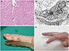
A 48-year-old male presented with a 6-month history of muscle weakness and facial discoloration. The facial discoloration was unlikely to be heliotrope rash since it appeared as a brownish color over the entire face. A neurologic examination revealed proximal muscle weakness, with MRC grade 4 in bilateral shoulder abduction and hip flexion. An electrodiagnostic study revealed early recruited myopathic motor-unit action potentials and positive sharp waves. The serum level of creatinine kinase was normal, at 185 units/L reference range 58–348 units/L). A muscle biopsy showed mild myopathic change without inflammatory cell infiltration (Fig. 1A). However, electron microscopy revealed tubuloreticular cytoplasmic inclusions of endothelial cells, which are mostly found in DM among inflammatory myositis3 (Fig. 1B). The classical dermatologic manifestation of DM was absent, but both hands showed cyanotic change and multiple digital ulcers (Fig. 1C). Chest CT revealed multiple subpleural ground glass opacities and consolidations in both lower lung fields. Perfusion scintigraphy of the hand showed a clear decrease in blood flow after cold stimulation. We investigated myositis specific autoantibodies for Mi-2, TIF1γ, MDA5, NXP2, SAE1, Ku, PM-Scl100, PM-Scl75, SRP, PL-7, PL-12, EJ, OJ, and Ro-52 using an immunoblot assay kit (Immunoblot-PreQ system, EUROIMMUN Co., Ltd., Luebeck, Germany), which showed strong positivity only against MDA5.
The patient was started on glucocorticoid therapy comprising 1 g of methylprednisolone for 5 days followed by 1 mg/kg oral prednisolone for 1 month. This treatment was effective against the muscle weakness, but it aggravated the digital ulcers and Raynaud's phenomenon. The intravenous administration of prostacyclin, nifedipine, and phosphodiesterase type 5 inhibitor had no effect. Considering the treatment choice for refractory digital ulcers in systemic sclerosis, oral ERA, bosentan (62.5 mg twice daily), and the injection of botulinum toxin [10 IU of Meditoxin (Medytox, Seoul, Korea) dissolved in 0.1 mL of saline] into the soft tissue of the palm proximal to the A1 pulley were attempted. Bosentan and botulinum toxin injections weekly for 3 weeks significantly improved the digital ulcers and Raynaud's phenomenon. The improvement was maintained for 12 weeks after the initial injection of botulinum toxin (Fig. 1D).
Muscle weakness is usually mild or absent and there is lower elevation of muscle enzymes in anti-MDA5-Ab-positive DM patients.4 The first-line therapy for digital ulcers and Raynaud's phenomenon are conservative care and vasodilators, with ERA and botulinum toxin injection considered in refractory cases.5 Endothelin is a potent vasoconstrictor, and ERA shows a preventive effect against digital ulcers in systemic sclerosis.5 Botulinum toxin injection also improved Raynaud's phenomenon and digital ulcers in a pilot study.6 Injecting botulinum toxin into the neurovascular bundle of the palm can attenuate vasospasm by blocking hyperactive vascular responses.6
In the present case, the digital ulcers combined with Raynaud's phenomenon were significantly improved after applying a combination of botulinum toxin injection and oral ERA. Although the pathophysiology of skin ulceration in anti-MDA5-Ab-positive DM has been not fully elucidated, the present case provides new insight into the possible mechanism of abnormal dermatologic manifestation in anti-MDA5-Ab-positive DM. Furthermore, we suggest novel treatment options for digital ulcers in anti-MDA5-Ab-positive DM patients. A future larger scale study is needed to confirm the therapeutic effects of botulinum toxin injection and ERA in dermatologic manifestations of anti-MDA5-Ab-positive DM patients.
 XML Download