

 PDF
PDF ePub
ePub Citation
 Print
Print


INTRODUCTION
Onychopapilloma is a rare benign tumor occurring in the nail bed and the distal matrix and found most frequently in the middle aged female1. It commonly affects a single nail, mostly on the thumb, with longitudinal erythronychia beginning from the nail matrix and extending to the onychocorneal band2. Since the naming of the disease was finalized in 2000 by Baran and Perrin3, there have been only a limited number of reports on the disease245, and its characteristics have not been fully understood yet. To the best of our knowledge, only four cases have been reported in the Korean literature67. Herein, we present an uncommon case of onychopapilloma presenting as a longitudinal erythronychia.
CASE REPORT
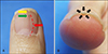
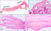
A 50-year old female was referred for the evaluation of 1-mm width of a single longitudinal erythematous streak on the left thumbnail, which was gradually widened for three years. The patient complained of a dull pain, especially when picking up small objects. Distal onycholysis and splinter hemorrhages were observed along the band with a solitary hyperkeratotic papule on the hyponychium (Fig. 1). To aid diagnosis and relieve the discomfort, the nail plate extraction and a longitudinal excisional biopsy from the nail bed to matrix were performed. Histopathologic examination revealed no significant changes in the nail matrix while the nail bed epithelium showed prominent papillomatosis and acanthosis. The upper layers of the nail bed consisted of epithelial cells with abundant eosinophilic cytoplasm reminiscent of the keratogenous zone of the matrix, which indicates matrix metaplasia of the nail bed. In addition, layered hyperkeratosis and focal hemorrhages were found beneath the nail plate (Fig. 2). Based on these clinical findings and histopathologic features, the patient was diagnosed with onychopapilloma.
DISCUSSION
Onychopapilloma is the most common cause of localized longitudinal erythronychia (LLE), which sometimes manifests as leukonychia48 or melanonychia69. Distal subungual hyperkeratosis is one of the most significant clinical signs of onychopapilloma; splinter hemorrhages, onycholysis and distal splitting also accompany the disease occasionally. The symptom varies from none to pain, but the tumor usually interferes in the grabbing motion2.
The pathogenesis of longitudinal erythronychia is still uncertain whether it is caused by the neoplastic or reactive process in the distal matrix which may create a defect in its function. A consequential nail plate thinning forms longitudinal groove in the ventral surface and accentuates a subtle reddish band with splinter hemorrhages due to engorged nail bed epithelium trapped in the furrow. The weakened plate sometimes leads to a triangular notch at the distal nail table and a subungual keratotic mass is exposed at the hyponychium1011.
Dermoscopically, many of the nail diseases presenting LLE have enlarged ends in the lunula, whereas the proximal verge of the streak in onychopapilloma is not completely clubbed, showing pointed edge shapes. Dermoscopic approach at the distal edge seeks out a thread-like papule underneath the plate corresponding to the red line, which is a highly suggestive sign for diagnosis2.
Histologically, papillomatous acanthosis and abundant eosinophilic cytoplasm in the upper layers of nail bed epithelium are commonly identified, which indicates matrix metaplasia12. Focal subungual hyperkeratosis with hemorrhages is also one of the diagnostic features, whereas the nail matrix usually suggests nonspecific changes.
Other benign nail tumors that exhibit LLE, such as glomus tumor and subungual wart can be differentiated by their distinct clinical signs2101314. The former shows a history of a painful nail, particularly exacerbated by cold, and is not associated with a free edge keratotic mass. The latter, on the other hands, presents multiple black specks on the surface and accompanies periungual lesions occasionally. Early stages of other nail disorders with polydactylous erythronychia such as Darier's disease and lichen planus should be also considered10, but typical cutaneous or mucosal involvement usually precedes the nail symptom. In addition, Bowen's disease and squamous cell carcinoma are two of the most frequent malignant nail tumors associated with LLE391516. As distal subungual keratosis, onycholysis and splinter hemorrhages are also found in Bowen's disease1314, it is often indistinguishable from onychopapilloma, if not confirmed by its histopathologic characteristics of atypical keratinocytes and dyskeratotic cells.
Some previous studies have proposed the algorithm to evaluate the patients with localized erythronychia.1011 Patients with stable band of LLE are asked to follow up at regular intervals to check the progress. When the involved lesion is painful or clinically uncertain due to its atypical features, surgical exploration or excision should be performed. Since the clinical patterns of LLE vary in terms of the width of the linear band, the degree of erythronychia, and the presence of onycholysis or splinter hemorrhages, it is hard to predict malignancy17. So, for rapidly evolving cases, it is recommended to conduct a longitudinal biopsy from the matrix to the nail bed. As a global recurrence rate of onychopapilloma was reported 20% recently1, complete excisional biopsy is mandatory not only to confirm the diagnosis but also to resolve the finger discomfort1017. We received the patient's consent form about publishing all photographic material.
 XML Download