

 PDF
PDF ePub
ePub Citation
 Print
Print


INTRODUCTION
Bowen's disease (BD) was first described by Bowen in 1912 as a cutaneous squamous cell carcinoma (SCC) in situ1. BD appears as a slowly enlarging, sharply demarcated erythematous plaque2. Histological examination reveals hyperkeratosis and parakeratosis, overlying an epidermis that is replaced by disorganized atypical keratinocytes and abnormal mitosis3. The risk of progressing BD to SCC is within 3%~8%, of which 13%~20% develop metastasis34. Different guidelines for BD recommend various treatments such as surgical excision, photodynamic therapy (PDT), imiquimod 5% cream, 5-fluorouracil cream, cryotherapy, laser therapy, and curettage4567. However, the most effective treatment is uncertain because of the lack of controlled trials48. The British guidelines state that no treatment modality is superior to others and that choosing a therapeutic option for a patient depends on the tumor location, expected efficacy, patient basal status, economic resources, and patient preference47. PDT is a frequently used treatment modality because of its noninvasive character and excellent cosmetic results. PDT has an advantage at body sites associated with poor wound healing and in the case of multiple lesions7910. Recently, ingenol mebutate (IMB), which is a drug approved by the US Food and Drug Administration for the treatment of actinic keratosis (AK), is being used off-label to treat BD. Unlike in AK treatment111213, no studies comparing the treatment outcomes of PDT and IMB are available in BD treatment. Also, no clinical or histological factors that affect the treatment outcome of IMB are reported.
This study aimed to assess the effectiveness of the PDT and IMB in the treatment of BD and to determine which clinical or histological factors affected the treatment outcome.
MATERIALS AND METHODS
Patient selection
Patients with histologically confirmed BD from January 1, 2006, to December 31, 2017, were identified from the histological database of CHA Bundang Medical Center. Of the selected patients, those treated with PDT or IMB were included. Patients with tumors found on the anogenital region were excluded because of their different etiologies and known high recurrence rates. Patients who lost to follow-up (FU) or with inadequate medical information in the patient electronic chart were also excluded from the analysis. Ethical approval was obtained from the ethic committee of CHA Bundang Medical Center (IRB no. 2019-07-039-001).
Procedure
Data on patient and tumor characteristics were retrospectively collected from the patient electronic chart. The parameters included in the analysis were as follows: age, sex, disease duration, tumor location, histological epidermal thickness, the number of treatment sessions and pretreatment with fractional CO2 laser, clinical result at the first FU visit, the date of any recurrences noted during later FU visits, and adverse events. In this study, complete response was defined as the absence of clinical evidence of BD at the first FU visit. Conversely, any clinical evidence of BD in the treatment area at the first FU visit indicated an incomplete response. Meanwhile, recurrence was defined as the presence of any clinical or histological evidence of BD during later FU visits.
Therapy
For patients treated with PDT, all BD lesions were treated with the same methyl aminolevulinate cream (Metvix®; Galderma Laboratories, Paris, France), light source (Omnilux; Photo Therapeutics, Inc., Altrincham, UK; wavelength: 633 nm), and light dose (126 J/cm2). The methyl aminolevulinate cream was applied to BD lesions and covered with plastic film to avoid light exposure. The cream was removed after 4 hours, and the lesion was irradiated with the light source. 10 BD lesions out of 40 BD lesions were treated with fractional CO2 laser (eCO2; Lutronics, Seoul, Korea; 50 mJ×200 spots/cm2) before PDT treatment. For patients treated with IMB, head and neck lesions were treated with 0.015% IMB (Picato®; Leo Pharma, Ballerup, Denmark) for 3 consecutive days, and trunk and limb lesions were treated with 0.05% IMB for 2 consecutive days. Patients were instructed on the modalities of IMB self-administration.
Statistical analysis
Distributions of patient characteristics were described by means of standard deviation.
To determine the presence of any potential factors associated with the treatment outcome after PDT or IMB, we performed logistic regression on the following parameters: age, sex, disease duration, tumor location, histological epidermal thickness, and the number of treatment sessions and pretreatment with fractional CO2 laser. Statistical significance was considered when the p-value was below 0.05. Statistical analyses were performed using IBM SPSS Statistics ver. 25.0 (IBM Corp., Armonk, NY, USA).
RESULTS
Patient and tumor characteristics
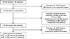
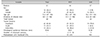
During the study period, 160 histologically proven cases of BD in 145 patients were found in the histological database of CHA Bundang Medical Center. Of the 160 BD lesions, 13 on the anogenital region were excluded because of their different etiologies and known high recurrence rates. The treatment modality was retrieved from the patient electronic chart. Of the 147 remaining BD lesions, 64 treated with surgical excision (56), cryotherapy (4), and imiquimod (4) other than PDT and IMB were excluded. We also excluded 15 lesions in 15 patients who lost to FU or with inadequate medical information in the electronic chart. Consequently, the 68 remaining lesions in 61 patients were included. Among them, 44 BD lesions in 44 patients were treated with PDT, whereas 24 BD lesions in 17 patients were treated with IMB. The average age was 71.4 years (47~93 years), and 45 patients were female. A flowchart of patient selection is presented in Fig. 1, whereas the demographics of the patients and the clinical, histological characteristics of BD lesions are shown in Table 1.
Evaluation of the treatment outcome
The mean time to the first FU visit of patients treated with PDT was 1.5 months (0.5~3.8 months), whereas that of patients treated with IMB was 0.9 months (0.5~1.5 months). The mean FU duration of patients treated with PDT was 7.6 months (1~24 months), whereas that of patients treated with IMB was 6.6 months (1~36 months). The mean number of treatment sessions for PDT is 2 sessions (1~5 sessions), while that of IMB was equally 1 session. Complete response was found in 29 lesions (66.7%) after PDT and 13 lesions (53.0%) after IMB (p=0.349). Of the 29 complete-response BD lesions treated with PDT, 3 lesions (10.3%) recurred. Meanwhile, 2 lesions (15.3%) recurred in the 13 complete-response BDs treated with IMB (p=0.993). Table 2 shows the number of BD lesions with a complete response and the recurrence rates noted after FU. In patients with BD treated with PDT, complete response was found in 8 out of 10 lesions (80%) with pretreatment with fractional CO2 laser and 20 out of 34 lesions (60%) without fractional CO2 laser (p=0.096; Table 3).
Factors associated with successful treatment
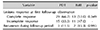
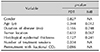
When analyzing the factors associated with successful treatment, no significant correlation was found in the success rates of PDT and IMB for BD and in any of the following parameters: sex, disease duration, tumor location, histological epidermal thickness, and the number of treatment sessions and pretreatment with fractional CO2 laser. Only older age significantly correlated with the lower complete-response rate of IMB (p=0.012; Table 3).
Adverse events
A description of the adverse events in 20 of the 68 cases was available in the patient electronic chart. Among these 20 lesions, 9 were reported from PDT (20.5%), and 11 were reported from IMB (45.8%). In PDT, the most common adverse event was crusting, which was observed in four cases. Other observations were itching (2), hyperpigmentation (2), and erythema (1). In IMB, the most common adverse event was irritation, which was observed in four cases. Other observations were crusting (2), stinging (2), and erythema (2). Nonetheless, no serious adverse events and discontinuation of treatment caused by treatment-related adverse events were reported.
DISCUSSION
Our results show that the complete-response rate of BD treated with PDT (66.7%) was higher than that of BD treated with IMB (53.0%). Meanwhile, the recurrence rates of BD treated with PDT (10.3%) was lower than of BD treated with IMB (15.3%). In a previous study, the clinical clearance and recurrence rates of BD cases treated with PDT were 76.1%~98% and 10.5%~31%, respectively, thereby comparable to our results714151617. Effective treatment with IMB in several cases of BD was also reported18192021222324, but studies on the clinical clearance and recurrence rates of BD treated with IMB are unavailable. The treatment outcome of PDT in BD was previously compared with other noninvasive treatments, mostly with cryotherapy and topical 5-fluorouracil cream. The results of these studies demonstrate that PDT is superior to cryotherapy and 5-fluorouracil cream in terms of therapeutic efficacy2526. Meanwhile, a comparison of the treatment outcomes of PDT and IMB in BD has not yet been reported. However, according to the results of studies comparing the treatment outcomes of PDT and IMB in AK, the clinical clearance rates of PDT and IMB were 67.1% and 62.9%, respectively, indicating to be nonsignificant13. In our study, the results demonstrate that PDT is an effective treatment option for BD, with a higher complete-response rate and lower recurrence rate than IMB. No statically significant difference of the complete-response rate and recurrence rate was found between PDT and IMB, but a larger study population and a longer FU are needed to validate these results.
The outcome of BD treatment can be variable according to several clinical and histological factors. In our study, we searched for different clinical and histological factors influencing the PDT and IMB treatment outcomes in BD. A peak incidence of BD is described between the age of 70 and 79 years2. The average age of patients with BD in our study was 71.4 years. Furthermore, a significantly low complete- response rate was found in elderly patients in the IMB treatment group. Advanced age is associated with a decreased function of the immune system27. Perhaps, the immune response induced by cytokine secretion by activated protein kinase C toward tumor cells during IMB is decreased in the elderly. Also, PDT is prescribed by the physician, and the treatment is performed by appropriately trained staff on a day-case basis. By contrast, IMB is applied by the patient at home. Possibly, the elderly patient has not been treated appropriately because of decreased compliance. Hence, further study is needed to compare the treatment outcomes of patients treated with IMB at the hospital and patients treated with self-application.
Meanwhile, the longer the time from lesion onset to diagnosis, the more likely it will affect biological behavior and reduce the clearance rate of treatment. However, the duration of disease in our results did not affect the treatment outcome in both PDT and IMB.
A study by Truchuelo et al.7 showed a decreased clearance rate on the lower extremities. Tyrrell et al.28 also showed that acral tumors exhibited a lower clearance rate after PDT than non-acral tumors. In our study, tumors were most commonly located on the head and/or neck in both PDT and IMB. However, the location of BD was not significantly associated with the treatment outcome. The proportion of female patients and that of tumors located on the head and/or neck were higher in the IMB than those in the PDT. This result is because female patients consider cosmetic aspects more and prefer a simpler and more rapid treatment than male patients.
In studies concerning basal cell carcinoma (BCC), tumor thickness was a substantial predictor of clinical clearance of PDT, with no response in BCC measuring more than 2 mm thick2930. Furthermore, invasive SCC, as well as nodular BCC, has low overall success rates after PDT3132. Therefore, increased epidermal thickness might also play an important role in BD. However, in our study, the epidermal thickness was not significantly associated with the treatment outcome. The mean epidermal thicknesses were 0.63 mm and 0.38 mm in PDT and IMB, respectively, and no BD was thicker than 2 mm. These findings suggest that protoporphyrin IX (PpIX) and IMB penetrated deep enough to reach the entire epidermis and might explain why the increased epidermal thickness did not influence the treatment outcome of PDT and IMB in BD.
Another possible influencing factor is pretreatment with fractional CO2 laser. In comparing the treatment outcomes according to the presence or absence of pretreatment with fractional CO2 laser in PDT, the pretreatment with fractional CO2 laser could increase the rate of complete response. The American Society of Dermatological Surgery recommends the pretreatment of hyperkeratosis before the application of photosensitizer to improve the treatment outcome33. A review by Bay et al.34 showed that pretreatment with fractional CO2 laser, rather than curettage/debulking, microdermabrasion, and microneedling, increases the accumulation of PpIX and the treatment outcome after PDT. Although not investigated in our study, Erlendsson et al.35 reported that combination therapy with fractional CO2 laser can increase the therapeutic effectiveness of IMB, enabling customized topical delivery and the potential use of IMB for the treatment of non-melanoma skin cancer.
The main limitation of our study is the retrospective study design with a small sample size. Another limitation is that the assessment of complete response and recurrence was mainly based on clinical findings.
This retrospective study of 68 BD lesions showed that both PDT and IMB are effective noninvasive treatment modalities for BD. However, PDT is a safer treatment modality than IMB, considering that PDT has fewer adverse events. Especially in patients with old age, which is a factor that reduces IMB treatment outcome, PDT can be considered as preferred treatments over IMB for BD. In patients with BD treated with PDT, pretreatment with fractional CO2 laser tends to increase the complete response rate. We recommend pretreatment with fractional CO2 laser to improve the treatment outcome in BD treatment with PDT and IMB. Further prospective studies with a large sample size are required to acquire even better and more reliable data.
 XML Download