

 PDF
PDF ePub
ePub Citation
 Print
Print


INTRODUCTION
Mycosis fungoides (MF) is the most common type of cutaneous T cell lymphoma, accounting for about 50% of all primary cutaneous lymphoma and 60% of cutaneous T cell lymphoma1. Classic mycosis fungoides is defined as an indolent non-Hodgkin lymphoma that characterized with patches and plaques preferentially located on the covered sites such as buttocks clinically in early stage and tumors and extracutaneous involvement with possibly fatal outcomes in the late stage. Numerous variants of MF have been reported in recent decades234. In 2018 update of the World Health Organization-The European Organisation for Research and Treatment of Cancer (WHO-EORTC) classification5 for primary cutaneous T cell lymphomas, folliculotropic MF (FMF), pagetoid reticulosis and granulomatous slack skin (GSS) are described as variants of MF due to their distinctive clinical and histopathologic features. Meanwhile, there are clinical variants of MF such as hypopigmented MF (hMF), ichthyosiform MF, and poikilodermatous MF, which have unique clinical but similar histopathologic features to classic MF.
The epidemiological features of mycosis fungoides and its variants may vary by geographical area. To date, few studies on MF from Asian678 did not focus on MF variants. Besides, few studies reported characteristics of mycosis fungoides and its variants in a Chinese population. Therefore, the present study aimed to evaluate the features of mycosis fungoides and its variants in China.
MATERIALS AND METHODS
Patient selection and clinical parameters
In this retrospective analysis, we searched our institutional clinical registry and pathology database between October 2012 and January 2018 for patients diagnosed with MF. Clinical course and diagnostic results were reviewed. Patients who were confirmed clinically and pathologically at our institution were included. MF was diagnosed according to the 2018 updated version of the WHO-EORTC classification for primary cutaneous T cell lymphomas system5. Patients who had diagnosed biopsy with hypopigmented skin lesions, ichthyosiform skin lesions or poikilodermatous skin lesion were defined as hMF, ichthyosiform MF, and poikilodermatous MF respectively. Patients were exclusion based on the criteria: clinical records were incomplete; the patients followed at our hospital for less than one year unless they died within that period. The following data were retrieved from the database: age at diagnosis, sex, duration between symptom onset and diagnosis, duration of follow-up (from the first diagnosis to the last follow-up record), tumor-node-metastasis-blood (TNMB) classification stage at the time of diagnosis, treatment modality, and follow-up data. This study was approved by the research ethics committee of the hospital (IRB no. S-K839).
Statistical analysis
All statistical calculations were performed using IBM SPSS ver. 23.0 (IBM Corp., Armonk, NY, USA). The date of the first diagnostic biopsy was considered as the date of diagnosis. Disease-specific survival (DSS) rate was calculated from the date of diagnosis to death from MF or last follow-up. For surviving patients, the study endpoint was 31 December, 2018. Kaplan–Meier plots were used for survival analysis. Comparison between curves was made by log-rank testing. Moreover, Mann–Whitney tests were used as appropriate. p values below 0.05 were considered significant.
RESULTS
Clinical characteristics of mycosis fungoides
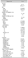
A total of 93 patients diagnosed with mycosis fungoides from October 2012 and January 2018. All patients were Chinese. The age at the diagnosis was 38.9±1.7 years (range: 6~77). Forty-five males (48.4%) and 48 females (51.6%) were included in this study, with male to female ratio 1:1.1. The range of duration of symptoms was six months to 360 months (median, 72 months). Moreover, the range of duration of follow-up was 8~120 months (median, 44.5 months). All patients were evaluated clinical stage by using the TNMB staging system. Of the 93 patients, the early stage (stage IA, IB, and IIA) included 75 cases (80.6%), and the advanced stage (stage IIB, III, and IV) included 18 cases (19.4%) (Table 1).
Patients were treated with the stage-directed treatment modalities. In general, patients with early-stage MF were mainly treated with skin-directed therapy, including topical corticosteroid, phototherapy. Psoralen with ultraviolet A (UVA) and UVA combination with ultraviolet B (UVB) are the common choice in phototherapy. Patients with advanced stage MF were treated with systemic modalities combined with skin-directed therapy. The systemic modalities included interferon α, methotrexate, systemic steroid, acitretin, and radiotherapy.
Among the 93 patients, 76 patients (81.7%) were classified as classic MF, 1 patient (1.1%) was FMF, 2 patients (2.2%) were GSS, 10 patients (10.8%) were hMF, 3 patients (3.2%) were ichthyosiform MF, and 1 patient (1.1%) was poikilodermatous MF.
Survival outcomes
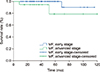
The follow-up period of all patients ranged from 8~120 months (median, 44.5 months). During the period of follow-up, three patients died from mycosis fungoides. Kaplan–Meier estimated of survival early stage and advanced stage of mycosis fungoides were calculated (Fig. 1). Of 93 mycosis fungoides patients, the DSS rate of all MF patients was 96.7%. In the early stage MF, the DSS rate was 98.6%, while in advanced stage MF, the DSS rate was 88.9% (p=00.042, log-rank test).
Epidemiology data of patients based on variants of mycosis fungoides
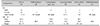
According to the several MF variants, the characteristics of patients were stratified. Among the classic MF (76 patients, 81.7%), there were 33 males and 43 females. The mean age of classic MF was 42.6±1.7 years. For hMF, there were 6 males and 4 females. The median age was 10.5 years (range: 6~28). In addition, there was a significant difference between the age of hMF and that of classic MF (p<0.05, Mann–Whitney test). The hMF patients were younger and had a significant difference than that of classical MF patients (Table 2).
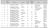
Clinical data of 10 patients with hypopigmented mycosis fungoides
From a population of 93 patients diagnosed with MF, ten patients were included in the sample as hMF patients: one adult man, one adult woman, five boys, and three girls. Two patients only had hypopigmented lesions. Eight patients had both hypopigmented lesions and erythematous lesions.
The clinical description is shown in Table 3. For the clinical stage, all patients were either stage IA or IB. Four patients had stage IA, and six patients had stage IB.
The primary treatment modalities employed were phototherapy, including narrowband UVB, UVB and UVB combination with UVA. Three patients underwent phototherapy with interferon α. All of these ten patients went into remission. Seven patients responded completely, and three patients responded partially. Moreover, no patient was with recurrences documented.
DISCUSSION
Mycosis fungoides usually has the disease progression as a slow evolution from patches, plaques to tumors and extracutaneous involvement with possibly fatal ends, which is by the long duration of symptoms and cutaneous lesions at presentation1. Besides classic MF, there are still several variants of MF. The wide range of clinical and histopathologic presentations of MF, especially early-stage MF, requires a broad differential diagnosis. It helps to establish a correct diagnosis of MF, particularly in the early stages of the disease, that being aware of the distinctive characteristics of this disease. Because of the rarity of the disease, there are few studies about subtype distribution of mycosis fungoides and its variants9. Although studies focused on specific subtype were published, relevant data in Chinese population are seldom reported.
In our study, the mean age at diagnosis of all MF patients and classic MF patients were 39.0 years and 42.6 years, respectively, which were both younger compared with other studies from western countries and Asian countries71011, while in accordance with the research from Singapore9. The possible reasons might be that patients could get access to dermatologists directly due to the imperfect referral system in China and that data, from a single center, might be biased. Also, there was not a male preponderance in the sex distribution of MF patients, in accordance with our previous study8 but in contrast to other studies1112.
The stage is related to prognostic significance in mycosis fungoides13. According to this study, the majority of our patients (75 patients, 80.6%) were in the early stage, and 18 patients (19.4%) were in an advanced stage. The prognosis of advanced stage MF was significantly worse than early-stage MF. The survival analysis of our study also confirmed the favorable prognosis of MF with a 96.7% DSS rate. Besides stage, older age was considered as a risk factor for MF, but it is controversial. Based on the study from Lebowitz et al.14, older age at diagnosis of MF does not predict worse disease-specific outcomes. We speculated the main reason is the difficulty of careful monitoring in daily life.
The therapy of mycosis fungoides should follow a stepwise, stage-adapted approach. The consensus recommendation from the European Organisation for Research and Treatment of Cancer-Cutaneous Lymphoma Task Force (EORTC-CLTF) is accepted and used widely.
In this study, although there were some patients in T1a stage, none of them chose a “watch and see” policy that recommended by consensus recommendation from EORTC-CLTF. The early stage MF patients were mainly treated with phototherapy, according to the standardized guidelines released by the United States Cutaneous Lymphoma Consortium15. Some of the patients in this study also used phototherapy and interferon α combined therapy. Safety and efficacy have been confirmed1617.
It has been reported that in large series, FMF accounts for approximately 10% of all patients with MF1318. Among 93 patients in our study, one was diagnosed in FMF without any other medical history, which is less than 10%. In two studies from Korea, the percentage of FMF in all MF patients accounted for around 2% and 4% respectively1920. In a study in Argentina21, the portion was about 6%. Those data from Asian countries might suggest that the relative frequency of FMF has geographical and racial differences. Our FMF patient was in an early stage with multiple lesions on her face and got complete remission after topical steroids treatment. It is suggested that FMF patients are received treatment based on stage, similar to the stepwise approach in classic MF22.
GSS is extremely rare. The two GSS patients presented with typical manifestation: pendulous folds of scaly atrophic skin, huge ulcer, and secondary bacterial infection. They were treated with chemotherapy and interferon α plus systemic steroids, respectively. Both of them got a partial response and were suggested to go under the long-term follow-up to exclude the increased risk of other malignant lymphomas23.
hMF, ichthyosiform MF, and poikilodermatous MF are not listed in the latest WHO classification of cutaneous lymphoma. However, they do have distinctive clinical manifestation to lead to narrow the differential diagnosis of MF. Ichthyosiform MF and poikilodermatous MF have the same typical pathological features as classic MF and similar developing process.
Besides clinical aspects, hMF is also distinct from the classic MF in epidemiological and prognostic aspects. It most commonly affects patients in childhood and adolescence. According to the recent study from Brazil24, the average age of 20 hMF patients was 43.85 years. Rodney analyzed 20 hMF patients with an average age of 32.2 years3. Tan et al.9 included 47 hMF patients in Singapore with a median age at diagnosis of 17. The present study demonstrated an average age of 13.4 years with age ranged from 6 to 28, in line with the previous findings. All patients in our study were Chinese. It is reported that white patients usually have hypopigmented lesions with an erythematous lesion. This “mixed” pattern is said to prevail in the Caucasian population25. However, eight patients among 10 hMF patients presented with hypopigmented lesions and erythematous lesions in our study. Two patients were only with hypopigmented lesions, showing that the “mixed” pattern is also common in Chinese hMF patients. Our data showed all the hMF patients, in stage IA or IB, treated by phototherapy or other skin-directed treatment and got remission. Until the end time of follow-up, no relapse was documented. As shown in the literature, the prognosis of hMF is good, and patients respond very well to skin-direct treatment26.
The limitations of this study include a single-center based experience and a small sample number. Despite the flaws, the study is meaningful as it gives more insight into mycosis fungoides and its variants among Chinese patients: early-stage MF has a better prognosis than advanced stage and hMF affects younger patients than that of classic patients among Chinese patients. This study provides a new insight of mycosis fungoides and its variants in Chinese population. To be knowledgeable of variants of MF could help to give an accurate and prompt diagnosis and initiate appropriate treatment.
 XML Download