

 PDF
PDF ePub
ePub Citation
 Print
Print


INTRODUCTION
Pruritus is an important symptom in psoriasis, and several studies have reported that patients with psoriasis suffer from chronic pruritus123. However, the reported prevalence of pruritus in psoriasis differs in previous studies, and the mechanism of pruritus in psoriasis remains unclear.
Various mediators have been suggested as responsible for pruritus in psoriasis. Histamine, and neuropeptides such as substance P (SP), calcitonin gene-related peptide (CGRP), vasoactive intestinal peptide (VIP), and neuropeptide Y (NPY), are known itch-associated mediators45. In addition, neuropeptides are known to have proinflammatory effects. Previous studies have reported increased expression of neuropepetides and their receptors in psoriatic lesions, and their association with pruritus678. Recently, interleukin-31 (IL-31) was suggested to play an important role in pruritus in human T-cell-mediated skin diseases such as atopic dermatitis910. Narbutt et al.11 observed a significantly higher concentration of IL-31 among patients with psoriasis compared to controls.
Moreover, it was reported that the level of serum inflammatory cytokines differs according to morphological phenotype (chronic stable [CS] vs. eruptive inflammatory [EI])12. Based on this report, we hypothesized that the prevalence and clinical characteristics of pruritus and pruritogenic mediators might differ according to morphological phenotype.
In this study, we investigated differences in clinical characteristics of pruritus and the levels of serum neuropeptides and IL-31 according to morphological phenotype.
MATERIALS AND METHODS
Subjects
Patients who were at least 18 years of age, diagnosed with psoriasis clinically and histologically, and not on systemic treatment for at least 4 weeks prior to participate in this study were included. The patient with erythrodermic and pustular type of psoriasis, history of other medical diseases were excluded. According to criteria used in a previous study, we divided patients into 2 subgroups (Table 1): EI and CS12. As a control, 10 healthy volunteers attending the same institution for the health medical examination with no significant medical history or underlying pruritogenic skin diseases were recruited. All participants were provided with detailed information on the purpose and methods of the study. Approval from the Institutional Review Board was obtained (IRB no. H-1508-001-032).
Methods
1) Clinical findings
Patient demographics and clinical findings of psoriasis were recorded. The severity of psoriasis was assessed by using the Psoriasis Area and Severity Index (PASI) score, and body surface area (BSA). The severity of psoriasis was classified as mild, moderate, or severe according to PASI score (<10, 10~20, and >20, respectively) and BSA (< 10%, 10%~30%, and >30%, respectively)13. To assess the characteristics of pruritus, participants were surveyed using a questionnaire if they currently suffered from pruritus, and were investigated for its location, frequency, diurnal variation, duration, aggravating factors, and intensity (determined by numeric rating scale [NRS]; 0 corresponds to no pruritus and 10 indicates the most severe pruritus). We further analyzed baseline demographics, characteristics and intensity of pruritus according to morphological phenotypes (EI vs. CS). The correlation between pruritus intensity and clinical parameters of psoriasis was analyzed in both subgroups.
In addition, psoriasis patients were categorized into pruritic (NRS 1~10) and non-pruritic (NRS 0) groups, and we analyzed the differences in baseline demographics and other clinical information.
Serum neuropeptides and IL-31 were measured in psoriasis patients and healthy controls. Differences in serum level were analyzed according to the presence of psoriasis (healthy controls vs. total psoriasis patients), morphological phenotype (EI vs. CS), and presence of pruritus (pruritic vs. non-pruritic). Correlations between clinical parameters (pruritus intensity, severity of psoriasis) and neuropeptides or IL-31 were also assessed.
2) Neuropeptides and IL-31 measurement
For quantitative measurements of serum levels of neuropeptides and IL-31, venous blood samples were collected from psoriasis patients and healthy controls. Fresh serum was obtained, spun in a centrifuge, immediately frozen, and stored at −70℃ until processed. An enzyme-linked immunosorbent assay was used for the measurement of serum IL-31 (Alpco, Salem, NH, USA), histamine (Alpco), and SP (Enzo Life Sciences, Farmingdale, NY, USA). Enzyme immunoassay was used for the measurement of serum NPY (Phoenix pharmaceuticals, Belmon, CA, USA), CGRP (Phoenix pharmaceuticals), and VIP (Phoenix pharmaceuticals) according to the manufacturer's instructions.
Statistical analysis
All data were analyzed using IBM SPSS Statistics ver. 21.0 (IBM Corp., Armonk, NY, USA). Fisher's exact test, chisquare test, and linear-by-linear association were used to test independence of categorical variables. Student's t-test was used to compare the mean/onset age, disease duration, mean PASI, and BSA, and serum level of neuropeptides of EI and CS groups. Correlation analysis was performed by calculating Spearman's coefficient, and p<0.05 were considered significant.
RESULTS
Clinical findings
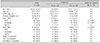
From July 2014 to August 2015, 50 consecutive patients (30 males, 20 females) with psoriasis were included in this study. The mean±standard deviation age was 45.7±17.5 years (range, 18~80). Of the 50 patients, 15 patients (30.0%) and 35 patients (70.0%) were classified as EI and CS, respectively (Table 2).
There was slight female predominance in EI (Male:Female= 0.9:1), and male predominance in CS (Male:Female=1.9: 1), but the difference was not statistically significant. There were no significant differences in mean age, onset age of psoriasis, disease duration, mean PASI, and scalp/nail involvement between the 2 subgroups. However, mean BSA was higher in EI group (EI, 22.7% vs. CS, 18.8%). Among 50 psoriasis patients, 40 patients (80.0%) complained of pruritus, and the prevalence of pruritus was not different between 2 subgroups.
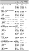
Among 40 psoriasis patients with pruritus, the mean intensity measured by NRS was not significantly different between the 2 subgroups (EI, 4.4 vs. CS, 5.3). The characteristics of pruritus including location, frequency, diurnal variation, duration, and aggravating factors also showed no significant differences between subgroups (Table 3). Moreover, pruritus intensity did not correlate with age, sex, disease duration, and severity of psoriasis in both subgroups (data not shown).
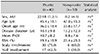
When comparing psoriasis patients with or without pruritus, the differences were also not significant in demographic and clinical parameters (Table 4).
Neuropeptides and IL-31 level according to morphological phenotype
Quantitative measurements of neuropeptides and IL-31 were performed in psoriasis patients and 10 healthy controls. Compared with healthy controls, psoriasis patients showed significantly lower serum SP and CGRP levels. However, EI and CS did not show significant differences in serum neuropeptide and IL-31 levels, with similar results in a comparison of pruritic and non-pruritic groups (Table 5). Serum neuropeptides and IL-31 levels did not correlate with severity of psoriasis in both EI and CS groups (data not shown).
DISCUSSION
Pruritus is an important symptom of psoriasis, with a reported frequency of 64% to 91%1415. Psoriasis manifests with various morphological phenotypes, including plaque-type, guttate, erythrodermic, pustular, and others13. However, there are limited data on the characteristics of pruritus according to morphological phenotype and pruritogenesis in psoriasis.
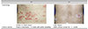
EI phenotype is characterized by rapidly spreading, small plaques with guttate morphology, and is frequently associated with pruritus, and sometimes with pustule formation. In contrast, CS phenotype is characterized by relatively large, stable plaques with lesser clinical activity12. Therefore, we hypothesize that morphological differences might affect the characteristics of pruritus, and assessed clinical differences, prevalence, and characteristics of pruritus according to morphological phenotypes.
We found no significant difference in male/female ratio between EI and CS groups like previous study12. In addition, morphological phenotypes did not show significant differences in other clinical parameters including age, onset age, disease duration, mean PASI and scalp/nail involvement, except for mean BSA (Table 2).
We found one previous study reporting the prevalence of pruritus according to morphological phenotype, and that study showed that pruritus was more common in plaque type (50% of guttate psoriasis vs. 65% of plaque psoriasis)14. However, prevalence of pruritus was same in the both groups in our study (80%).
Pruritus intensity also did not correlate with severity of psoriasis in both EI and CS groups (data not shown). The reported correlation between pruritus intensity and severity of psoriasis has shown conflicting results; some reports have shown a lack of correlation between pruritus intensity and severity of psoriasis416, whereas positive correlations between the PASI score and pruritus intensity were shown in other reports217. Our results support the former position.
In psoriasis, neuropeptides are known to be responsible for the induction and maintenance of the inflammatory process181920. Pruritus in psoriasis is reportedly associated with abnormal expression of neuropeptides. Chang et al.6 reported that keratinocytes in psoriatic plaques of patients with pruritus showed higher expression of receptors for SP and CGRP. Higher CGRP plasma levels in patients with pruritus and a positive correlation between CGRP and pruritus intensity were reported21. IL-31 is a novel T-helper-lymphocyte-derived cytokine that plays an important role in human T-cell-mediated skin diseases. Serum IL-31 was previously found to be elevated in atopic dermatitis9. IL-31 mRNA expression of psoriatic lesion was not upregulated compared with healthy skin22, but higher serum IL-31 levels in patients with psoriasis were observed compared with a control group11. However, in our study, there were no significant differences in prevalence and characteristics of pruritus, serum level of neuropeptides, and IL-31 according to psoriasis clinical phenotype (Table 3). Moreover, pruritic and non-pruritic psoriasis patients showed insignificant differences in the clinical features (Table 4). In addition, there were no significant differences in serum levels of neuropeptides and IL-31 between pruritic and non-pruritic groups (Table 5). We assume that these results might reflect the complex pathogenesis of pruritus in psoriasis. Neuropathic inflammation is an important mechanism linked with pruritus in psoriasis, and other mechanisms have been suggested, including abnormal activation or function of the opioid system in the skin23, gamma-aminobutyric acid and its receptor24, endothelial leukocyte adhesion molecule 1, and E-selectin25. In addition, serum neuropeptides and IL-31 levels did not correlate with severity of psoriasis in both EI and CS groups (data not shown), in accordance with previous reports showing no correlations between psoriasis severity and plasma level of SP, CGRP, VIP, and NPY15.
Skin barrier dysfunction is important pathogenesis of psoriasis. Psoriatic lesions have decreased stratum corneum hydration, increased transepidermal water loss and reduced levels of moisturizing factors and fatty acids26. Since intraepidermal sensory nerve fibers penetrate nearly the full thickness of the epidermis, such barrier defects would be expected to lower the itch threshold of sensory afferent nerve fibers27. Cathepsin S is overproduced by keratinocytes in patients with psoriasis relative to healthy controls28. In response to damage or stress, keratinocytes produce epidermal proteases such as cathespin S, so it might directly stimulate neuronal pathways to trigger itch. In our study, psoriasis patients showed significantly lower SP and CGRP serum levels than healthy controls. It could be speculated that the reduced serum concentrations may reflect an increased level of consumption or peripheral enzymatic degradation of these substances. It is known that neuropeptides are rapidly degraded in the tissue after release from nerve endings29. However, results from previous studies are conflicting. Reich et al.15 reported that SP levels did not differ between patients with psoriasis and controls, and that CGRP was elevated in patients with psoriasis.
This study has some limitations. Firstly, we used PASI, the most widely used tool for measurement of severity of psoriasis. However, PASI has limited ability to assess disease severity in small-plaque type psoriasis patients (which is common in Koreans), because PASI poorly reflects the speed of spread, and it is difficult to assess the extent of numerous small lesions1230. Secondly, our study included a relatively small sample size. Third, all neuropeptides and cytokines were measured in the serum which does not necessarily reflect their levels in the skin.
This study is unique in that it describes the characteristics of pruritus according to morphological phenotypes, and investigates the association with serum neuropeptides related to pruritus and IL-31. In conclusion, the morphological phenotype does not seem to be an important factor affecting the prevalence and characteristics of pruritus in psoriasis.

 XML Download