

 PDF
PDF ePub
ePub Citation
 Print
Print


INTRODUCTION
Metabolic syndrome (MetS) is a chronic disease with inflammatory and hypercoagulable states [1]. This chronic inflammatory condition is called metaflammation or para inflammation, having increased levels of inflammatory cytokines, including interleukin-6 (IL-6) [23]. MetS patients are also vulnerable to oxidative stress due to excessive production of reactive oxygen species (ROS) and a weak antioxidant defense system. Oxidative stress is associated with insulin resistance and chronic inflammation. Nowadays, MetS is one of the most important public health issues with increasing prevalence rates [45]. Considering the association between the components and complications of MetS such as inflammation, excessive ROS, and endothelial and fibrinolysis dysfunction [67], recent studies have proposed to add some important items such as subclinical inflammatory factors to MetS components because of their potential roles in cardiovascular disease (CVD) and type 2 diabetes mellitus (T2DM) pathogenesis [8].
Studies have shown that functional foods have beneficial effects on MetS status and other health issues. In fact, one or more food items in a regular diet can change metabolic features or cell signaling pathways [910]. Currently, flaxseed is one of the most attractive functional foods. Flaxseed oil is a major source of alpha-linolenic acid (ALA) (50%–60% of whole fatty acids composition), which has higher bioavailability in comparison to its milled or whole seed [111213]. Eicosapentaenoic acid (EPA) and docosahexaenoic acid (DHA) from ALA possess anti-inflammatory and anti-platelet aggregation properties, while arachidonic acid (AA(, the end product of linoleic acid (LA) metabolism, has the opposite effects [10]. Other useful components of flaxseed oil are phenolic acid and phenolic compounds (p-coumarin acid and pherolic acid) [12]. It also contains vitamin E in form of γ-tocopherol and thus protects cellular membrane lipids from oxidation [13].
According to the results of some clinical studies, omega-3 fatty acids and ALA had a potential role against inflammatory mediators [51314]. Indeed, flaxseed might be able to reduce the risk of CVD due to its antioxidant, anti-platelet adhesion, and other bioactive properties [31415]. Considering the conflicting results [1415], this study aims to compare the effects of flaxseed oil as a good source of ALA to those of sunflower oil as a common vegetable oil in the family food basket on the total antioxidant capacity (TAC), IL-6, and coagulation score.
MATERIALS AND METHODS
Study design
In this randomized controlled clinical trial, 60 participants were randomly assigned to the control (n = 30, receiving 25 mL/day sunflower oil) or the intervention group (n = 30, receiving 25 mL/day flaxseed oil) using the block method. Sample size was based on the previous study in this field and based on the formula of mean difference between the 2 groups with α = 0.05 and 80% power and mean difference equal to 0.48 and standard deviation of 0.6. The sample size was determined at least 25. Considering a dropout rate of 20%, the number of samples increased to 30 in each group.
After a 3-week washout period, the participants were re-examined in the 2 groups. In order to increase accuracy, a measuring spoon was given to each participant. According to weight and estimated energy requirement equation, individualized diets were designed such a way to contain 55% carbohydrate, 30% fat, and 15% protein. The participants were asked not to change their physical activity levels and to report any changes in their diets or medications and any adverse effects following the consumption of oil throughout the study. The compliance of the participants with the study procedure was monitored via phone and interviews, twice a month. The study followed the Declaration of Helsinki guidelines and was approved by the local Ethics Committee of Shiraz University of Medical Sciences (SUMS) (IR.SUMS.REC.1394.108). It was also registered in the Iranian Registry of Clinical Trials (No. IRCT2015012020737N1).
Study population
The participants were 30–60 years old and were diagnosed with MetS. According to the National Cholesterol Education Program's Adult Treatment Panel IV report criteria (NCEP ATP IV), MetS was diagnosed by the medical doctor in healthy heart Shiraz home based on the presence of three or more of the following criteria: waist circumference ≥ 102 cm or 40 inches for males and ≥ 88 cm or 35 inches for females, fasting blood sugar (FBS) ≥ 110 mg/dL, high-density lipoprotein cholesterol (HDL-C) < 40 mg/dL for males and < 50 mg/dL for females, fasting triglyceride (TG) level ≥ 150 mg/dL, and systolic blood pressure (SBP) ≥ 130 mmHg or diastolic blood pressure (DBP) ≥ 85 mmHg. The patients were recruited from a screening program in healthy heart Institute in Shiraz from April to July 2017. Written informed consent was obtained from each participant before the study. The exclusion criteria of the study were suffering from any chronic diseases such as cancer, autoimmune or inflammatory diseases, and kidney and liver problems, taking lipid, blood pressure, or glucose-lowering medications, consuming dietary supplements containing omega-3 fatty acid, using aspirin, propranolol, and non-steroidal anti-inflammatory drugs or any form of steroids, participating in other studies, and having a history of hospitalization or surgery, pregnancy, and lactation. The participants who reported any adverse effects or started taking any of the aforementioned medications were also excluded from the study.
Dietary intakes and blood sampling
All participants filled out 3-day food records (2 weekdays and 1 weekend) at the beginning and after the study. At the beginning and at the end of the study, 2 mL venous blood samples were taken after a 12-hour overnight fasting. Blood clot tubes were centrifuged at 2,500 rpm at 4°C for 10 minutes. Serum TAC was measured using ELISA kit (ZellBio GMBH, Ulm, Germany). IL-6 level was also assessed using ELIZA kit (IBL International GMBH, Hamburg, Germany).
In order to measure the coagulation score, blood samples were obtained from the patients via venipuncture. The blood was decalcified (by collecting it into a tube with oxalate or citrate ions) to prevent the clotting process from starting before the test. Then, blood cells were separated from the liquid part of the blood (plasma) by centrifugation. The prothrombin time (PT) test was performed by adding the plasma to some sources of tissue factor, such as protein or thromboplastin from homogenized brain tissue that converts prothrombin to thrombin. The mixture was then kept in a warm water bath at 37°C for 1 or 2 minutes. Afterwards, calcium chloride (excess quantities of ionized calcium) was added to the mixture in order to counteract the sodium citrate and allow clotting to start. The PT test was determined by timing from the addition of calcium chloride until the plasma clotted.
In order to carry out partial thromboplastin time (PTT) test, decalcified blood was used to prevent clotting before the test. At the beginning, the plasma was separated via centrifugation. After that, ionized calcium and activating substances were added to the plasma to start the intrinsic pathway of the coagulation cascade. These substances included kaolin (hydrated aluminum silicate) and cephalin. Kaolin served to activate the contact-dependent factor XII, and cephalin substituted for platelet phospholipids. The PTT refers to the time it takes for a blood clot to form, measured in seconds. Normally, the sample clots in 35 seconds.
Data analysis
The data were analyzed using the SPSS statistical software, version 19.0 (SPSS Inc., Chicago, IL, USA). Normal distribution of the variables was verified by Shapiro-Wilks test. Student's t-test and paired t-test were used to assess between-group and within-group changes, respectively. The p < 0.05 was considered to be statically significant.
RESULTS
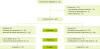
The flow chart of participants through the trial is presented in Figure 1. Eighty-five patients with MetS were primarily assessed against the inclusion and exclusion criteria. Sixty participants met the criteria and were randomly assigned into the flaxseed oil or sunflower oil group. Five participants were excluded from the flaxseed oil group during follow-up phase due to an unwillingness to continue the study. In addition, 3 participants were excluded from the sunflower oil group during follow-up phase due to travel and personal problems. Therefore, 52 subjects completed the trial and included to final analysis. No side effects were reported in participants during the intervention.
Oils specifications have been presented in Table 1. Accordingly, the amounts of saturated fatty acids (SFAs), monounsaturated fatty acids (MUFAs), and polyunsaturated fatty acids (PUFAs) were equal in flaxseed oil and sunflower, but the percentages and kinds of PUFAs were different.
Table 1
Flaxseed oil and sunflower oil fatty acids specifications
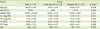
| Fatty acids | Sunflower oil | Flaxseed oil |
|---|---|---|
| C16 | 7.8 | 7.1 |
| C18 | 4.9 | 8.3 |
| C20 | 0.4 | 0 |
| C22 | 0.9 | 0 |
| C18:1 | 27.6 | 27.5 |
| C18:2 | 58.0 | 16.1 |
| C18:3 | 0 | 41.0 |
| C20:1 | 0.4 | 0 |
| SFA | 14.0 | 15.4 |
| MUFA | 28.0 | 27.5 |
| PUFA | 58.0 | 57.1 |
All values represent the percentage of total fatty acids.
SFA, saturated fatty acid; MUFA, monounsaturated fatty acid; PUFA, polyunsaturated fatty acid.
![]()
Demographics
According to Table 2, no significant differences were observed between the flaxseed oil and sunflower oil groups regarding demographic and biochemical characteristics at the baseline. Sex and age distributions were also similar in the 2 groups.
Table 2
The participants' baseline characteristics

Data have been presented as mean ± standard deviation.
IL-6, interleukin 6; TAC, total antioxidant capacity; PT, prothrombin time; PTT, partial thromboplastin time; INR, international normalized ratio.
*The p value was obtained from Independent samples t-test.
![]()
Dietary intake
Based on the 24-hour recall assessment, no statistically significant difference was seen between the 2 groups in terms of dietary intakes of energy, fat and omega-3 fatty acids (Table 3).
Table 3
The participants' dietary intakes at the baseline and during the intervention

Data have been presented as mean ± standard deviation.
SFA, saturated fatty acid; PUFA, polyunsaturated fatty acid.
*Paired t-test (within groups before and after the intervention).
![]()
Findings recorded in the 2 groups of participants before and after intervention
The results depicted in Table 4 illustrated a reduction in serum IL-6 level in comparison to the beginning of the study. Changes in IL-6 level were also significant within and between the 2 study groups (p = 0.017). Nonetheless, no significant differences were found in the two groups concerning the TAC and coagulation scores compared to the baseline (p > 0.05).
Table 4
Comparison of changes in TAC, IL-6, PT, PTT, and INR between and within the 2 groups

All values represent mean ± standard deviation.
IL-6, interleukin 6; TAC, total antioxidant capacity; PT, prothrombin time; PTT, partial thromboplastin time; INR, international normalized ratio.
*The p value was obtained from paired t-test; †p value was obtained from analysis of variance.
![]()
DISCUSSION
The study results revealed that flaxseed oil had favorable effects on reducing the inflammatory status, including serum IL-6 concentration, in MetS patients. The adjusted diet for the participants altered the n-6: n-3 ratio. n-6 and n-3 compete for the main enzymes in the fatty acid metabolism pathway, namely lipoxygenase and cyclooxygenase. Consumption of the two fatty acids leads to a decline in the production of prostaglandin E2 and leukotriene B4. It has been proposed that the reduction of these two eicosanoids could decrease the production of inflammatory markers, such as IL-6 [16].
Su et al. [17] and Nelson et al. [18] reported that consumption of 5% of total energy from (ALA) had no significant effects on reduction of serum IL-6 level in healthy abdominally obese adults. Indeed, receiving 3 g/day ALA caused no significant changes in IL-6 levels among patients with type 2 diabetes mellitus. In another study, the low and high doses of flaxseed oil were unable to change IL-6 and some other inflammatory parameters in MetS patients [19]. In the present study, the patients were required to consume approximately 10 g/day ALA. However, inflammatory status is different in various diseases. For instance, CVD has an elevated level of inflammatory markers. On the other hand, MetS is a proinflammatory status and has different conditions in comparison to abdominal obesity. These discrepancies may result from the baseline levels of inflammatory markers as well as habitual diets, which are the main influential factors. Consequently, replacement of various fatty acids in the diet may be accompanied with different outcomes, indicating the importance of a habitual diet. On the other hand, it seems that replacement of n-3 with n-6 fatty acids was not effective in healthy individuals or those with low levels of inflammatory markers [20]. In this regard, observational studies have shown conflicting results [2122]. Rallidis et al. [16] reported that ALA from flaxseed oil (15 mL/day) reduced C-reactive protein (CRP), serum amyloid A (SAA), and IL-6 in dyslipidemic patients. In another trial, taking 8.1 g/day ALA decreased IL-6 and other inflammatory markers, such as CRP, SAA, and soluble vascular cell adhesion molecule type 1 (sVCAM-1) in dyslipidemic men [20]. In that study, the participants' dietary pattern was high in saturated fatty acids. Thus, some other factors, such as dietary background, total fat, or other sources of n-3 received during the study, might have interfered with their results.
The current study results indicated no changes in the TAC among the participants who received flaxseed oil. In the research performed by Mirfatahi et al. [23] on hemodialysis patients, consumption of 6 g/day flaxseed oil had no significant effects on oxidative stress factors, such as soluble intercellular adhesion molecule type 1, sVCAM-1, sEselectin, and malondialdehyde. However, a significant decline was detected in high-sensitive CRP in the flaxseed oil group [23]. In another study, Rahmani et al. [24] reported that 1,000 mg omega-3 fatty acids (400 mg α-linolenic acid plus 400 IU vitamin E) improved oxidized low-density lipoprotein and lipoprotein gene expression among women with polycystic ovary syndrome [24]. In that study, adding vitamin E to omega-3 fatty acids improved oxidative status. PUFAs containing more than three double bonds can produce hydroperoxy epidioxides as the primary oxidation products are easily oxidized in our tissues or cell membrane because of the high degree of unsaturation [25]. Other antioxidants, such as vitamin E or C, are required to protect PUFAs or help their activity.
In another experimental study on rats with non-alcoholic fatty liver disease, flaxseed oil and lipoic acid increased superoxide dismutase, catalase, and glutathione peroxidase activities as well as glutathione levels [26]. It seems that enriched flaxseed oil with an antioxidant agent has a protective effect and increases the antioxidant defense.
It is worth mentioning that 2 or more antioxidant factors were evaluated in the above-mentioned studies. On the other hand, one of the strong points of the current study was the measurement of TAC, which indicates power and change in all antioxidant systems in the body.
The present study results revealed no significant differences between the two groups regarding PT or PTT at the end of the study. Evidence has shown that n-3 fatty acid improved endothelial function and platelet aggregation markers in patients with coronary heart disease as well as in healthy cigarette smokers [27]. It seems that inflammatory status affects endothelial dysfunction in patients with MetS, and eventually increases the coagulation factors. However, details and mechanisms of the antithrombotic effects of ALA are unclear. Therefore, further studies on the issue are warranted. ALA can inhibit thrombosis formation, tissue expression, and platelet activation. Although the coagulation mechanism is well known in some diseases, it is unknown in MetS. For example, obesity is a moderate risk factor for venous thromboembolic events, but limited evidence is available for MetS [2829]. Since MetS and CVD are closely related, in addition to the traditional MetS identification factors, efforts to identify the common causes of MetS and CVD are worthwhile. Many factors contribute to blood coagulation or thrombosis, some of which are affected by inflammatory factors or vascular atherosclerosis. Thus, comprehensive studies are needed to measure all relevant factors.
Although the positive effects of flaxseed on health have been reported, a particular concern about flaxseed in some populations including pregnant women and men in their reproductive ages should be considered. Especially in chronic use, this possible adverse effect due to hormonal action of the lignans must not be neglected [30].
This study has some limitations that should be addressed. The main limitation of the present study is the lack of an appropriate placebo for the flaxseed oil. In addition, other known inflammatory and oxidative stress markers were not measured. Finally, relatively short intervention period and small sample size are considered as other limitations of the study.
CONCLUSIONS
Dietary intake of flaxseed oil as the main source of dietary fat could improve the inflammatory status among MetS patients. However, no significant changes were observed in serum levels of TAC and coagulation score. With regard to the baseline level of inflammation in MetS that leads to CVD and T2DM and the importance of dietary fat in these patients, flaxseed oil might be helpful. However, further studies are needed to confirm the veracity of our results.
 XML Download