

 PDF
PDF ePub
ePub Citation
 Print
Print


INTRODUCTION
Non-alcoholic fatty liver disease (NAFLD), which has attracted much attention in recent years, is a chronic liver disease and is known as the major cause of liver enzymes disorder in recent decades. The disease is characterized by an increase in fat accumulation by about 5%–10% of the liver's weight in patients without a history of significant consumption of alcohol [1], steatogenic medication or hereditary disorders [2]. This clinicopathological condition is related to a spectrum of liver disorders ranging from steatosis, steatohepatitis and cirrhosis which can be developed to hepatocellular carcinoma over time. Symptoms of nonalcoholic steatohepatitis are presented in 5%–10% of NAFLD cases, and 1.3% of them are developed to cirrhosis [3]. Risk factors of NAFLD include central obesity, type 2 diabetes, dyslipidemia, high blood pressure, sedentary life-style and unhealthy dietary pattern [4]. The prevalence rate of NAFLD is about 33.9% in the general population of Iran [5]. From the precautionary medical point of view, the early diagnosis of this disease can prevent subsequent liver dysfunctions through screening and following appropriate interventions [67].
Quercetin (3-3-4-5-7 pentahydroxy flavones, C15H10O7) [8], contains 5 hydroxyl groups and numbers of derivatives including galactoside, glucoside, xylose, rhamnose, and glucuronide [9]. It is a polyphenolic compound, which is widely distributed in edible vegetables such as caper, onion, tea, and apple [8]. Quercetin absorption is mainly mediated using the sodium-glucose transporter or lactase phlorizin hydrolase in the enterocytes (mostly in the upper part of the small intestine) [10].
It is almost totally confirmed that oxidative stress plays a crucial role in the pathogenesis of NAFLD [11]. Studies have demonstrated that red blood cell (RBC) destruction and death are mainly mediated by oxidative stress. The continuous exposure to reactive oxygen species (ROS) might cause RBC membrane to be fragile and make them susceptible to damage [1213]. This oxidative damage is largely caused by the reaction of ROS with membrane lipids and proteins, which can affect membrane structure and function. One of the involved possible mechanisms is that oxidative stress has shown to inhibit Ca-ATPase, which can result in elevation of intracellular calcium concentration and subsequent leakage of potassium from the RBC and finally shrinkage of the cell and impaired deformability [14]. Previous studies have also indicated that one-third of NAFLD patients are iron deficient, and red blood cell distribution width (RDW) is significantly increased in NAFLD cases mainly because of inflammatory conditions of this disease [1516]. We hypothesized that quercetin as a potent antioxidant, could protect RBC membrane against oxidative damage, and improve the hematological parameters [171819]. To the best of our knowledge, this is the first study, which evaluates the effect of quercetin supplementation on hematological parameters in NAFLD patients, in the form of a placebo-controlled trial.
MATERIALS AND METHODS
Recruitment and selection of subjects
This work was conducted on 90 men and women, with the ages of between 18–65 years old with NAFLD as approved by ultrasonography recruited from radiology clinic of Imam Khomeini hospital in Kermanshah, Kermanshah province, Iran. From a total of 120 interviewed patients, 90 of them were qualified based on inclusion criteria including: Body mass index between 25–40 kg/m2, no history of alcohol abuse, lack of acute and chronic disease such as liver disorders (hepatitis B and C), Wilson's disease, cirrhosis, autoimmune disease, and thyroid disorders plus no previous exposure to pesticides and insecticides and not taking steatogenic medications, which potentially interact with quercetin (anticoagulant agents, inhibitors of CYP3A4, etc.). We also selected subjects who were not pregnant and lactating, and also had no either weight loss surgery in the recent year, neither a moderate nor severe weight loss within three months before sampling. Exclusion criteria were as follows: unwilling to continue cooperation, developing diseases that require special treatment and being pregnant during follow up, migration and not consuming more than 10% of supplements in each follow-up period.
Study design and randomization
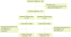
All eligible patients with NAFLD were enrolled in this study in November 2017. The study protocol was approved by the Ethics Committee of the School of Public Health (KUMS.REC.1395.79) in Kermanshah University of Medical Sciences and was with respect to the declaration of Helsinki of the world medical association. This study was also registered in the Iranian Center for Clinical Trials (IRCT2016060628299N1). The patients signed an informed consent form after a full review of the inclusion and exclusion criteria and explaining the risks and benefits of the study. From a total of 120 participants who were enrolled, 90 subjects were randomly allocated into 2 groups of placebo (n = 45) and quercetin (Sigma Q4951 ≥ 95% purity; Sigma-Aldrich, St. Louis, MO, USA) (n = 45) as shown in Figure 1. The placebo (Avicel PH101; Sigma-Aldrich) produced with the same size, color and smell as quercetin capsules. Random assignment was performed using computer-generated random numbers. Each patient received 250 mg capsules twice daily. Any compliance was checked through weekly telephone interviews and monthly session meetings in the school of public health.
Clinical, paraclinical assessments
All participants completed a demographic questionnaire at the beginning and the week 12 of the study. In addition, all other measurements including anthropometric and hematological parameters, were conducted at baseline and week 12. Fasting blood samples were obtained at the baseline and week 12 after a 12-hour overnight fasting. Hematological parameters were measured using Sysmex kx21n. Ferritin was measured by Pishtazteb ELIZA kits (Pishtazteb, Tehran, Iran).
Statistical analysis
All statistical analyses were performed using the SPSS version 20 statistical software (IBM Corp., Armonk, NY, USA). Kolmogorov-Smirnov test was used to assess the normality distribution of variables. Quantitative variables were expressed as a mean ± standard deviation and qualitative variables were expressed in terms of frequency (number and percentage). Paired-samples t-test was used to compare intra-group changes variables and independent t-test was applied to compare the inter-group changes. In all cases, a value for p < 0.05 was considered significant.
RESULTS
Baseline characteristics of the patients
During the intervention, 12 patients (6 people in each group) failed to complete the trial mainly because of immigration and non-adherence (Figure 1). Seventy-eight patients completed the study, 39 individuals in each group. Average age of all patients were of 44.67 ± 10.20 years old. The level of baseline physical activity and demographic data were not significantly different between these two groups (Table 1).
Table 1
Baseline characteristics of patients
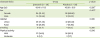
Data presented as number (%) or mean± standard deviation
Statistical tests were used *independent-sample t-test, †χ2 test unless indicated.
![]()
Hematological parameters
As shown in Table 2, There was no significant difference in considered parameters between the two groups at baseline. Between-group comparisons also revealed that the level of RBC was significantly higher (p = 0.025), and the level of mean corpuscular volume (MCV; p = 0.004), mean corpuscular hemoglobin (MCH; p = 0.002), and ferritin (p = 0.013) were significantly lower in quercetin group at the end of the trial. However, the values still remained in normal range.
Table 2
Change of hematological parameters from baseline to end of treatment
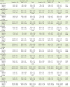
Data presented as mean ± standard deviation. Bold-faced p values indicate statistical significance (p < 0.05).
WBC, white blood cell; HGB, hemoglobin; RBC, red blood cell; HCT, hematocrit; MCV, mean corpuscular volume; MCH, mean corpuscular hemoglobin; MCHC, mean corpuscular hemoglobin concentration; RDW-cv, red blood cell distribution width-coefficient of variation; PLT, platelet; MPV, mean platelet volume; PDW, platelet distribution width.
Statistical tests were used *independent-sample t-test (total), †paired t-test.
![]()
End of trial values for RBC (p = 0.002), mean corpuscular hemoglobin concentration (MCHC; p = 0.029), and mean platelet volume (p = 0.017), significantly increased by 0.32 106/ul, 0.69 g/dL, and 0.48 FL, respectively, and the levels of MCV (p = 0.023), RDW-coefficient of variation (p = 0.005), platelet distribution width (p = 0.015), and ferritin (p = 0.002) significantly decreased by 2.05 FL, 0.57%, 1.08 ng/mL, and 46.52 ng/mL, respectively, compare to the baseline in group receiving quercetin. None of the abovementioned variables showed significant changes in placebo group. None of the patients had reported any side-effects, which indicated tolerance to the treatment.
DISCUSSION
Flavonoids and in particular quercetin have controversial effects on hematological parameters. Reviewing the publications over last years, we could not find any related paper, which have been exclusively focused on this topic. In this study, quercetin supplementation significantly increased RBC in intervention group. Flavonoids can increase the production of RBCs by stimulating the secretion of erythropoietin [2021]. Erythrocytes are highly exposed to oxygen and their membrane has high concentration of polyunsaturated fatty acids, which can make them susceptible to oxidative stress [19]. RBCs can also bind polyphenols to their surfaces and play a crucial role in the distribution of polyphenols and protect the erythrocytes membrane against oxidative damage [22]. Quercetin has a great potential in restoring the content of endogenous glutathione in erythrocytes that was depleted by radical generators [18]. In our study, patients had normal ferritin level at baseline. Ferritin level had been significantly decreased in the quercetin group from the baseline to the end of treatment. In enterocytes, quercetin acts as an electron donor and facilitates inorganic iron absorption. However, quercetin can also repress the expression of ferroportin and hephaestin, and subsequently inhibits basolateral iron absorption [2324]. Another possible mechanism is that quercetin may be able to chelate iron in the enterocytes and the quercetin iron complex being retained in the cytosol [25]. Therefore, the reduction in ferritin level may be as the result of functional cellular iron deficiency [26]. Inconsistent with other findings of our study, including increased levels of RBC and hemoglobin, the levels of MCV and MCH significantly decreased in quercetin group. It is possibly due to erythrocytosis which occur in 3% of patients at the early stage of anemia [27].
Supporting our findings in a study conducted by Keskin et al. [20], supplementation with quercetin in streptozotocin induced diabetic rats has not shown any significant changes on MCV, MCH, and MCHC, but RBC count significantly increased in group receiving quercetin. Similarly in another animal study, rabbits receiving 3 different doses of guercetin for 90 days 3 times a week, exhibited slight changes in some hematological parameters including: RBC, hemoglobin, and hematocrit; however, the differences were not significant between these two groups [28]. In contrast with our results, Kasmi et al. [17] demonstrated that co-administration of quercetin (20 mg/kg) improved the levels of platelet and white blood cells in rats received different doses of difenoconazole to liver injury induction [17]. In similar manner, quercetin supplementation (50 mg/kg) had a significant favorable effect on polychlorinated biphenyls induced hematological changes in adult male Wistar rats [29]. Mahmoud et al. [21] indicated that MCV, MCH, and MCHC increased to near normal amounts in diabetic rats receiving citrus flavonoids.
CONCLUSION
The effect of flavonoids on hematological profile can be simulated to a 2-edged sword. As we described mechanisms previously, flavonoids might either cause or cure anemia. In this work, RBC increased and ferritin decreased significantly in group receiving quercetin compared to those in placebo group but the values of RBC and ferritin still remained in normal range throughout the study period. It is worth mentioning that none of the patients had reported any side-effects and this indicate that the quercetin treatment was well-tolerated by the patients.
 XML Download