

 PDF
PDF ePub
ePub Citation
 Print
Print


INTRODUCTION
Hemifacial microsomia (HFM) is the second most common facial anomaly, with a broad spectrum of phenotypes.123456 Although it usually affects one side of the face, ears, mouth, and mandible, both sides are sometimes affected. HFM has heterogeneous etiological factors related to the alteration of mesodermal development as well as the increased susceptibility to vascular disruption.789 Most patients with HFM require long-term multidisciplinary medical and dental care to rehabilitate occlusal function and improve facial aesthetics.1011121314
Vento et al.15 proposed the orbit, mandible, ear, nerve, soft tissue (OMENS) classification system, which grades 5 anatomic features of HFM according to severity on a scale from 0 to 3. Horgan et al.16 refined the OMENS classification system into the OMENS-plus classification system to account for the presence of extra-craniofacial anomalies (e.g., central nervous system, cardiac, pulmonary, renal, gastrointestinal, and vertebral deformities). Tuin et al.,17 in their association study between structures, suggested that deformities of structures that mainly developed from the first branchial arch (i.e., the orbit, mandible, and soft tissue) show a similar degree of severity. This also holds true for structures mainly derived from the second branchial arch (i.e., the facial nerve and ear). However, studies have shown that mandibular and ear deformities are the major features of the HFM classification system.1819
The degree of mandibular deformity evaluated using the Pruzansky–Kaban classification system10112021 might be one of the most important factors in treatment planning and prognosis prediction of orthodontic treatment using unilateral functional appliances or surgical interventions, including mandibular distraction osteogenesis, orthognathic surgery, and costochondral graft.42223 If patients with HFM show compensatory growth of the mandibular body despite hypoplasia of the condyle/ramus complex on the ipsilateral side, surgical intervention should be delayed until the completion of growth because of the possibility of severe chin point deviation to the non-affected side. In addition, Angle's classification is another important issue in orthodontic diagnosis and treatment planning. If patients with HFM show a Class III molar relationship, distraction osteogenesis of the mandible cannot be performed because of the possibility of anterior crossbite.
The Korean National Health Insurance Service (KNHIS) plans to provide insurance benefits for orthodontic treatment to patients with HFM in the near future. Basic data on the distribution and phenotypes of patients with HFM can provide useful information for diagnosis, treatment planning, and prognosis evaluation. Several previous studies conducted in other countries have investigated the clinical characteristics of patients with HFM from the perspective of plastic surgery.1924252627 However, no clinical demographic studies have investigated the distribution and phenotypes of Korean patients with HFM from an orthodontic perspective. Therefore, the purpose of this retrospective study was to investigate the distribution and phenotypes of HFM and its association with other anomalies using clinical data.
MATERIALS AND METHODS
The study sample comprised 249 Korean patients with HFM (mean age at first consultation, 7.07 ± 3.99 years), who visited the Multi-disciplinary Clinic, Department of Pediatric Plastic and Reconstructive Surgery, Seoul National University Children's Hospital (SNUCH) and the Department of Oral and Maxillofacial Surgery and Department of Orthodontics, Seoul National University Dental Hospital (SNUDH) during 1998–2018. The inclusion criteria were as follows: (1) patients diagnosed with HFM and followed-up or treated and (2) patients whose charts, clinical photographs, cephalometric and panoramic radiographs, and/or three-dimensional computed tomography (3D-CT) images were available. HFM prevalence according to sex, side involvement, degree of mandibular deformity, compensatory growth of the mandibular body, and Angle's classification, and its association with other anomalies were investigated. This study was reviewed and approved by the Institutional Review Board of the SNUDH (ERI19025).
Since Vento et al.15 confirmed the concept that mandibular deformity is the cornerstone of the anomaly, the degree of mandibular deformity has been evaluated using the Pruzansky–Kaban classification system. In this system, Type I indicates that the ramus/condyle complex has a normal shape but small size. In Type IIa, although the ramus/condyle complex is hypoplastic and abnormally shaped, the glenoid fossa is placed at the right position and the temporomandibular joint is functional. Type IIb indicates that the glenoid fossa is placed at the inferiorly, medially, and anteriorly altered positions with a severely hypoplastic ramus/condyle complex. Type III indicates the complete absence of the ramus/condyle complex and the glenoid fossa.1120
Compensatory growth of the mandibular body was assessed using panoramic radiographs, posteroanterior cephalometric radiographs, and/or 3D-CT images. Compensatory growth of the mandibular body was identified if the lower dental midline was deviated to the unaffected side in patients with unilateral HFM (UHFM) and to the less severely deformed side in patients with bilateral HFM (BHFM). Angle's classification was assessed via molar relationships by using intraoral photographs or orthodontic models.
Descriptive statistics, chi-square goodness-of-fit test, chi-square independence test, and Fisher's exact test were used for statistical analysis using SPSS for Windows, ver. 12.0 (SPSS Inc., Chicago, IL, USA). A p-value of less than 0.05 was considered significant.
RESULTS
Distribution of sex and sidedness
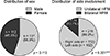
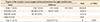
No difference was observed in HFM prevalence between male and female patients (n = 137 [55.0%] vs. n = 112 [45.0%], p > 0.05; Figure 1). UHFM was more prevalent than BHFM (n = 215 [86.3%] vs. n = 34 [13.7%], p < 0.001; Figure 1). Right-sided UHFM (n = 113) was the most common, followed by left-sided UHFM (n = 102) and BHFM (n = 34) (Figure 1). However, patients with UHFM showed no difference in occurrence between the right and left sides (n = 113 [52.6%] vs. n = 102 [47.4%], p > 0.05; Table 1).
Distribution of the Pruzansky–Kaban types in patients with unilateral hemifacial microsomia
A significant difference was observed in the distribution of the Pruzansky–Kaban types in these patients (Type I, n = 114 [53.0%]; Type IIa, n = 40 [18.6%]; Type IIb, n = 53 [24.7%]; and Type III, n = 8 [3.7%]; p < 0.001; Table 1).
Distribution of the Pruzansky–Kaban types in patients with bilateral hemifacial microsomia
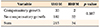
Patients with different Pruzansky–Kaban types on the right and left sides were more common than were those with the same type on both sides (n = 23 [67.6%] vs. n = 11 [32.4%], p < 0.05; Table 2).
Compensatory growth of the mandibular body
Despite hypoplasia of the condyle/ramus complex, compensatory growth of the mandibular body on the ipsilateral side occurred in 35 patients (14.1%; Table 3). No difference in prevalence of compensatory growth of the mandibular body was observed between patients with UHFM and BHFM (n = 33/215 [15.3%] vs. n = 2/34 [5.9%], p > 0.05; Table 3).
Angle's classification
Class I and II molar relationships were more prevalent than were Class III molar relationships (n = 232 [93.2%] vs. n = 17 [6.8%], p < 0.001; Table 4). No difference in the prevalence of Class III molar relationships was observed between patients with UHFM and BHFM (n = 12/215 [5.6%] vs. n = 5/34 [14.7%], p > 0.05; Table 4).
Association with other craniofacial and extra-craniofacial anomalies
Forty-nine patients with HFM (19.7%) had other anomalies (n = 32 with UHFM and n = 17 with BHFM; Table 5). The association with other anomalies was more prevalent in patients with BHFM than in those with UHFM (n = 17/34 [50.0%] vs. n = 32/215 [14.9%], p < 0.001; Table 5).
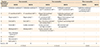
Eight kinds of craniofacial anomalies were observed in 42 patients (n = 42/48, 87.5%) and six kinds of extra-craniofacial anomalies were observed in 9 patients (n = 9/48, 18.8%) (Table 6). Three patients had concomitant craniofacial and extra-craniofacial anomalies (Table 6). In terms of the number of associated anomalies, one anomaly was the most common (n = 37/49, 75.5%), followed by two anomalies (n = 11/49, 22.4%) and three anomalies (n = 1/49, 2.0%) (Table 6). Among the recorded anomalies (n = 61 in total; Table 6), relatively high associations were found with Goldenhar syndrome (n = 14/61, 23.0%), Tessier no. 7 craniofacial clefts (n = 10/61, 16.4%), missing teeth (n = 8/61, 13.1%), plagiocephaly (n = 8/61, 13.1%), and vertebral anomaly (n = 7/61, 11.5%; 3 with Klippel–Feil syndrome, 3 with scoliosis, and 1 with torticollis). A relatively low degree of association was found with oral cleft (n = 3/61, 4.9%; 2 with cleft palate and 1 with unilateral cleft lip and palate), cardiac anomaly (n = 3/61, 4.9%), Parry–Romberg syndrome (n = 3/61, 4.9%), micrognathia (n = 1/61, 1.6%), microstomia (n = 1/61, 1.6%), digit anomaly (n = 1/61, 1.6%), and chromosome anomaly (n = 1/61, 1.6%).
DISCUSSION
Distribution of sex and sidedness
The lack of a difference in the prevalence of HFM between male and female patients was in agreement with the results of previous studies, despite differences in geographical and ethnic factors (Table 7).1519252728
In the present study, the prevalence of UHFM was higher than that of BHFM. Moreover, no difference was observed in occurrence between the right and left sides among patients with UHFM. These results were also consistent with those of previous studies despite differences in geographical and ethnic factors (Table 7).1524252728
Distribution of the Pruzansky–Kaban types in patients with unilateral hemifacial microsomia
The distribution pattern of the Pruzansky–Kaban types in patients with UHFM was similar to that reported by Park et al.24 However, these results differed from those of previous studies conducted in other countries (Table 7).151927 The reasons for this difference could be the following. First, the subjects of this study were Korean patients with HFM. Second, in this study, patients with HFM who had skin tags or microtia and insignificant skeletal deformity were considered as showing the M1 (Type I) pattern. Park et al.24 suggested that patients with microtia could be considered as those with HFM Type I, because all patients with microtia tend to have minimal mandibular deficiency. Therefore, the total number of patients with the M0 (normal) and M1 (Type I) patterns in previous studies (51% in Vento et al.15; 50.1% in Caron et al.27; Table 7) would be similar to that in our study (53%, Table 7). Third, Takahashi-Ichikawa et al.29 reported that panoramic radiography was a reliable modality for evaluating patients with HFM Type I, while 3D-CT was preferred for evaluating patients with HFM Type II. Therefore, we recommend the use of two modalities for the clinical assessment of HFM types.
Distribution of the Pruzansky–Kaban types in patients with bilateral hemifacial microsomia
No previous studies have reported the differences in the Pruzansky–Kaban types between the right and left sides in patients with BHFM. According to the results of this study, different Pruzansky–Kaban types on the right and left sides were more prevalent than was the same type on both sides. This is a novel finding that should be considered, especially when planning distraction osteogenesis of the mandible on both sides.
Compensatory growth of the mandibular body
Despite hypoplasia of the condyle/ramus complex, compensatory growth of the mandibular body on the ipsilateral side occurred in 14.1% of patients with HFM (Table 3). This novel finding should be considered during diagnosis, treatment planning, and prognosis prediction for orthodontic and surgical interventions. If compensatory growth of the mandibular body occurs at the affected side, the lower dental midline and chin point would be shifted to the unaffected side. Therefore, when orthodontic treatment using unilateral functional appliances or surgical interventions, including mandibular distraction osteogenesis or costochondral graft, is performed, matching the lower dental midline and chin point to the facial midline is difficult.
Angle's classification
The finding that Class III molar relationships occurred in 6.8% of patients with HFM has clinical implications. Since patients with HFM usually have simultaneous hypoplasia of the condyle/ramus complex and mandibular body, the origin of the Class III molar relationships should be investigated in future studies. In addition, when patients with HFM show Class III molar relationships, distraction osteogenesis of the mandible cannot be performed at preadolescent and adolescent ages owing to the possibility of anterior crossbite. Therefore, orthognathic surgery should be considered after the completion of growth.
Association with other craniofacial and extra-craniofacial anomalies
Since the association with other craniofacial and extra-craniofacial anomalies was more prevalent in patients with BHFM than in those with UHFM, attention should be paid to the general medical condition of patients with BHFM for ensuring proper treatment planning and prognosis prediction.
In the present study, eight kinds of craniofacial anomalies were observed in 42 patients (87.5%) and six kinds of extra-craniofacial anomalies were observed in 9 patients (18.8%) (Table 6). Moreover, Goldenhar syndrome was the most commonly associated craniofacial anomaly. However, Goldenhar syndrome tends to be subjectively over diagnosed in patients who show more severe HFM features.30 The percentage of patients with HFM and extra-craniofacial anomalies in the current study was lower than that reported by Horgan et al.16 (55.4%), Park et al.24 (41.4%), and Caron et al.27 (35.0%). Park et al.24 reported no significant correlation between the total OMENS score and extra-craniofacial anomalies, despite significant correlations between the orbit and mandible, mandible and soft tissue, and facial nerve and soft tissue.
Despite the potential limitations of data collected from a single university hospital, we obtained meaningful basic information on the distribution and phenotypes of Korean patients with HFM who were diagnosed and followed-up or treated during the last two decades (1998–2018). The results of this study might serve as the basis for setting guidelines on health care utilization for the orthodontic treatment of patients with HFM. Nevertheless, nationwide multicenter studies with long-term follow-up and systematic statistical analyses have to be conducted to obtain information necessary for providing the sophisticated insurance plans of the KNHIS for orthodontic treatment of patients with HFM.
CONCLUSION
Patients with UHFM show a significant difference in the distribution of the Pruzansky–Kaban types. In patients with BHFM, different Pruzansky–Kaban types on the right and left sides were more prevalent than was the same type on both sides. Compensatory growth of the mandibular body on the ipsilateral side occurred in 14.1% of patients with HFM. Class III molar relationships occurred in 6.8% of patients with HFM. Other anomalies were more prevalent in patients with BHFM than in those with UHFM. Because of the diverse phenotypes of HFM and its association with other anomalies, patients with HFM require individualized diagnosis and treatment planning.


 XML Download