Journal List > Yonsei Med J > v.61(2) > 1141545

Kwon, Shin, Moon, Lee, and Lee: Arthrodesis Using Bilateral Dual Iliac Screws with Autologous Iliac Bone Transfer for the Treatment of Pyogenic Sacroiliitis

Abstract

Pyogenic sacroiliitis is a relatively rare condition that often leads to surgical treatment, including debridement and arthrodesis. Here we introduce a new surgical technique using bilateral dual iliac screws to secure early ambulation and maximal fusion success rate for the treatment of pyogenic sacroiliitis. We retrospectively reported a case and technical reports of pyogenic sacroiliitis treated by a new bilateral dual iliac screw fixation arthrodesis technique using radiologic outcomes, including plain X-rays and MRI scans, as well as outcomes based on the visual analogue scale for pain measurement. This technique improved uncontrolled pyogenic sacroiliitis with immediate stability that enabled ambulation and secured firm fixation for extensive evacuation of infected debris and subsequent autograft bone arthrodesis. In conclusion, we recommend bilateral dual iliac screw fixation for the treatment of pyogenic sacroiliitis, as this technique can improve uncontrolled pyogenic sacroiliitis with immediate stability.

INTRODUCTION

Pyogenic sacroiliitis is a relatively rare condition, ranging from 1–2% of all cases of septic arthritic conditions.123 Considering the complexity of its anatomical and biomechanical characteristics,4 conservative treatment with antibiotics alone is often unsuccessful.5 As a result, surgical treatment is often necessary; such treatment includes debridement/drainage and/or arthrodesis.678 Therefore, we have introduced a surgical technique using bilateral dual iliac screws to secure early ambulation and a maximal fusion success rate for the treatment of pyogenic sacroiliitis. This study has been reviewed by the appropriate ethics committee, and has therefore been performed in accordance with the ethical standards laid down in the 1964 Declaration of Helsinki.

CASE REPORT

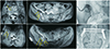
A healthy 23-year-old female patient suffered from hip pain for 3 weeks. The patient had no history of recent illness and no significant past medical history. The pain was constant and severe (VAS 9–10), and was not relieved by resting or analgesics. Initial laboratory tests at a local clinic showed a leucocyte count of 8180/mm3, C-reactive protein (CRP) level of 67 mg/L (normal range 0–5 mg/L), and an erythrocyte sedimentation rate (ESR) of 75 mm/hour. Blood cultures were positive for Streptococcus agalactiae. The patient was initially treated at the local clinic with bed rest and intravenous antibiotics (ciprofloxacin 400 mg IV for 14 days, ceftriaxone 2 g IV for 28 days, along with oral metronidazole 500 mg for 14 days). Radiographs of the pelvis and sacroiliac joints were normal. MRI of sacroiliac joints after 42 days of intravenous antibiotics showed a focus of infection on the right side along with intramuscular abscess in the iliacus muscle, which connected into the right sacroiliac joint. Then, the patient was transferred to Severance Hospital for interventional treatment. Initial laboratory tests in the emergency room at Severance Hospital showed a leucocyte count of 5990/mm3, CRP level of 6.29 mg/L, and an ESR of 65 mm/hour. Then, percutaneous abscess drainage was done on the right intramuscular abscess in the iliacus muscle using a pig-tail catheter. The total amount of drained abscess was 22 mL for 8 days. As the patient's right sacroiliac pain symptoms did not improve after abscess drainage, she had difficulty in turning over on the bed. MRI was performed again to confirm abscess pocket shrinkage, but it revealed the remaining right sacroiliac arthritis (Fig. 1).
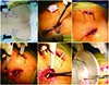
Finally, the patient underwent right sacroiliac arthrodesis with curettage and autologous iliac bone transfer mixed with ceftriaxone and vancomycin powders using the bilateral dual iliac screw fixation technique. An oblique skin incision was made bilaterally over the dual iliac screw entry points. The proximal iliac screw (Legacy pedicle screw system, Medtronic Sofamor Danek, Inc., Memphis, TN, USA) was inserted using the guidance of a C-arm, from the posterior superior iliac crest, to maximize the fixation force in terms of the larger screw diameter and longest screw length. Considering the sacroiliac joint anatomy, arthrotomy and curettage drainage of the sacroiliac joint were performed in that order. After copious irrigation (preferentially 10000 mL of normal saline), antibiotics mixed with autologous iliac bone were packed into the arthrotomized sacroiliac joints. Submuscular dual rod was inserted and assembled with an adequate amount of compressive force on the affected sacroiliac joint (Fig. 2).
The patient was able to sit up at a 45 degree angle from postoperative day 1. Her pain decreased to VAS 2 on postoperative day 5. Ambulation in a wheelchair or with walking aids was permitted 5 days after surgical treatment, with the patient wearing a back brace. CRP was normalized to 3.55 mg/L at 2 weeks after surgery.

DISCUSSION

Conservative treatment for pyogenic sacroiliitis using antibiotics alone is often unsuccessful in osteomyelitis of the pelvic bones.6910 In pyogenic sacroiliitis, these anatomical characteristics could be broken, and may lead to refractory infection and chronic painful joints. Therefore, proper and timely decisions to convert conservative treatment to surgical treatment are mandatory. The patient showed immediate pain improvement from preoperative VAS 9–10 to postoperative VAS 2 at postoperative day 5, which was enough to try ambulation and toileting activities using walking aids without the assistant of caregivers. Therefore, we recommend the bilateral dual iliac screw fixation method combined with incision drainage and arthrodesis using antibiotics mixed with autologous bone graft for the treatment of pyogenic sacroiliitis.

Figures and Tables

Fig. 1

Initial MRI scan demonstrates intramuscular abscess in the right iliacus muscle, which connects into the right sacroiliac joint (indicated by yellow arrows). A pig-tail catheter was inserted to drain the intramuscular abscess. Finally, right sacroiliac arthrodesis using autologous bone and antibiotics mixture was done to relieve uncontrolled pyogenic sacroiliitis causing persistent and severe pain in the right buttock. R, right.

ymj-61-198-g001
Fig. 2

Using preoperative marking of anatomic landmarks, minimal dissection was done to expose the posterior superior iliac spine. After bilateral dual iliac screw insertion with the guidance of a C-arm, curettage, debridement, irrigation, and autologous iliac bone graft mixed with antibiotics was performed. Then, two rods were assembled to apply moderate compression to the right sacroiliac joint.

ymj-61-198-g002

Notes

The authors have no potential conflicts of interest to disclose.

AUTHOR CONTRIBUTIONS

  • Conceptualization: Ji-Won Kwon, Byung Ho Lee, Seong-Hwan Moon, and Hwan-Mo Lee.

  • Data curation: Ji-Won Kwon, Byung Ho Lee, and Hwan-Mo Lee.

  • Investigation: Ji-Won Kwon, Byung Ho Lee, and Jong-Kwan Shin.

  • Methodology: Ji-Won Kwon, Byung Ho Lee, Hwan-Mo Lee, and Jong-Kwan Shin.

  • Supervision: Ji-Won Kwon, Seong-Hwan Moon, and Hwan-Mo Lee.

  • Validation: Byung Ho Lee and Jong-Kwan Shin.

  • Writing—original draft: Ji-Won Kwon and Byung Ho Lee.

  • Writing—review & editing: Ji-Won Kwon and Byung Ho Lee.

  • Approval of final manuscript: All authors.

References

1. Mancarella L, De Santis M, Magarelli N, Ierardi AM, Bonomo L, Ferraccioli G. Septic sacroiliitis: an uncommon septic arthritis. Clin Exp Rheumatol. 2009; 27:1004–1008.
pmid
2. Hermet M, Minichiello E, Flipo RM, Dubost JJ, Allanore Y, Ziza JM, et al. Infectious sacroiliitis: a retrospective, multicentre study of 39 adults. BMC Infect Dis. 2012; 12:305.
crossref
3. Wu MS, Chang SS, Lee SH, Lee CC. Pyogenic sacroiliitis–a comparison between paediatric and adult patients. Rheumatology (Oxford). 2007; 46:1684–1687.
crossref
4. Vleeming A, Schuenke MD, Masi AT, Carreiro JE, Danneels L, Willard FH. The sacroiliac joint: an overview of its anatomy, function and potential clinical implications. J Anat. 2012; 221:537–567.
crossref pmid pmc
5. Dmitriev AE, Shepilov GF, Kriukov BN, Trushin IuA. [Osteomyelitis of the sacrum]. Khirurgiia (Mosk). 1989; (8):3–7.
6. Giannoudis PV, Tsiridis E. A minimally-invasive technique for the treatment of pyogenic sacroiliitis. J Bone Joint Surg Br. 2007; 89:112–114.
crossref pmid
7. Chan DS, Saklani A, Shah PR, Haray PN. Laparoscopic drainage of retroperitoneal abscess secondary to pyogenic sacroiliitis. Ann R Coll Surg Engl. 2010; 92:W32–W34.
crossref
8. Kanakaris NK, Psarakis S, Chalidis B, Kontakis G, Giannoudis PV. Management of pelvic instability secondary to chronic pyogenic sacroiliitis: case report. Surg Infect (Larchmt). 2009; 10:353–358.
crossref pmid
9. Bindal M, Krabak B. Acute bacterial sacroiliitis in an adult: a case report and review of the literature. Arch Phys Med Rehabil. 2007; 88:1357–1359.
crossref pmid
10. Doita M, Yoshiya S, Nabeshima Y, Tanase Y, Nishida K, Miyamoto H, et al. Acute pyogenic sacroiliitis without predisposing conditions. Spine (Phila Pa 1976). 2003; 28:E384–E389.
crossref
TOOLS
ORCID iDs

Ji-Won Kwon
https://orcid.org/0000-0003-4880-5310

Jong-Kwan Shin
https://orcid.org/0000-0002-0119-3460

Seong-Hwan Moon
https://orcid.org/0000-0003-1121-2730

Hwan-Mo Lee
https://orcid.org/0000-0002-5405-3832

Byung Ho Lee
https://orcid.org/0000-0001-7235-4981

Similar articles