

 PDF
PDF ePub
ePub Citation
 Print
Print


INTRODUCTION
It was previously shown that allergen contact via the nasal mucosa leads not only to allergic inflammation, but also increases systemic allergen-specific immunoglobulin E (IgE) levels.1
Accordingly, the impairment of the barrier function of the airway epithelium has been recognized as important for the pathogenesis of allergic airway diseases.2 Using in vivo experiments with labelled protein antigens, it was shown already in 1975 that the nasal mucosa of patients with allergic rhinitis shows greater permeability than that of non-allergic individuals.3 Several studies using in vitro cultured respiratory epithelial cells demonstrated that certain mediators and pro-inflammatory cytokines of allergic inflammation can indeed reduce barrier function45 and that this leads to increased penetration of allergen molecules.6 Taken together, these studies suggested that respiratory allergic inflammation in the tissue leads to reduced barrier function in allergic patients. Recently, it was even reported that barrier function in allergic patients is not only disturbed through allergic inflammation, but that there may be genuine differences between respiratory epithelial cells from allergic and non-allergic subjects to build up barrier even when they are cultivated ex vivo, i.e., outside the context of the host tissue.7 The latter study suggested that ex vivo cultured primary nasal epithelial cells (pNECs) from allergic subjects may show impaired barrier function as compared to those from non-allergic subjects.7 In this study, we reinvestigated if there are intrinsic differences between cultured pNECs from allergic and non-allergic individuals regarding barrier function and production of pro-inflammatory cytokines.
MATERIALS AND METHODS
Tissue samples from patients and ethical considerations
With the approval of the Ethics Committee of the Medical University of Vienna (EK Nr. 1476/2013) and valid written informed consent from patients undergoing routine nasal surgery, human nasal tissue samples were used for epithelial cell isolation.8
Nasal biopsy specimens were taken under general anesthesia. No local anesthesia (e.g., 5% cocaine) was applied, which could affect the cells. Patients were excluded if nasal corticosteroids were used up to 2 months before the surgery. Surgery was performed due to anatomical reasons (i.e., septal deviation, turbinate hypertrophy). None of the subjects had a history of acute or chronic diseases. All experiments were conducted in accordance with guidelines and regulations of the Declaration of Helsinki. IgE ImmunoCAP ISAC (ThermoFisher Scientific, Uppsala, Sweden) measurement was performed to obtain total IgE levels9 and patients were asked by the International Study of Asthma and Allergies in Childhood (ISAAC) questionnaire regarding the presence or absence of allergic symptoms. Additionally, allergen-specific IgE levels were measured by allergen microarray technology (ImmunoCAP ISAC; ThermoFisher Scientific).10 Total and allergen-specific IgE levels were obtained from serum samples. Peripheral blood was drawn from patients during surgery from venous access that was already available with informed consent.
Until surgery was completed the samples were stored in physiologic saline solution (0.9% NaCl). Thereafter, tissue samples were transferred to the laboratory on ice and were processed immediately. Samples were washed with Minimum Essential Medium (MEM) medium containing deoxyribonuclease (10 μg/mL; Sigma-Aldrich, St. Louis, MO, USA), dithiothreitol (0.5 mg/mL; Sigma-Aldrich) and antibiotics (20 μg/mL streptomycin, 20 U/mL penicillin [Gibco; ThermoFisher Scientific], 50 μg/mL gentamicin [Gibco; Thermo Fisher Scientific] and 0.25 μg/mL amphotericin B [Sigma-Aldrich]).
Thereafter, samples were transferred to MEM medium containing antibiotics and a protease (1% w/v; Sigma-Aldrich)/DNase (0.01% w/v; Sigma-Aldrich) mixture overnight. By scraping the epithelial surface with a scalpel, cells were collected and were subsequently centrifuged. The cell pellet was resuspended in MEM medium containing 0.25% (w/v) trypsin/EDTA mix (Sigma-Aldrich) for 3 minutes. In collagen/fibronectin-coated flasks and bronchial epithelial growth medium (BEGM; BulletKit medium; Lonza Group LTD, Basel, Switzerland) cells were cultured at 37°C and 5% CO2 to reach confluence (7-10 days of culturing). Every 2-3 days the medium was changed. The morphological features of the cultured epithelium which after 7-10 days of submerged culture was cultured for 21 days in the air-liquid interface (ALI) were confirmed as previously described.8
Measurements of barrier function with the xCELLigence real-time cell analysis system and in the transwell system
The measurements were made in a carefully established experimental set-up.8 These pNECs were transferred into collagen-fibronectin coated permeable 0.4 μm pore polyester transwell supports (Costar, Corning, NY, USA) at an ALI to mimic the physiological environment of the nasal epithelium. Primary cells were cultured in BEGM BulletKit medium (Lonza Group LTD). BEGM medium was removed from the upper well to establish the ALI after reaching confluence. Trans-epithelial electrical resistance (TER) measurements were made with the transwell system by using a Volt-Ohm meter (Millicell ERS-2; Merck Millipore, Darmstadt, Germany) to assess epithelial barrier function. Each value represents the mean value of independent experiments done in triplicate. The xCELLigence Real-Time Cell Analysis (RTCA) dual-purpose (DP) system (ACEA Biosciences, San Diego, CA, USA) was used to measure impedance-based cell responses (Cell Index).8 Primary epithelial cells were subcultured from tissue culture flasks in collagen-fibronectin-coated wells of E-plates of the xCELLigence RTCA DP system (ACEA Biosciences). Cell aliquots were cultured per well at a concentration of 1× 10⁵ cells/mL, i.e., 2 × 10⁴ cells/well. With the real-time cell electronic sensing, a label-free technique for continuous and automatic electronic monitoring of living cells was utilized. Confluent cell monolayers were ascertained by phase-contrast microscopy (Olympus IX73; Olympus, Tokyo, Japan) which corresponded to a Cell Index of 13–15 after 24 hours of incubation (i.e., plateau phase). Cell Index values are calculated from impedance measurements between sensors of the xCELLigence system and the cells. Each value represents the mean value of independent experiments done in triplicate.
Cytokine measurements
The analysis was performed for the cytokines granulocyte-macrophage colony-stimulating factor (GM-CSF), interleukin (IL)-33, regulated on activation, normal T cell expressed and secreted (RANTES), IL-1α and thymic stromal lymphopoietin (TSLP) in the culture supernatants with the Procarta Plex Human Basic kit (Cat#EPX010-10420-901; ThermoFisher Scientific) according to the manufacturer's instructions. Briefly, standards were reconstituted to a total volume of 250 µL in MEM with 10% FBS, 1.9% HSA and incubated for 10 minutes on ice before preparation of the standard curve with a 4-fold dilution series. Beads were diluted in a total of 5 mL wash buffer at ambient temperature and 50 µL were added to each well of a flat-bottom 96-well plate. Beads were washed with a handheld magnetic plate washer before the addition of samples and standards in duplicate. Plates were incubated on a plate shaker for 2 hours at 500 rpm in the dark. Biotinylated antibodies were diluted in a total volume of 3 mL, plates were washed twice and 25 µL antibodies were added per well, incubated for 30 minutes in the dark on the plate shaker. Beads were washed twice, 50 µL Streptavidin-PE were added to each well, plates were sealed and incubated on the shaker for 30 minutes in the dark. Beads were washed twice and 120 µL of reading buffer was added to each well, beads were resuspended for 5 minutes on the shaker in the dark and plates were read with Luminex Minimum bead count per well was set to 50, gates were set at 5,000 and 25,000. Standard curves were checked for fit and points below (Observed/Expected × 100) were excluded from the curve.
Statistical analysis
Statistical significance analyses were performed with the unpaired t-test. Standard deviations are visualized as error bars. The significance level has been set at α = 0.05 and showed in all analyses that there is no significance between the 2 study groups. The confidence interval was set at 95%.
RESULTS
We isolated primary epithelial cells from nasal mucosa tissue samples from the inferior turbinates of 9 allergic and 13 non-allergic individuals undergoing routine surgery for anatomical reasons. The characterization of our study cohorts is summarized in Supplementary Tables S1 and S2. The 2 study groups were comparable regarding age, smoking habits, female/male ratio, and none of them used intranasal steroids or suffered from asthma. Symptoms of allergy were recorded by ISAAC questionnaires, allergen-specific IgE levels were determined by allergen chip microarrays and total IgE levels by ImmunoCAP ISAC technology (see Supplementary Table S2). Subjects were defined as being non-sensitized if they had negative allergen chip microarray results. None of the non-allergic subjects reported symptoms characterized by allergic disease. All allergic patients suffered from allergic symptoms, i.e., rhinitis. Three out of 9 allergic patients additionally named conjunctivitis as a burden/symptom and had elevated allergen-specific IgE levels matching the reported symptoms. All the allergic subjects were sensitized to house dust mite allergens and all of them were polysensitized (see Supplementary Table S2). In total 4 smoking individuals were included in the cohorts (i.e., 2 smokers in each group) and they showed no differences in building up barrier function compared to non-smokers (see Fig. 1, labeled in red). These pNECs were grown for 21 days in ALI culture and showed normal morphology, i.e., columnar epithelial cells with apical cilia and normal healthy ciliary beat frequency (12–14 Hertz) in both groups. Furthermore, we observed confluent cell monolayer formation in allergic and non-allergic patient samples within 6–8 days.
Fig. 1
Transepithelial resistance and impedance of cultured primary human nasal epithelial cells from non-allergic and allergic subjects. (A) Epithelial barrier function was measured by analyzing TER after 21 days of ALI cultured cells. (B) Impedance-based values were obtained after 7 days in submerged cultures. Samples were extracted from inferior turbinates from allergic (right panel; squares) and non-allergic (left panel; dots) patients. Smoking subjects are labeled in red. Statistical significance analyses were performed with the unpaired t-test. Standard deviation values are visualized as error bars.
TER, trans-epithelial electrical resistance; ALI, air-liquid interface.
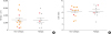
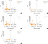
Impedance-based cell responses (Cell Index) and TER were measured to investigate epithelial barrier function (Fig. 1) and showed no differences between the 2 study groups. Cytokine levels in supernatants from cultured pNECs were measured using ProcartaPlex immunoassays. Proinflammatory cytokine levels were measured after 7, 14, 21 and 28 days of ALI culture. The 2 study groups showed similar cytokine levels (Fig. 2).
Fig. 2
Cytokine levels of supernatants from cultured primary human nasal epithelial cells obtained from non-allergic and allergic subjects. On the left are the results from 13 non-allergic individuals (dots). To the right are the results from 9 allergic patients (squares). Proinflammatory cytokines that were measured after 7, 14, 21 and 28 days of culture include GM-CSF (A), IL-33 (B), RANTES (C), IL-1α (D) and TSLP (E). A different scale is used in A as a higher level of GM-CSF is expressed compared with the other cytokines.
GM-CSF, granulocyte-macrophage colony-stimulating factor; IL, interleukin; RANTES, regulated on activation, normal T cell expressed and secreted; TSLP: thymic stromal lymphopoietin.
DISCUSSION
In this study, we reinvestigated whether ex vivo cultured primary nasal cells from allergic and non-allergic subjects differ regarding their ability to build up barrier as recently suggested.7 The barrier function of nasal epithelial cells from 9 well-characterized allergic patients and 13 confirmed non-allergic individuals were studied. Interestingly, we found no differences in barrier function, as assessed by 2 different methods, i.e., TER measurements performed in a 2-chamber model and determination of Cell Index in an impedance-based system. Cultured primary respiratory epithelial cells from allergic and non-allergic subjects also did not differ regarding pro-inflammatory cytokine secretion.
This is in contrast to a previous study, which reported that primary nasal respiratory epithelial cells from allergic patients when cultivated ex vivo showed impaired barrier function as compared to those from non-allergic subjects.7 However, in the latter study, no detailed clinical and serological characterization of the patients was performed to confirm that they suffered from allergy. By contrast, allergic patients in our study were meticulously characterized by recording both symptoms of allergy and by measurement of allergen-specific and total IgE levels. We thus have discriminated allergic from non-allergic subjects thoroughly. Importantly, we collected specific information about sensitization and symptom severity at the time of removing the tissue samples, which we then used for examination of nasal barrier function by TER with the 2-chamber system and by impedance-based cell responses (Cell Index) when using the xCELLigence system. In both systems we found no difference of barrier integrity in allergic individuals compared to non-allergic controls (Fig. 1). We thought that the strength of our study was that we addressed the key question whether ex vivo cultured nasal epithelial cells from allergic and non-allergic subjects have barrier function, by 2 different methodologies, i.e., the well-established TER method and one of the most updated research tools for measuring barrier function, i.e., the xCELLigence system and obtained comparable results in both models, whereas measuring only the expression of proteins like occludin and ZO-1 provides only indirect evidence for barrier function. On the other hand, we must acknowledge as a limitation of our study and of the earlier study7 that only a limited number of subjects could be analyzed. However, the number of subjects in our study and in the previous study7 were comparable. Nevertheless, it is possible that the differences between the latter study7 and our study may be due to differences in the cell culture protocols.
Since it has been shown that cytokines can affect the epithelial barrier integrity811 we measured cytokine levels in supernatants from cultured pNECs isolated from nasal biopsy specimens every 7 days as we sought to investigate a possible influence of the culturing time of the cells, i.e., after 7, 14, 21 and 28 days of ALI culture. Our 2 study groups showed similar cytokine levels at all time-points (Fig. 2). Therefore, we did not find any evidence of differences between the cells from allergic and non-allergic subjects to produce inflammatory cytokines.
In conclusion, we did not find any relevant intrinsic differences of ex vivo cultivated primary nasal epithelial cells from allergic and non-allergic subjects regarding barrier formation and inflammatory cytokine production. Differences regarding nasal mucosa barrier integrity between allergic and non-allergic individuals, therefore, seem to be rather due to other factors which come into play in the in vivo situation such as allergic inflammation in the surrounding tissue than due to intrinsic differences of respiratory epithelial cells.
 XML Download