

 PDF
PDF ePub
ePub Citation
 Print
Print


Abstract
Purpose
To compare the classification and severity of congenital color vision deficiency using a Nagel anomaloscope and Farnsworth Munsel 100-hue Test (FM 100-hue).
Methods
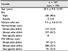
A total of 394 eyes of 197 patients diagnosed with congenital color vision deficiency were included. Examinations using a Nagel anomaloscope and FM 100-hue were performed, and color vision abnormalities were classified as a protan color defect or deutan color defect by each test, and the degrees of color vision abnormalities were compared.
Results
The tests showed 64.3% (p < 0.001) agreement in the classification of color vision deficiencies. The Nagel anomaloscope was able to classify all cases, whereas 143 eyes (36.3%) could not be classified using the FM 100-hue test. In the case of the same type of color vision abnormality in both eyes, 196 cases (99.5%) using the Nagel anomaloscope and 111 cases (56.3%) using the FM 100-hue were observed. Regarding the degree of color defect, there was a moderate positive correlation between the two tests (r = 0.43; p < 0.001). There were no significant differences in the total error scores between mild anomalous trichromacy and severe anomalous trichromacy as assessed using FM 100-hue (p = 0.087).
Conclusions
The Nagel anomaloscope was a more appropriate test for discerning the degree of color defect and binocular classification. In severity assessments, there was a moderate positive correlation between the two test methods. However, there were no significant differences in the total error scores between mild anomalous trichromacy and severe anomalous trichromacy as assessed using FM 100-hue. Therefore, it was difficult to perform severity classification using the Nagel anomaloscope based on the total error score of the FM 100-hue test.
Figures and Tables
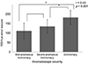
Figure 1
Correlation between Anomaloscope severity & FM 100-hue score. Bars indicate averaged FM 100-hue score in each group. Error bars indicate standard deviations. *Statistically significant difference between parameters (p < 0.05). Spearman rank correlation (r = 0.43, p < 0.001).

References
1. Swanson WH, Cohen JM. Color vision. Ophthalmol Clin North Am. 2003; 16:179–203.


2. Pokorny J, Smith VC, Verriest G, Pinckers AJLG. Congenital and Acquired Color Vision Defects. New York: Grune & Stratton;1979. p. 120–125.
3. Um BS. Eye exam. 3rd ed. Goyang: Naewae-haksool;2013. Vol. 1:p. 105–114.
4. Nam MH, Son MS. Nagel's anomaloscope examination for subdivision of 100 cases of congenital color defects. J Korean Ophthalmol Soc. 1980; 21:511–515.
5. Cooper H, Bener A. Application of a laserjet printer to plot the Farnsworth-Munsell 100-hue color test. Optom Vis Sci. 1990; 67:372–376.
6. Smith VC, Pokorny J, Pass AS. Color-axis determination on the Farnsworth-Munsell 100-hue test. Am J Ophthalmol. 1985; 100:176–182.
7. Foster DH. Vision and visual dysfunction: inherited and acquired colour vision deficiencies. 1st ed. London: Pan Macmillan;1991. Vol. 7:p. 32–45.
8. Thuline HC. Color-vision defects in american school children. JAMA. 1964; 188:514–518.
9. Sato S. Statistical observations on congenital abnormalities in colour vision in Japan. Acta Soc Ophthalmol Jpn. 1935; 38:2227–2230.
10. Jeong HW, Ahn CS. A comparison of the types of color defects measured by the hahn color vision test and neitz anomaloscope OT. J Korean Ophthalmol Soc. 1990; 31:1084–1088.
11. Vingrys AJ, Atchison DA, Bowman KJ. The use of colour difference vectors in diagnosing congenital colour vision deficiencies with the Farnsworth-Munsell 100-hue test. Ophthalmic Physiol Opt. 1992; 12:38–45.
12. Birch J. Use of the Farnsworth-Munsell 100-hue test in the examination of congenital colour vision defects. Ophthalmic Physiol Opt. 1989; 9:156–162.
13. Shin YJ, Choi SY, Park KH, et al. The classification of congenital color vision deficiency by SNU computerized color test. J Korean Ophthalmol Soc. 2004; 45:2099–2104.
14. Shin YJ, Choi SY, Park KH, et al. The discriminatiuon between congenital and acquired color vision defects by computerized color vision test. J Korean Ophthalmol Soc. 2005; 46:125–132.
15. Rabin J, Gooch J, Ivan D. Rapid quantification of color vision: the cone contrast test. Invest Ophthalmol Vis Sci. 2011; 52:816–820.
16. de Fez D, Luque MJ, Matea L, et al. New iPAD-based test for the detection of color vision deficiencies. Graefes Arch Clin Exp Ophthalmol. 2018; 256:2349–2360.
 XML Download