

 PDF
PDF ePub
ePub Citation
 Print
Print


INTRODUCTION
Left ventricular (LV) function is important in the assessment and management of patients with cardiac disease as well as in prediction of outcomes.1)2)3) Echocardiography is the most widely used tool for assessment of LV function; however, it is operator dependent and is limited by a variable acoustic window in patients with obesity, chronic obstructive pulmonary disease, narrow rib intercostal spaces, or prior cardiothoracic surgery.4)5) Although cardiac magnetic resonance imaging is considered the gold standard for LV function assessment, it is relatively expensive and cannot be performed in patients with claustrophobia and those with implanted pacemakers or defibrillators.6)
Cardiac computed tomography angiography (CCTA) can be used to detect coronary artery disease in patients with chest pain. It also provides information about LV volume and LV ejection fraction (LVEF) using the same dataset without the need for additional contrast or additional scanning.7) Therefore, in patients with a poor acoustic window undergoing CT imaging for coronary artery disease without previous ventricular assessment, especially those with a history of acute coronary syndrome or heart failure, simultaneous evaluation of LV contractile function by CCTA can be considered.5) Furthermore, LV function and volume assessed using CCTA can be used to improve risk stratification and identification of patients at risk for incident mortality.3)
Several studies have demonstrated that LV functional parameters measured using CCTA correlated well with those obtained using cardiac magnetic resonance imaging or echocardiography.8)9)10)11)12)13)14) However, radiation exposure remains a concern owing to the wide exposure window, because retrospective electrocardiography gating is used for LV end-diastolic volume (LVEDV) and LV end-systolic volume (LVESV) measurements.12)15) Several dose-reduction techniques have been implemented, such as use of a lower tube voltage, lower tube current, electrocardiography-based tube current modulation, prospective electrocardiography-gated acquisition, high pitch, and iterative reconstruction.16)17)18) A newly marketed third-generation dual-source computed tomography (CT) scanner (Somatom Force; Siemens Medical Solution, Forchheim, Germany) is equipped with automated attenuation-based tube voltage selection (CARE kV, Siemens Medical Solution) from 70 kV to 150 kV at 10-kVp increments, electrocardiography-based tube current modulation, and model-based iterative reconstruction (advanced modeled iterative reconstruction, ADMIRE; Siemens Medical Solutions), enabling cardiac imaging with reduced noise and radiation dose. Moreover, owing to the high temporal resolution (up to 66 ms) of this CT scanner, it can obtain coronary artery images at high heart rates (HRs). Accordingly, we reasoned that use of this third-generation dual-source CT scanner would allow assessment of both coronary artery disease and LV function with reduced radiation exposure.
Thus, the purpose of the present study was to assess LV function and coronary artery simultaneously by third-generation dual-source CT using a low radiation dose.
METHODS
This study was approved by the ethical review committee of our institution. The requirement for informed consent was waived due to the retrospective nature of this study.
Patients
We retrospectively searched a database of CCTA examinations that were performed from June 26, 2017, to January 4, 2019. During this period, a total of 84 consecutive patients underwent both CCTA using a third-generation dual-source CT scanner and echocardiography. During the study period, prospective gating CT angiography was performed in patients who had no chest pain with risk factors such as hypercholesterolemia or hypertension; these patients were excluded (n = 20) from this study. Patients with an interval of > 30 days (n = 16) between CCTA and echocardiography were also excluded from the study. A total of 48 patients were included in the final analyses. Among these patients, seven patients had typical chest pain, 28 patients had atypical chest pain, and 13 patients had dyspnea. Patient data including age, sex, height, weight, and HR during the CCTA examination were collected from electronic medical records.
Cardiac CT scan protocol and image reconstruction
CCTA examinations were performed using a third-generation dual source CT scanner (Somatom Force, Siemens Medical Solution). No beta-blockers were given to the patients before the scan. Nitroglycerin spray (0.4 mg, Nitrolingual; Mckesson Medical-Surgical Inc., Richmond, VA, USA) was used sublingually 5 min before the examination to dilate the coronary arteries. CCTA scan was started by continuous injection of a bolus of 80 mL iopromide (370 mg/mL, Ulravist; Bayer, Berlin, Germany) followed by 30 mL saline solution into an antecubital vein via an 18-gauge catheter (injection rate 5 mL/s). Contrast agent application was controlled by bolus tracking. A region of interest was placed into the aortic root, and image acquisition started 5 s after signal attenuation reached the predefined threshold of 100 Hounsfield units. Data acquisition was performed from the level of the carina to the heart base in a cranio-caudal direction with a detector collimation of 2 × 192 × 0.6 mm using a flying focal spot technique and a gantry rotation time of 250 ms. Tube voltage was selected semiautomatically (70–90 kVp) by automated tube voltage selection (CARE kV), and automatic exposure control was used for the tube current based on body size. Electrocardiography-based tube current modulation (Mindose, Siemens Medical Solution) was applied in all patients for a reduced radiation dose. In patients with a mean HR < 65 bpm, full tube current was applied from the 60% to 70%, and then to the 4% phase throughout the remainder of the cardiac cycle. In patients with a HR ≥ 65 bpm, full tube current was applied from the 30% to 40% phase, and then to the 4% throughout the remainder of the cardiac cycle.
First, the best phase was automatically chosen during the 60% to 70% phase or 30% to 40% phase to obtain optimal coronary artery images. Then, other images were reconstructed at 10%–100% of the R-R interval in 10% increments to assess LV function. Reconstruction parameters included a section thickness of 0.75 mm with 0.3-mm increments, a Bv40 of medium-smooth kernel, and a model-based iterative reconstruction strength level of 5 (ADMIRE, Siemens Medical Solutions). Image datasets were imported into a dedicated workstation (AquarisNet Viewer 1.8.0.3; TeraRecon, Foster City, CA, USA) and picture archiving communicating system (Maroview 5.4; Infinitt, Seoul, Korea).
Image analysis
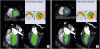
LV function was measured by three independent, blinded radiologists with 13, 8, and 3 years' experience of performing CCTA, respectively. LV function parameters were evaluated using dedicated software (AquarisNet Viewer 1.8.0.3, TeraRecon) (Figure 1). Observers manually delimited the correct mitral plane in the horizontal and vertical long-axis planes once. Then, the threshold of Hounsfield units was adjusted for segmentation of the LV cavity and the endo- and epicardial contours. Papillary muscles and trabeculae were excluded in the volumetric analysis. LVEDV, LVESV, and LVEF were calculated using software. Average values of the three observers were used for analyses.
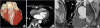
Figure 1
CT images of a 71-year-old male patient in end diastole (A) and end systole (B), which were obtained to assess left ventricular function. Mean heart rate during CT scan acquisition was 72 bpm. After the mitral valve plane was manually determined in the horizontal and vertical long-axis planes, the software automatically defined the blood pool of the LV by using a fixed Hounsfield unit threshold. Hounsfield unit threshold was adjusted to exclude the papillary muscles and trabeculae when calculating the LV volume. LVEDV, LVESV, and LVEF were automatically calculated. CT-derived LVEDV, LVESV, and LVEF of the patient were 72.0 mL, 33.7 mL, and 53.2%, respectively. Echocardiography-derived LVEDV, LVESV, and LVEF of the patient were 74.3 mL, 30.0 mL, and 59.6%, respectively. CT: computed tomography, LV: left ventricle, LVEDV: left ventricular end-diastolic volume, LVEF: left ventricular ejection fraction, LVESV: left ventricular end-systolic volume.
The quality of coronary artery images was analyzed by two independent, blinded radiologists with 13 and 3 years of experience performing CCTA, respectively. Images for coronary artery analysis were displayed at a fixed window setting (window level, 300 HU; width, 800 HU) and evaluated on a separate picture archiving communicating system (Maroview 5.4; Infinitt). Axial and curved multiplanar reformation images of the coronary artery were used to analyze the quality of the coronary artery images. In accordance with the scheme proposed by the American Heart Association,19) the coronary artery tree was divided into 16 segments. The intermediate artery, if present, was designated as a segment. All vessel segments with a diameter > 1.5 mm were included in the analysis. Image quality was graded using a 4-point scale on a per-segment level as follows: 1, excellent (no motion artifacts); 2, good (minor blurring artifacts); 3, fair (moderate blurring artifacts); and 4, poor (significant blurring or doubled appearance of the structure).20) Grade 4 was considered non-diagnostic image quality. In case of disagreement among the observers, a final decision was made by consensus.
Two-dimensional transthoracic echocardiography was performed using commercially available equipment (IE33; Philips, Andover, MA, USA) according to the American Society of Echocardiography recommendations.21) Images were acquired in standard apical and parasternal 2- and 4-chamber views using a 3.5-MHz transducer in the left lateral decubitus position. One cardiologist (with 14 years' experience of performing echocardiography) measured the chamber and wall dimensions according to the standard recommendations for chamber quantification. Manual tracing of the endocardial borders at end systole and end diastole was performed, and the modified Simpson's rule was used to calculate LVEDV and LVESV. Papillary muscles were excluded from the myocardium for these calculations. The EF was calculated as follows: EF = (EDV - ESV) ⁄ EDV.
Radiation dose analysis
Volume CT dose index and dose-length product were displayed on the dose report on the CT scanner, and recorded. The effective radiation dose of CCTA was calculated by multiplying the dose-length product by a conversion coefficient for the chest and coronary arteries (k = 0.014 mSv / [mGy × cm]).22)
Statistical analysis
Statistical analysis was performed using SPSS version 12.0 (SPSS, Chicago, IL, USA). Continuous data were expressed as means ± standard deviations and categorical data were expressed as numbers (percentages). Inter-observer reproducibility was calculated using intra-class correlation analysis in which intra-class correlation coefficients of < 0.5, between 0.5 and 0.75, between 0.75 and 0.9, and > 0.90 indicate poor, moderate, good, and excellent reliability, respectively.23) LV functional parameters measured using CCTA and echocardiography were compared using paired Student's t-test. Image quality was compared between the low HR group (≤ 65 bpm) and high HR group (> 65 bpm) and low BMI group (< 25 kg/m2) and high BMI group (≥ 25 kg/m2) using Student's t-test. The strength of correlations between LVEDV, LVESV, and LVEF measured using CCTA and echocardiography were determined using Pearson's correlation analysis (Pearson correlation coefficient: “r”; 0 = poor, 0.01–0.20 = slight, 0.21–0.41 = fair, 0.41–0.60 = moderate, 0.61–0.80 = good, 0.80–1.00 = excellent correlation), and the distribution of mean differences was evaluated using Bland-Altman analysis. A p value of less than 0.05 was considered statistically significant.
RESULTS
A total of 48 patients (36 men, 12 women; mean age 57.0 ± 9.5 years) were enrolled in this study. Table 1 shows the baseline characteristics of the enrolled patients. Mean HR during the CT examination was 59.9 ± 9.9 bpm (range 38–79 bpm). There was one patient with an irregular HR (HR variability >10 bpm). During the scan, a tube voltage of 90 kVp was used in one patient, 80 kVp in 18 patients, and 70 kVp in 29 patients.
Table 1
Baseline characteristics of enrolled patients
LV function analysis
Inter-observer reliability of the LVEDV, LVESV, and LVEF measurements among the three observers was excellent (intra-class correlation coefficient = 0.966, 0.945, 0.910, respectively; all p < 0.001). Mean LVEDV calculated using CCTA (113.5 ± 22.4 mL) was significantly higher than that calculated using echocardiography (104.7 ± 29.0 mL, p = 0.040), although mean LVESV (38.2 ± 11.3 mL vs. 36.0 ± 11.7 mL, p = 0.188) and mean LVEF (66.3% ± 6.9% vs. 65.5% ± 6.8%, p = 0.393) calculated using CCTA and echocardiography were not significantly different (Table 2). LVEDV, LVESV, and LVEF measured using CCTA and echocardiography demonstrated a fair to moderate correlation (r = 0.395, p = 0.005 for LVEDV; and r = 0.509, p < 0.001 for LVESV; r = 0.551, p < 0.001 for LVEF) (Figure 2). Bland-Altman analysis showed no systematic errors and acceptable limits of LV function measured using CCTA, with echocardiography as the reference test (Figure 3). Mean differences in LVEDV, LVESV and LVEF measured by CCTA and echocardiography among BMI groups are compared in Table 3. Mean BMI was 22.7 ± 2.1 kg/m2 (n = 26; range, 17.0–24.9) for the low BMI group and 26.6 ± 1.1 kg/m2 (n = 22; range, 25.0–29.4) for the high BMI group. The mean difference in LV function parameters was not significantly different between the low and high BMI groups except for LVEDV (LVEDV CCTA-Echo, 16.5 ± 23.3 mL in the low BMI group vs. -0.4 ± 32.4 mL in the high BMI group, p = 0.042). Table 4 shows a comparison of the mean differences in LVEDV, LVESV and LVEF measured using CCTA and echocardiography according to heart rate (low HR group vs. high HR group). Mean HR was 55.1 ± 7.3 bpm (n = 34; range, 38–65) in the low HR group and 70.8 ± 4.4 bpm (n = 14; range, 66–79) in the high HR group. Mean differences in LV function parameters were not significantly different between the low and high HR groups.
Table 2
Comparison of left ventricular function parameters between CCTA and echocardiography

| CCTA | Echocardiography | p-value | |
|---|---|---|---|
| LVEDV (mL) | 113.5 ± 22.4 | 104.7 ± 29.0 | 0.040 |
| LVESV (mL) | 38.2 ± 11.3 | 36.0 ± 11.7 | 0.188 |
| LVEF (%) | 66.3 ± 6.9 | 65.5 ± 6.8 | 0.393 |
Figure 2
Linear regression plots showing the correlation between LVEDV (A), LVESV (B), and LVEF (C) measured using CCTA and echocardiography. CCTA: cardiac computed tomography angiography, echo: echocardiography, LVEDV: left ventricular end-diastolic volume, LVEF: left ventricular ejection fraction, LVESV: left ventricular end-systolic volume.
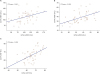
Figure 3
Comparison of CCTA and echocardiography assessments of left ventricular function. Bland-Altman plots of LVEDV (A), LVESV (B), and LVEF (C) showing the difference between each pair plotted against the average value of the same pair and the mean value of differences ± 2 SDs. Two of the 48 points (4.2%) lie outside the limits of agreement in the Bland-Altman plots of LVEDV and LVEF and three of the 48 points (6.3%) lie outside the limits of agreement in the Bland-Altman plot of LVESV. CCTA: cardiac computed tomography angiography, echo: echocardiography, LVEDV: left ventricular end-diastolic volume, LVEF: left ventricular ejection fraction, LVESV: left ventricular end-systolic volume, SD: standard deviation.
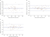
Table 3
Comparison of mean differences in left ventricular function parameters between CCTA and echocardiography according to patients' BMI

| Low BMI (n = 26) | High BMI (n = 22) | p-value | |
|---|---|---|---|
| LVEDV CCTA-Echo (mL) | 16.5 ± 23.3 | −0.4 ± 32.4 | 0.042 |
| LVESV CCTA-Echo (mL) | 4.1 ± 11.9 | 0 ± 10.6 | 0.222 |
| LVEF CCTA-Echo (%) | 1.5 ± 7.5 | 0 ± 5.1 | 0.409 |
Table 4
Comparison of mean differences in left ventricular function parameters between CCTA and echocardiography according to patients' HR

| Low HR (n = 34) | High HR (n = 14) | p-value | |
|---|---|---|---|
| LVEDV CCTA-Echo (mL) | 10.4 ± 28.2 | 4.9 ± 31.0 | 0.557 |
| LVESV CCTA-Echo (mL) | 2.6 ± 9.5 | 1.2 ± 15.5 | 0.702 |
| LVEF CCTA-Echo (%) | 0.8 ± 6.5 | 0.9 ± 6.6 | 0.931 |
Image quality analysis
In total, there were 791 coronary artery segments with a diameter greater than 1.5 mm, and all of them were assessable. Inter-observer reliability of image quality assessment among the two observers was excellent (intra-class correlation coefficient 0.838; p < 0.001). Average image quality score was 1.0 ± 0.1 (range 1–2). A total of 99.0% (783 of 791) of segments had an excellent image quality score, and 1.0% (8 of 791) of segments had a good score (Figure 4). No segments had a fair or poor score. There was no significant difference between the low HR group (n = 34) and high HR group (n = 14) according to the mean coronary artery image quality score (1.01 ± 0.09 vs. 1.01 ± 0.11, p = 0.599). Mean coronary image quality score in the high BMI group (1.00 ± 0.00) was superior to that in the low BMI group (1.02 ± 0.13, p = 0.005).
Figure 4
Cardiac computed tomography angiography images of a 47-year-old male patient referred because of atypical chest pain (average heart rate 77 bpm). Volume rendering image (A), axial image (B), and curved multiplanar reformation image (C) showing no relevant motion artifacts in the main coronary arteries. All coronary artery segments were scored 1 (excellent image quality). LAD: left anterior descending artery, LCX: left circumflex artery, RCA: right coronary artery.
DISCUSSION
In the present study, measurements of LV function obtained using third-generation dual-source CT with low kVp, electrocardiography-based tube current modulation, and advanced model-based iterative reconstruction were compared with measurements obtained using echocardiography. There was fair to moderate agreement in LVEF, LVEDV, and LVESV measured by CCTA and echocardiography. The excellent inter-observer reliability suggests that CCTA, an operator-independent technique, is suitable for LV function measurement. Furthermore, our findings suggest that the coronary artery can be imaged with diagnostic quality without an HR control.
Accurate measurement of LVEF is important in the care of patients with various cardiac conditions as it has prognostic value and can guide patient management.1)2)3)24) Among the various imaging modalities available for LV function measurement, CCTA is a unique modality that can simultaneous evaluate LV function and coronary artery anatomy without additional image acquisition. However, radiation exposure is one of the limitations of CCTA. In the present study, the effective radiation dose was 2.2 ± 0.7 mSv, which is much lower than that reported in previous studies using 64-slice CT12)25)26) (13.7–16.2 mSv) and 128-slice CT (16.8 mSv).13) This lower radiation dose for CCTA in the present study was because of the use of low kV, electrocardiography-based tube current modulation with iterative reconstruction.
In the present study, CCTA overestimated LVEDV relative to the echocardiography measurement with statistical significance. This finding is consistent with those of previous studies.12)27) Overestimation of LV volume in CCTA may be explained by several factors28): (i) foreshortening of the LV apex could result in underestimation of the LV volume on echocardiography; (ii) the two techniques have different approaches for LV volume calculation (modified Simpson's method with a modified biplane apical view for 2-dimensional echocardiography vs. 3-dimensional threshold segmentation algorithm for CCTA); (iii) variable intrathoracic pressures during different respiratory phases (inspiration for CCTA vs. free breathing for echocardiography); and (iv) HR changes related to beta-blocker use during CCTA can influence the absolute values of ventricular volumes. However, the last factor can be excluded in the present study because no beta-blockers were used for CCTA.
However, LVESV did not differ between CCTA and echocardiography, which is inconsistent with previous studies.11)25) This is probably due to differences in the LV volume measurement method among studies. We excluded papillary muscles from the LV cavity for volume analysis in the present study. The papillary and trabecular muscles constitute a significant percentage of LV mass and volume (mean LV papillary and trabecular muscles = 23% ± 3% of LVEDV).29) Therefore, a method excluding the papillary muscles yields a lower LV volume than the volume obtained using the traditional CCTA method (including papillary muscles)30) and cardiac magnetic resonance imaging.29) In a previous study that used CCTA, a significant difference (5.6% –30.1% for LV volume, 5.8% – 9.4% for LV mass, and 4.3% – 6.0% for LVEF) was observed in LV function parameters between papillary muscle-included and papillary muscle-excluded groups.30)
Interestingly, no coronary artery segments were scored as nondiagnostic in the present study. This result is presumably due to improved temporal resolution secondary to increased gantry rotation speed, resulting in reduced motion artifacts. The larger z-axis coverage yields fewer potential slab-to-slab misregistration and banding artifacts.31) Additionally, using a low tube voltage has the advantage of a substantially higher contrast-to-noise ratio for contrast-filled vessels, owing to the lower mean photon energy that is closer to the K-edge of iodine.32) Another interesting result was that the image quality of coronary artery segments was not significantly different between the low HR group and the high HR group. In the present study, there was no patient with an HR > 80 bpm and there were only 14 patients in the high HR group despite the fact that no beta-blockers were used. Lastly, in contrast to a previous study,33) mean coronary image quality score in patients with a high BMI (1.00 ± 0.00) was superior to that in patients with a low BMI (1.02 ± 0.13, p = 0.005). Unidentified errors while scanning and cardiac motion artifacts in patients with an intermediate HR (69 bpm) may have contributed to the poorer image quality in the low BMI group in our study.
The present study has several limitations. First, due to its retrospective nature, there is an inherent risk of selection bias. Second, measurements of LV functional parameters were compared with those obtained using echocardiography instead of cardiac magnetic resonance imaging, which is the current gold standard for volumetric measurement. Third, no patient had an HR > 80 bpm and one patient had high HR variability (> 10 bpm) despite not using a beta-blocker. Therefore, we could not analyze the effect of high HR or HR variability on LV function measurement. Finally, we did not analyze the diagnostic accuracy of coronary artery stenosis, because evaluating the diagnostic accuracy of CCTA was not the primary objective of the present study and only four patients underwent coronary artery angiography.
In conclusion, third-generation dual-source CT using a low radiation dose simultaneously provides information regarding LV function and coronary artery disease. Therefore, it is an alternative option for functional assessment, particularly in cases where other imaging modalities are inadequate.
 XML Download