

 PDF
PDF ePub
ePub Citation
 Print
Print


INTRODUCTION
Since their discovery over a century ago, sphingolipids have been increasingly acknowledged as key signaling molecules that regulate essential cell functions, with implications for a wide variety of diseases. In addition to their historically recognized function as essential components of eukaryotic cell membranes, modern research has elucidated their roles as signaling molecules that regulate apoptosis, autophagy, nutrient transport, organ homeostasis, and protein synthesis, as well as modulating classical signaling pathways by regulating kinases and phosphatases.1 Aberrancies in these processes due to perturbed sphingolipid metabolism have been reported in diseases including obesity, type 2 diabetes mellitus (T2DM), neurodegenerative disorders, liver disease, cancer, and cardiovascular disease.23 Numerous studies have demonstrated that dysregulated sphingolipid metabolism induces alterations in cardiomyocyte structure and function.456 Moreover, sphingolipids are known to regulate crucial cell processes involved in cardiac structure and function, including apoptosis, autophagy, cell differentiation, and mitochondrial metabolism, suggesting that sphingolipids likely contribute to cardiomyopathy through these mechanisms. Therefore, the aims of this review are to summarize current knowledge regarding the contribution of sphingolipids to various cardiomyopathies and to discuss the current and future clinical potential of targeting sphingolipid metabolism.
Sphingolipids are defined by their sphingoid base, which is generated by condensation of an amino acid with an acyl-CoA. The sphingoid base serves as a ‘backbone’ upon which all sphingolipids are built, including ceramides, sphingosine-1-phosphate (S1P), sphingomyelins (SM), and glycosphingolipids. The sphingoid base (Fig. 1A, leftmost structure) can vary in structure depending on the amino acid and/or acyl-CoA used as substrate; for example, using serine and palmitoyl-CoA yields an 18-carbon sphingoid base (Fig. 1A), while utilization of serine and myristoyl-CoA or stearoyl-CoA yields sphingoid bases with 16- or 20-carbon bases (Fig. 1A), respectively. Additionally, recent studies have addressed variations in the amino acid used to form the sphingoid base, finding that replacing serine with alanine or glycine gives rise to structurally aberrant sphingoid bases.78910 Serine palmitoyltransferase (SPT) is a multi-subunit enzyme that catalyzes this initial reaction, which is the rate-limiting step in sphingolipid synthesis. The typical SPT enzyme is a heterodimer composed of serine palmitoyltransferase long chain base subunit (SPTLC) 1 and SPTLC2 subunits and condenses the 2-carbon serine with the 16-carbon palmitoyl-CoA to synthesize d18:0-dihydrosphingosine (DHS), an 18-carbon sphingoid base (Fig. 1A).10 Palmitate-derived d18-base-containing sphingolipids are the most abundant, as several complex downstream sphingolipids including ceramide, hexosylceramides, SM, and S1P largely are built upon this 18-carbon backbone. In contrast, a heterodimer composed of SPTLC1 and SPTLC3 can also utilize the 14-carbon myristoyl-CoA, generating a subset of d16-based sphingolipids (Fig. 1B).9 Although these sphingolipids are abundant in the myocardium, they remain understudied.11 Acyl-CoA utilization can also be influenced by 2 SPT small subunits (ssSPTa and ssSPTb), which enable SPT to utilize stearoyl-CoA as a substrate, resulting in a subset of d20-backboned sphingolipids.1213
Fig. 1
Overview of sphingolipid structure and metabolism. (A) From left to right; DHS d18:0 sphingoid base, ceramide d16:1/16:0, DHC d18:0/16:0 and S1P d18:1. (B) The blue arrow indicates sulfatides, green arrows indicate the globosides and the orange arrows indicate the gangliosides.
DHS, dihydrosphingosine; DHC, dihydroceramide; S1P, sphingosine-1-phosphate; DHS1P, dihydrosphingosine 1-phosphate; GalCer, galactosylceramide; SM, sphingomyelin; GlcCer, glucosylceramide.
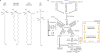
Ceramide, which have been implicated in apoptosis, senescence, autophagy, and other cell processes, are generated from N-acylation of the sphingoid base with an additional acyl-CoA. The acyl-CoA molecules utilized vary widely, including medium-chain fatty acids (12–14 carbons), long-chain fatty acids (16–20 carbons), and very-long-chain fatty acids (22–26 carbons) in mammals. Ceramide exist in all of these variations and can even include ultra-long-chain fatty acids (≥26 carbons). In highly specialized organs, such as the skin, ceramide undergo numerous structural modifications, including hydroxylation, O-acylation, branching, methylation, and others. Ceramide can also be catabolized yielding a sphingoid base, which can be either re-acylated to ceramide (sometimes with a change in the N-acyl chain length) or phosphorylated to generate S1P, an important sphingolipid. Most sphingolipid metabolic pathways have enzymes that catalyze the forward and reverse steps, and altering the sphingolipid profile occurs through highly dynamic regulation of these processes. Additionally, once a sphingoid base is synthesized, the only way to catabolize sphingolipids to non-sphingolipid components is reduction of S1P by S1P lyase, yielding phosphoethanolamine and a fatty aldehyde. Depletion of the S1P lyase leads to a dramatic accumulation of sphingolipids, usually with deleterious effects. These pathways are presented in depth in Fig. 1B.
The de novo pathway is initiated in the endoplasmic/sarcoplasmic reticulum by SPT, a dimer composed of SPTLC1 and either 2 or 3 subunits. Each of these complexes can utilize palmitoyl-CoA, yielding d18:0 DHS. Additionally, the SPTLC1/3 complex can use serine and myristoyl-CoA to synthesize d16:0 DHS. The SPT complex can be regulated by small subunits to alter CoA utilization, including by enabling stearoyl-CoA to be used to generate d20:0 DHS. In terms of amino acid variations, alanine or glycine can be used instead of serine to generate aberrant sphingoid bases. Alterations in amino acid utilization result from mutations of SPT subunits and are largely pathological. DHS is the primary substrate for ceramide synthases, which generate dihydroceramides (DHCs) that are then reduced by DHC desaturase, yielding ceramide, which is used as the substrate for the synthesis of complex sphingolipids, including SM, galactosylceramide (GalCer), glucosylceramide (GlcCer), and ceramide 1-phosphate (not shown) via the enzymes indicated above. De novo synthesis of ceramide occurs in the endoplasmic reticulum, and then ceramide is modified in the Golgi apparatus. GlcCer is the predominant precursor of the ganglioside and globoside glycosphingolipids. Lactosylceramide (LacCer) is synthesized from GlcCer to form gangliosidosis and Gaucher disease in the ganglioside and globoside series. Diseases that have been observed in humans due to deficiencies in enzymes in these pathways are shown in parentheses, underlined, and in bold. GalCer is the predominant precursor of the sulfatide glycosphingolipids. Sphingolipid catabolism occurs in lysosomes and at the plasma membrane. SM is hydrolyzed by lysosomal acid sphingomyelinase (aSMase) or plasma membrane neutral sphingomyelinase (nSMase), GalCer by β-galactocerebrosidase, and GlcCer by β-glucocerebrosidase (both lysosomal), and each of these steps yields ceramide. Catabolism of ceramide by ceramidases yields sphingosine, which is phosphorylated by sphingosine kinases 1 and 2 (SphK1-2), yielding S1P. The salvage pathway of ceramide synthesis occurs when ceramide is cleaved by ceramidase and the resulting sphingosine is re-acylated by ceramide synthases.
SPHINGOLIPIDS IN PRIMARY CARDIOMYOPATHIES
In primary (idiopathic) cardiomyopathy, there is no secondary cause such as hypertension or diabetes; however, cardiomegaly, endocardial thickening, mural thrombosis, myocardial scarring, or other lesions may be present. Primary cardiomyopathies are classified as genetic or acquired and historically have been classified according to cardiac morphology. Genetic primary cardiomyopathies can be further subdivided into hypertrophic cardiomyopathy (HCM), dilated cardiomyopathy (DCM), restrictive cardiomyopathy (RCM), arrhythmogenic right ventricular cardiomyopathy (ARVC), and unclassified cardiomyopathy. Acquired primary cardiomyopathies are categorized as ischemic cardiomyopathy (ICM), peripartum cardiomyopathy (PPCM), or myocarditis.14 Dysregulated sphingolipid metabolism has been observed and/or implicated in the following primary cardiomyopathies.
1. DCM
DCM is defined by the presence of left ventricular (LV) dilatation and systolic dysfunction or reduced ejection fraction; it is the end result of many cardiac insults, but a genetic etiology is also associated with DCM. Right ventricular dilation and dysfunction may be present, but are not necessary for the diagnosis of DCM.15 The excessive accumulation of ceramide and SM within and around the heart has been associated with DCM.161718 A DCM hamster model demonstrated elevated levels of ceramide and SM in the LV.19 Depletion of the antioxidant enzymes manganese superoxide dismutase and glutathione peroxidase-1 resulted in DCM, as well as findings of impaired mitochondrial function, increased ceramide levels, and increased levels of reactive oxygen species.2021
2. ICM
ICM is a disease of the heart muscle induced by narrowing of the coronary arteries, most commonly in patients with a history of myocardial infarction, which is usually a manifestation of atherosclerotic coronary artery disease (CAD). Regardless of the cause, ICM ultimately leads to congestive heart failure. Risk factors for ICM include hyperlipidemia, diabetes mellitus, hypertension, obesity, age, sex (with females demonstrating cardioprotective effects), and genetics.
In addition to the ischemic injury itself, some damage occurs upon reperfusion. An ischemia-reperfusion (IR) injury occurs when blood supply returns to the heart tissue after ischemia. Patients with IR injuries and a mouse model of IR injury demonstrated decreased plasma DHS, sphingosine, S1P, and DHS 1-phosphate levels; moreover, the levels of circulating lysosomal aSMase and plasma membrane nSMase decreased, suggesting less ceramide production from catabolic pathways. Of note, the distribution of S1P, which circulates either bound to ApoM on high-density lipoprotein (HDL) or on serum albumin, was altered such that HDL-bound S1P was reduced and non-HDL-bound S1P was elevated in IR injury patients.22232425 According to current thinking, some beneficial effects attributed to HDL may be mediated by S1P bound to HDL.26
3. Myocarditis
Inflammation of the myocardium is termed myocarditis, which often causes arrhythmias and contractility issues that gradually weaken the myocardium over time. The most common causes of myocarditis are adenovirus infection (the common cold) or coxsackievirus B3 (CVB3) infection. 293031 Viral myocarditis caused by dengue, the West Nile virus, or hepatitis C leads to altered host sphingolipid metabolism, which is thought to be advantageous for viral propagation.323334 An acute viral myocarditis mouse model showed higher levels of DHS, d18:0/14:0 ceramide, and d18:1/22:0 and d18:0/16:1 SM than observed in the control group, suggesting that the CVB3 virus alters sphingolipid metabolism to enhance its propagation in the host.3536 Furthermore, evidence also suggests that autoimmunity plays an important role in myocarditis.373839 Previous studies have shown that CD4 T cells, macrophages, and inflammatory cytokines accumulate in cases of myocarditis.4041 As S1P is a potent pro-inflammatory lipid, multiple studies have used either S1P receptor (S1PR) agonists or SphK1 inhibitors to treat myocarditis, as further discussed below.
SPHINGOLIPIDS IN SECONDARY CARDIOMYOPATHIES
Secondary cardiomyopathies are diagnosed as a comorbidity of many primary diseases that affect the endocrine system, lysosomal storage, endomyocardial function, or neuromuscular function. Hypertension and tachycardia can also cause secondary cardiomyopathies.
1. Endocrine and metabolic disorders
Diabetic cardiomyopathy (DbCM)
DbCM, which develops in many patients with T2DM, manifests as cardiac hypertrophy in the absence of traditional heart disease risk factors such as hypertension, valve disease, and tachycardia and can lead to heart failure.4243444546 T2DM has a characteristic pattern of dyslipidemia including increased low-density-lipoprotein cholesterol levels, hypertriglyceridemia, and reduced HDL cholesterol concentrations.47 It is thought that these changes promote the accumulation of lipid intermediates in the heart, a phenomenon known as cardiac lipotoxicity. Several animal models of myocardial lipotoxicity have demonstrated protective effects of inhibited sphingolipid synthesis, suggesting that sphingolipids play a major role in DbCM. For example, in a lipotoxic cardiomyopathic mouse model with cardiac-specific overexpression of glycosylphosphatidylinositol-anchored human lipoprotein lipase, it was observed that ceramide levels increased by 45% compared to control mice.17 Importantly, correcting sphingolipid profiles using an SPT inhibitor or SPTLC1 heterozygous mice improved cardiac function and prolonged survival through reduced ceramide and SM levels.174849 Moreover, our laboratory demonstrated that high saturated-fat feeding induced a DbCM-like condition in mice, which was completely ameliorated by pharmacological inhibition of sphingolipid synthesis. In primary cardiomyocytes, cell hypertrophy in response to saturated fatty acids was shown to require ceramide synthase 5 (CerS5), which generates medium- and long-chain (e.g., C14–C16) ceramides.5051525354 This was the first identification of a specific ceramide synthase and ceramide species involved in cardiac hypertrophy, which in this case occurred through autophagy-dependent mechanisms. In additional support of a role for specific fatty acids in cardiac hypertrophy, an intriguing study in a Burmese python model demonstrated that adaptive post-prandial cardiac hypertrophy was mediated by a combination of myristate (C14:0), palmitate, (C16:0), and palmitoleate (C16:1).55 Importantly, the myocardium contains detectable levels of sphingolipids with a C16:1 base, derived from the utilization of myristate by the SPTLC1/3 complex; however, we demonstrated that, in contrast to canonical d18-based sphingolipids, this class of sphingolipids with a shorter sphingoid base caused apoptosis, suggesting a critical need for precise regulation of these lipids, of which the constitutive roles remain unknown.11
Studies have demonstrated additional roles for myocardial ceramides in diabetes, including mitochondrial dysfunction and AKT inhibition, presumably via increased oxidative stress and inhibition of insulin signaling, respectively. The glycosphingolipid GlcCer acts independently from ceramide in impairing insulin signaling, which leads to insulin resistance, although the mechanisms have not been fully elucidated.56575859606162 In models of DbCM, ceramide has been shown to activate classic protein kinase C (PKC) isoforms to attenuate insulin-stimulated AKT translocation; in addition, we and others have shown that aberrant ceramide production induces reactive oxygen species and impairs mitochondrial function. Additionally, ceramide was shown to activate p38, thereby inhibiting AKT and c-Jun N-terminal kinase (JNK), and resulting in activation of pro-apoptotic B-cell lymphoma 2 associated X protein (BAX) signaling and hypertrophy.56575859606162
Hyperthyroidism
The diagnosis of hyperthyroidism is confirmed by elevated secretion of the thyroid hormone, triiodothyronine (T3) and/or thyroxine (T4), in the blood. T4 is converted into the more active form T3 in multiple organs including the thyroid, liver, gut, and skeletal muscles. Cardiomyopathy associated with hyperthyroidism is not unique, and mostly mimics cardiac hypertrophy or in rare cases, DCM.636465 T3 is a key regulator of cardiac physiology that also modulates sphingolipid metabolism. Prolonged treatment with T3 led to a prominent increase of DHS, sphingosine, ceramide and SM levels, but decreased S1P levels, in cardiomyocytes taken from the LV of male mice.66 Consistent with this, data demonstrated that T3 treatment reduced the activity of nSMase, but caused no change in ceramidase activity, which would be required to generate sphingosine as a substrate for SphKs.66
2. Lysosomal storage
Many lysosomal storage diseases arise from impaired catabolism of complex sphingolipids including SM and glycosphingolipids; these conditions are also termed ‘sphingolipidoses.’ Many of these diseases demonstrate a cardiac phenotype; however, there is currently little to no mechanistic information linking specific sphingolipids to these observed disease phenotypes.
Gaucher disease
Gaucher disease, the most common sphingolipidosis, is an autosomal recessive lysosomal storage disorder caused by deficiency of the enzyme acid-β-glucosidase or glucocerebrosidase. This leads to accumulation of GlcCer, a metabolic intermediate derived from the turnover of ganglioside and globosides, in the lysosomes of macrophages of many organs. In the most extreme cases, GlcCer accumulation occurs in the heart. Gaucher disease is divided into 3 types (GD1-3), depending on the severity and onset of neurological symptoms. Gaucher disease can cause severe congestive cardiomyopathy (Gaucher disease cardiomyopathy, GDC), which has only been observed in adults with GD3.676869 GDC is associated with cardiac hypertrophy and mitral and aortic valve calcification.707172737475 In a macrophage model of Gaucher disease, it was shown that although the lysosome is the primary site of GlcCer accumulation, at very high levels of GlcCer, it is distributed among various subcellular locations. This cellular saturation of GlcCer leads to increased levels of ceramides and other glycosphingolipids,76 which interfere with other biochemical pathways and lead to cell dysfunction and the pathologies observed in Gaucher disease. Generally, enzyme replacement therapy has been successful in Gaucher disease, as further discussed in the section of this review focusing on the therapeutic modulation of sphingolipids.
Anderson-Fabry disease
Anderson-Fabry disease is an X-linked recessive genetic disorder caused by deficiency of lysosomal enzyme α-galactosidase A, which catabolizes globotriaosylceramide, GalCer, LacCer, and other neutral glycosphingolipids. This causes progressive intracellular lysosomal accumulation of these lipids, primarily in the skin, kidneys, and heart.77 Fabry disease cardiomyopathy (FC) has been reported in up to 6% of men and 12% of women with Fabry disease, and in some individuals FC may be the sole manifestation of the disease.687879 FC is characterized by progressive symmetrical or concentric left ventricular hypertrophy (LVH), with progressive evolution towards heart failure.80818283 Most patients with FC are previously misdiagnosed with primary HCM, specifically LVH.84 A 2006 study found that endomyocardial glycosphingolipid deposition caused enlarged myocytes. Furthermore, ventricular walls became progressively more rigid, impeding ventricular filling. Ultimately, FC patients suffer from heart failure with preserved ejection fraction.45858687 Fabry disease has also been associated with RCM, though this link has not been thoroughly studied.15 Many therapeutics are available to treat Fabry disease, as discussed in a later section.
Gangliosidosis
The terminal β-galactosyl residues of GM1 gangliosides are hydrolyzed by β-galactosidase. In GM1 gangliosidosis, which is heritable, a lack of β-galactosidase leads to massive accumulations of the GM1 ganglioside, mainly in the central nervous system, but also in the heart. Cardiomyopathy has been reported to be present in 34% of patients with infantile (type I) GM1 gangliosidosis and in 38% of patients with juvenile (type II) or adult (type III) GM1 gangliosidosis. 90919293949596
There are 2 types of GM2 gangliosidosis. Type I, Tay-Sachs disease, results from deficiency of hexosaminidase A, and type II, Sandhoff disease, from a deficiency of hexosaminidase A and β-N-acetyl hexosaminidase. Patients with Tay-Sachs disease have accumulations of GM3/GD3 gangliosides, mainly in the brain and rarely in the heart.9798 In Sandhoff disease, also called GM2 gangliosidosis, the β-galactosidase A and B enzymes are deficient, leading to accumulation of GM3/GD3 gangliosides. The GM2-ganglioside storage disorders are heritable diseases, which are essentially neurodegenerative diseases of early infancy.9599 If cardiomyopathy is present, the cardiological symptoms are very similar to those described above for GM1-gangliosidoses.100101
As stated above, little is known about the mechanisms linking sphingolipidoses to the cardiac pathophysiology observed in these disorders. However, several other storage disorders—Krabbe disease; Niemann-Pick disease types A, B, and C; Farber disease, and hemochromatosis—cause cardiomyopathy very infrequently, if at all. This suggests that the mechanisms of sphingolipidosis-associated cardiomyopathies are linked to specific metabolic pathways and do not result from non-specific mechanisms, such as overall alterations in cell ultrastructure.
3. Neuromuscular disorders
Muscular dystrophy
There are 9 types of muscular dystrophy, of which only 5 cause secondary cardiomyopathy. Duchenne muscular dystrophy (DMD), Becker muscular dystrophy (BMD), Emery-Dreifuss muscular dystrophy, Limb-Girdle muscular dystrophy (LGMD), and trinucleotide/tetranucleotide-repeat muscular dystrophy (DM1 and DM2) all exhibit DCM as a cardiac complication. Additionally, DMD, BMD, and LGMD may also cause ARVC, whereas DM1 and DM2 may also result in cardiac hypertrophy.102 Upregulation of S1P and/or the SPT enzyme, genetically or through a pharmaceutical agent, reduced muscle degeneration or prevented muscle wasting in a dystrophin mutant (DMD or BMD) Drosophila model; however, it was not tested whether these interventions could reverse the pathology of DCM. Reducing the expression of the S1P transporter, spinster 2 (Spns2), which transports intracellular S1P to the extracellular compartment, thereby reducing intracellular S1P, also suppressed dystrophic muscle degeneration, suggesting that intracellular S1P suppresses muscle degeneration.
Mutations in caveolin-3 (CAV3) underlie LGMD, which can cause secondary cardiac arrhythmias or ARVC. CAV3 is a muscle-specific caveolin, unlike CAV1 and CAV2, found in the skeletal and cardiac muscles; it also organizes and concentrate glycosphingolipids within the caveolar membrane.103104 Therefore, in LGMD it is likely that sphingolipids are highly dysregulated due to CAV3 mutations, which could open new avenues for therapeutic interventions aimed at modulating membrane sphingolipids.
OTHER DISORDERS
Hypertension
Hypertensive cardiomyopathy (HTNCM) is characterized by concentric LVH and is prevalent in 20%–100% of cases, proportional to the severity of hypertension.105 Persistent HTNCM can lead to congestive heart failure. Interestingly, both the severity of hypertension and the severity of LVH in a patient have been shown to be proportional to the plasma ceramide level.106107108 HTNCM is difficult to distinguish from primary HCM, FC, and cardiomyopathy induced by acromegaly.108 The renin-angiotensin-aldosterone system (RAAS), which regulates blood pressure, is the central regulator of LVH induced by hypertensive cardiomyopathy, and sphingolipids are a central regulator of the RAAS system.109110111 Moreover, numerous links have been established between sphingolipids and regulation of vascular tone. Therefore, it is unsurprising that hypertension may arise from aberrant sphingolipid synthesis, ultimately precipitating cardiac outcomes. This is further discussed below.
Tachycardia
Tachycardia-induced cardiomyopathy (TIC) is caused by prolonged tachycardia or arrhythmia, termed tachyarrhythmias, and eventually leads to heart failure. Upon treatment of the causative tachyarrhythmia, TIC is generally a reversible disease.112113 TIC is characterized by LV dysfunction leading to DCM, and is usually only diagnosed after the recovery of LV function with normalization of the heart rate. Atrial fibrillation is the most common cause of TIC, although other tachyarrhythmias associated with TIC include atrial flutter, incessant supraventricular tachycardia, ventricular tachycardia, and premature ventricular depolarizations.114115116117118119 In a study of a TIC model, plasma and ventricle sphingolipid levels were altered. The LV showed reduced ceramide and S1P levels, stable levels of DHS, and increased levels of sphingosine, whereas the right ventricle (RV) showed increased DHS and sphingosine levels, but reduced ceramide and S1P levels. This suggests that ceramide catabolism was increased and the conversion of sphingosine to S1P was inhibited in the LV, while de novo synthesis was unaffected. In the RV, the former pattern was also found, but the data suggest an increase in de novo synthesis.120121 Long QT syndrome is a condition that affects repolarization of the heart after a heartbeat, resulting in an increased risk of irregular heartbeats. LQT9 is a genetic subtype of long QT syndrome involving mutations in the membrane structural protein, CAV3. LQT9 has been shown to lead to ventricular tachycardia. Ceramide regulates the hERG current (IHERG), which in turn regulates contractions and electrical activity of the heart via K+ ion channels.122123124 Ceramide and S1P also seem to play opposite roles in TIC, as in other pathologies (e.g., IR injury) as discussed above.
SIGNALING MECHANISMS AND NOVEL POTENTIAL ROLES FOR SPHINGOLIPIDS IN CARDIOMYOPATHIES
In addition to experimentally supported roles for sphingolipids in cardiomyopathies, the multitude of signaling pathways that sphingolipids regulate suggests that they may play additional roles in cardiac pathologies and also provides hints as to mechanism(s) for these roles. Sphingolipids are involved in regulating or are regulated by hypoxia, tumor necrosis factor (TNF)-α, Ca2+ signaling, K+ signaling, reactive oxygen species, endothelial nitric oxide synthase (eNOS), apoptosis, autophagy, necrosis, and many other cell processes implicated in cardiac pathophysiology.125126127128129130
Among signaling sphingolipids, S1P is perhaps the best mechanistically characterized. S1P has known intracellular signaling functions, and its most solidly established activities arise from its autocrine, endocrine, and paracrine activities that are mediated by binding to its receptors (S1P1-5). Cumulative data point towards S1P being involved in cardioprotective signaling pathways such as eNOS production, which is pro-angiogenic, as well as inhibition of class II histone deacetylase (HDAC) complexes, involved in repressing gene expression. It has in fact been proposed that S1P represses HDAC1 and HDAC2 to activate the transcription factor KLF4, thereby inhibiting hypertrophy in HCM.131132133134135136137 AKT activates Bcl2 to induce cell survival and increases eNOS activity to support vasodilation. The phosphoinositide 3-kinase (PI3K)-Rac pathway also induced by S1P promotes cell migration. S1P production may also be activated by the hypoxic transcription factors HIF1α and HIF2α and/or shuttled by TNF-α into the cell to activate pro-survival signaling pathways such as AKT, PI3K, Pak1, and Rac in HCM, DCM, Takotsubo cardiomyopathy, and ICM with IR injury.123138139140141142143144 These activated pathways in in vitro and in vivo IR models reduced infarct size, endothelial cell migration, and angiogenesis, while increasing the viability of isolated cardiomyocytes.123140141142143 In addition, S1P can activate Pak1 and protein phosphatase 2 (PP2A) in cardiomyocytes through its interactions with its receptors. This signaling pathway is an important mechanism involved in cardioprotection in ICM and IR injury, and could also potentially be involved in Takotsubo cardiomyopathy.141144 In HTNCM, S1P mediates renin release and stimulates aldosterone hormone secretion in the RAAS to maintain electrolyte and fluid balance, thereby maintaining blood pressure in HTNCM.145146 Additionally, S1P stimulates aldosterone secretion in a manner dependent on PKC and phospholipase D to maintain electrolyte and fluid balance, thereby maintaining cardiovascular homeostasis.145 S1P binds the S1PR1 present on cardiac mast cells and mediates the inhibition of cell degradation and renin release.146 Contrary to these findings, other studies have shown that elevated levels of S1P and SphK1 are associated with negative effects in hypertension.147148
The recent literature has shown that vascular endothelial growth factor (VEGF) induces expression of the S1PR1 receptor in the myocardial vascular endothelium, an instance of crosstalk that is required for proper angiogenic balance.149 In later stages of gestation, the placenta secretes VEGF inhibitors, often inducing angiogenic imbalance in PPCM.150 Upon treatment with pro-angiogenic therapies, such as VEGF, PPCM has been entirely reversed in PPCM mouse models.149 S1P has not been used to counteract the VEGF inhibitors secreted in the late gestational stages, but it seems likely that it would be just as efficacious as VEGF treatment. It seems likely that HTNCM could be reversed by treatment with S1P agonists that directly affect the RAAS. Spns2 transports intracellular S1P out of the cell, but in DM1 and DM2, there is a significant reduction of Spns2 that results in increased levels of intracellular S1P, which suppresses dystrophic muscle degeneration. In hyperthyroidism, T3 inhibits S1P while activating ceramide to induce cardiomyopathy. Cardiomyopathy caused by acromegaly results from increased intracellular Ca2+ and involves roughly 500 times more apoptosis. Because S1P has been demonstrated to serve these signaling functions in other contexts, it seems plausible that S1P signaling should be further elucidated in the context of cardiomyopathies involving perturbations of these signaling pathways.
In the context of cardiomyopathy, increased ceramide production and/or alterations in the ceramide profile (e.g., changes in the ratios between medium/long-chain and very-long-chain ceramides) are generally considered toxic. Ceramide has been shown to increase cytosolic Ca2+ levels, but not intramitochondrial Ca2+ levels, promoting hypertrophy or contractile function in IR by activating p38 MAPK and PKC. This inhibits AKT and JNK and activates pro-apoptotic BAX signaling. Reactive oxygen species are then increased, affecting the ryanodine receptor in the sarcoplasmic reticulum to stimulate Ca2+ release into the cytoplasm.151152153154155156
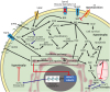
The role of ceramide in apoptosis has been established, and consistent with this, it induces the pro-apoptotic p38 MAPK signaling pathway, which is activated in Takotsubo/stress cardiomyopathy and other cardiomyopathies.157158159 This pro-apoptotic pathway is also induced in the RAAS by ceramide.160161 Ceramide is also a possible vasodilator via the RAAS through the PKC, cAMP, and PP2A signaling pathways.162163164165 Angiotensin II in the RAAS induces SM and intracellular ceramide synthesis via angiotensin II type 2 (AT2) receptors to inhibit cell growth and induce apoptosis in vascular and cardiac tissue.160161 Ceramide is also a possible vasodilator through the PKC, cAMP, and PP2A signaling pathways via AT2 and inhibiting AT1 action.162163164165 Many of these potentially relevant signaling pathways are depicted in Fig. 2.
Fig. 2
Summary of the signaling pathways of S1P and ceramide in the various types of cardiac hypertrophy. Activation is indicated with arrows, inhibition by perpendicular lines, black indicates known pathways, blue or red indicate potential pathways.
S1P, sphingosine-1-phosphate; VEGF, vascular endothelial growth factor; Spns2, spinster 2; T3, triiodothyronine; S1PR1, sphingosine-1-phosphate receptor 1; TNFR, tumor necrosis factor receptor; eNOS, endothelial nitric oxide synthase; PP2A, protein phosphatase 2; Pak1, p21-Activated kinase 1; PKC, protein kinase C; HDAC, histone deacetylases; ASK1, apoptosis signal-regulating kinase 1; JNK, c-Jun N-terminal kinase; BAX, B-cell lymphoma 2 associated X protein; KLF4, Kruppel-like factor 4; HCM, hypertrophic cardiomyopathy; SphK, sphingosine kinase; NOX2, NADPH oxidase 2; ROS, reactive oxygen species; DbCM, diabetic cardiomyopathy; RYR1, ryanodine receptor 1.
THERAPEUTIC MODULATION OF SPHINGOLIPIDS
Cardiomyopathy and heart failure remain the leading cause of morbidity and mortality worldwide despite the vast strides made in therapeutic interventions in recent years. Since enzymes, mediators, and inhibitors of the de novo sphingolipid pathway are directly involved in some primary and secondary cardiomyopathies, many have emerged as potential therapeutic targets.62166167 Therapeutics that directly or indirectly affect sphingolipid levels in cardiomyopathies are summarized in Table 1. The inhibition or ablation of the enzymes involved in ceramide biosynthesis has generally been shown to be cardioprotective.1749168 Studies assessing sphingolipid levels following the treatment of primary cardiomyopathies are rare. The S1P antagonist FTY-720 has been used to prevent the initiation of cardiac hypertrophy. Most intriguing was the profound reversal of existing hypertrophy and/or fibrosis in neonatal rat cardiomyocytes.169170 The proposed mechanism for the reversal of hypertrophy and/or fibrosis by FTY-720 is thought to be a dual-mechanism system whereby Pak1 and NFAT cells are negatively regulated to diminish periostin expression in the extracellular matrix of cardiomyocytes.171 In 2013, the FDA approved FTY-720 (fingolimod; Novartis, Basel, Switzerland) for treating relapsing multiple sclerosis.172 Carvedilol, an anti-arrhythmic drug used by ARVC patients, affects the expression of genes encoding enzymes involved in sphingolipid synthesis and increasing levels of carnitine palmitoyltransferase.173174175 A pharmaceutical agent, Shenfu infection, which is widely used in China for treatment of patients with acquired primary cardiomyopathy and acute viral myocarditis, was recently shown to target and significantly reduce SPTLC2, aSMase, DHS, d18:0/14:0 ceramide, and d18:1/22:0 and d18:0/16:1 SM to prevent viral replication in viral myocarditis.35176 A study showed that treatment with a S1PR receptor agonist (KRP-203) before or even after the onset of autoimmune myocarditis in a rat model markedly reduced inflammation by reducing the amounts of CD4 T cells, macrophages, inflammatory cytokines, while also improving lifespan and decreasing the ratio of heart weight to body weight.177 Another similar study was conducted using FTY-720 and showed similar results.178 While it is known that both KRP-203 and FTY-720 act at S1P receptors, their downstream mechanistic actions have not been completely elucidated. However, it is known that they are highly effective in attenuating the progression of myocarditis by reducing T cell infiltration and subsequent inflammatory cytokine activation in the inflamed myocardium. Additionally, in another study, FTY-720 prevented arrhythmias induced by IR, in which ischemia suppressed Ca2+ release from the sarcoplasmic reticulum and myofilaments, thereby activating the pro-arrhythmic Pak1 and AKT pathways.144 FTY-720 has also been shown to decrease the infiltration of pro-inflammatory eosinophils and T cells into the airway mucosa and bone marrow in hypereosinophilic syndrome.179180181
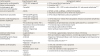
Table 1
List of sphingolipid drug or therapeutic agent in the various cardiomyopathies
| Cardiomyopathy | Drug/therapeutic agent | Sphingolipids targeted |
|---|---|---|
| Hypertrophic cardiomyopathy | - FTY720 (S1P antagonist) | ↓ S1P to reverse fibrotic hypertrophy169170171 |
| Dilated cardiomyopathy | - LVAD | ↑ Ceramide, DHS213214 |
| Arrhythmogenic right ventricular cardiomyopathy | - Carvedilol | ↓ Ceramide, ↓ S1P, ↑ CPT to reduce intracellular Ca2+ and prevent arrhythmias173174175 |
| Acute viral myocarditis | - Shenfu | ↓ SPTLC2, ↓ DHS, ↓ aSMase, ↓ d18:0/14:0 ceramide, ↓ d18:1/22:0 and d18:0/16:1 SM to inhibit viral replication35176 |
| Autoimmune myocarditis | - KRP-203 (S1PR agonist) | ↓ S1P to reduce inflammatory cells and cytokines177178 |
| - FTY720 (S1P antagonist) | ||
| Ischemic cardiomyopathy and ischemic reperfusion | - FTY720 (S1P antagonist) | - ↓ S1P to reduce Ca2+ release from sarcoplasmic reticulum and prevent arrhythmias |
| - LVAD | - ↑ Ceramide, DHS | |
| - Preconditioning mimetic TNF-α | - Mediated by cell-permeable (C2) ceramide, SphK1 and S1P to ↓ ceramide144213214215 | |
| Gaucher disease cardiomyopathy | - Recombinant β-lucocerebrosidase | - ↑ Functional β-glucocerebrosidase enzyme |
| - Eliglustat tartrate | - Ceramide glucosyltransferase inhibitor | |
| - Zavesca® imiglucerase | - GlcCer synthase enzyme inhibitor182183184188189190198 | |
| - Ambroxol | ||
| Anderson-Fabry disease cardiomyopathy | - α-galactosidase A | - ↑ Functional α-galactosidase A enzyme to reduce LV thickness |
| - Migalastat | - Binds and stabilizes α-galactosidase A, ceramide glucosyltransferase inhibitor191192193194195196197 | |
| Sandhoff/Tay-Sachs disease cardiomyopathy | - Ambroxol | - Chaperone of β-hexosaminidase A and improves autophagy by restoring lysosomal calcium release |
| - Pyrimethamine | - Improves cardiac function201202203 | |
| - Migalastat + ketogenic diet | ||
| Diabetic cardiomyopathy | - Fumonisin B1 | - ↓ DHC and ceramide to reduce apoptosis |
| - Fenofibrate | - ↓ Deoxysphingolipids | |
| - Restricted caloric intake | - ↓ DHC and ceramide to reduce apoptosis1162204 | |
| Hypertensive cardiomyopathy | - Myriocin | ↓ Ceramide levels to reduce blood pressure62106 |
| - Losartan | ||
| - Hydralazine |
S1P, sphingosine-1-phosphate; LVAD, left ventricular assist device; DHS, dihydrosphingosine; CPT, carnitine palmitoyltransferase; aSMase, acid sphingomyelinase; SM, sphingomyelin; TNF-α, tumor necrosis factor-α; SphK1, sphingosine kinase 1; GlcCer, glucosylceramide; LV, left ventricular; DHC, dihydroceramide.
Enzyme replacement or enhancement therapy has emerged as an effective treatment for the secondary cardiomyopathies observed as a result of lysosomal storage disorders, and is therefore a likely strategy for treating the resulting cardiomyopathies. Gaucher disease is treated by infusion of recombinant acid β-glucosidase or a ceramide glucosyltransferase inhibitor (eliglustat tartrate). Both treatments reversed the disease-related accumulation of complex sphingolipids in clinical trials.182183184 More recent technology has allowed for high-throughput screening to identify small-molecule therapeutics, such as chaperones to restore defective enzyme activity and compounds to clear accumulating substrates for many lysosomal storage diseases including Gaucher disease, Fabry disease, and gangliosidoses.185186187 As additional pharmaceutical agents, Zavesca® (miglustat), Cerezyme™ (imiglucerase), and VPRIV™ (velaglucerase) have been approved to treat all 3 types of Gaucher disease by reducing the accumulation of GlcCer through inhibition of GlcCer synthase.188189190 Anderson-Fabry disease has also been treated by replacement with recombinant α-galactosidase, which reduced LV wall thickness, improved regional myocardial function, and cleared microvascular endothelial deposits of globotriaosylceramide from the heart.191192193194195196 Another clinical trial evaluated the long-term effects of a small-molecule pharmacological chaperone, migalastat, that binds and stabilizes α-galactosidase A. Patients showed reduced cardiac mass and stable levels of globotriaosylceramides.197 Ambroxol is an FDA approved drug used to treat Gaucher and Tay-Sachs diseases, though whether it reduces cardiac symptoms has not yet been addressed.198199200 Pyrimethamine is another chaperone of β-hexosaminidase A in Tay-Sachs disease which mechanistically improves autophagy by restoring lysosomal calcium release.201202 Migalastat in combination with a ketogenic caloric restriction diet led to improved cardiac function and improved seizure control in a patient with Sandhoff disease, although the mechanism of action remains to be determined.203
Myriocin, a fungal toxin and specific inhibitor of SPT, has been shown to prevent cancer cell migration, insulin resistance, and cardiomyopathies in mouse models including SPT2 heterozygous mice, T2DM, hypertension, and atherosclerosis by reducing ceramide levels. However, myriocin has not yet been and is unlikely to be approved through clinical trials, as there are numerous toxic and off-target effects in humans and mice.174862204205206207 The ceramide synthase inhibitor fumonisin B1 (FB1), which inhibits ceramide synthases and thereby prevents synthesis of DHCs from DHS, has been shown to improve insulin sensitivity in rodents and isolated muscles that are lipid-infused; the effects of FB1 are mediated by decreased ceramide levels, which increase activation of PP2A and the PI3K/AKT signaling pathway. This inhibition attenuates apoptosis and other cellular signaling pathways observed in DbCM.62204 Fumonisin B1 has not been tested in a primary or secondary cardiomyopathic model, but it was shown that a specific ceramide synthase, CerS5, induced cardiomyocyte hypertrophy; therefore, targeting this enzyme could potentially have therapeutic benefits.51 To date, however, few inhibitors of specific ceramide synthases have been identified, and this is an area under intense investigation due to its potential benefits in these and other pathologies. In contrast to these largely deleterious roles of ceramide, it also exhibits antiproliferative effects that may be beneficial. In fact, stents have been developed that are coated with a cell-permeable Cer analogue, leading to reduced restenosis.208209210
Exogenously administered S1P accelerates neovascularization and blood flow recovery in ischemic limbs, suggesting its usefulness for angiogenic therapy. Baseline concentrations of S1P measured in peripheral blood samples in ischemic patients were more than 4-fold higher in patients with documented CAD undergoing a percutaneous coronary intervention than in healthy controls. By 5 minutes, coronary sinus and peripheral levels of S1P levels increased by 720% and 792%, respectively. Where troponin T was detectable at 12 hours, a strong correlation was found with peak S1P levels in ischemia.211 A recently patented approach assesses the total amount of SMs and SMase as a parameter to diagnose heart failure due to ICM or non-ischemic DCM.212 Left ventricular assist devices (LVADs), which provide mechanical support for advanced heart failure in patients with end-stage heart failure as a result of ICM or DCM, decreased the levels of numerous myocardial ceramide in patients after implantation compared to patients that did not receive an LVAD.213214
Since ceramide has been implicated in hERG and reactive oxygen species overproduction, as previously mentioned, ceramide may contribute to long QT syndrome and therefore could be targeted as a therapeutic strategy in this context.122123 Ischemic preconditioning (IPC) is an experimental technique used to produce resistance against acute IR injury, and may be regulated by ceramide, SphK1, and S1P.215 S1P is a mediator of IPC, as S1P binds S1PR2 and S1PR3 in myocardial ischemia, causing cardiomyopathy. These results provide evidence for S1P receptor subtype-specific pharmacological interventions as a novel therapeutic approach to myocardial diseases.216 In patients with ICM, S1P and sphingosine have been shown to be reliable predictors of CAD when a patient is undergoing coronary angiography.211217 Another potential therapeutic intervention is targeting aSMase, which hydrolyzes SM to generate ceramide, to reduce the cardiac production of ceramide in ischemic reperfusion after ICM.218 DbCM model animals fed with low amounts of unsaturated fatty acids and high amounts of saturated fatty acids, especially myristate (C14:0), had more severe phenotypes. This may contribute to the associations between saturated fatty acid consumption and cardiovascular disease, and furthermore suggests that changes in diet to reduce the conception of foods containing myristate and/or other medium-chain fatty acids would benefit patients.
The treatment of hypertension with losartan and/or hydralazine in spontaneously hypertensive rats significantly decreased blood pressure. This was associated with a concomitant lowering of vascular ceramide levels, although this most likely occurred due to decreased blood pressure and not the mechanisms of the drugs themselves.106
CONCLUSIONS
In this review, we highlighted the involvement of multiple sphingolipid species in various cardiomyopathies, including direct roles for sphingolipids in DCM, acute right ventricular cardiomyopathy, ICM with IR, AVMC, DbCM, GDC, FC, GM1 gangliosidosis GM1, both forms of GM2 gangliosidosis, DMD, BMD, DM1 and DM2, and cardiomyopathy in hyperthyroidism. In addition to these direct roles, the potential for identification of other direct roles is suggested by the numerous overlaps between signaling pathways known both to regulate cardiac pathophysiology and to be sphingolipid-regulated. This possibility may be relevant for conditions including HCM, Takotsubo disease with LQTS, TIC with LQTS syndrome, HTNCM, and cardiomyopathies in acute MC, PPCM, and Friedreich ataxia. To date, no sphingolipids have been associated with primary RCM, unclassified primary left ventricle non-compaction cardiomyopathy, and ICM without IR. Also discussed are the interventional studies of sphingolipids in various animal models, which show remarkable potential for various drugs, as well as the sphingolipid therapeutics currently in clinical trials or already approved for use in humans.
A major advance in understanding sphingolipid biology in general is the recent appreciation that chemically distinct sphingolipid species often have specific functions. While many studies have used genetic or pharmacological strategies to prevent or reduce sphingolipid synthesis in general, mass spectrometry-based strategies have enabled the discovery that different sphingoid bases (e.g., d16:0 vs. d18:0), distinct N-acyl chain lengths and degrees of saturation (e.g., C14:0 vs. C20:0, C18:1, C24:1, etc.), and other structural differences among sphingolipid species lead to distinct biological effects. Therefore, approaches taking these variations into account are leading to a better understanding of mechanisms underlying the pathology of cardiomyopathy, as well as providing more specificity in the identification of novel therapeutic targets.219
In summary, targeting enzymes involved in sphingolipid synthesis could have enormous—and thus far, untapped—therapeutic potential. Furthermore, the delivery of beneficial sphingolipids may also be feasible. For example, recently developed nano-liposomal lipid delivery systems are currently being used in a multitude of disease contexts.220221222 Further studies of sphingolipids in cardiac pathophysiology will undoubtedly continue to provide opportunities for novel therapeutic strategies.
 XML Download