

 PDF
PDF ePub
ePub Citation
 Print
Print


Abstract
Purpose
In this study, we evaluated changes in the anterior chamber structure and lens position before and after phacoemulsification in eyes grouped by axial length (AL).
Methods
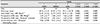
This study included 65 eyes (16 short eyes [AL < 22.5 mm], 33 normal eyes [22.5 mm < AL < 25.0 mm], and 16 long eyes [AL > 25.5 mm]) that underwent cataract surgery. Pre- and postoperative anterior chamber depth (ACD) was measured using Pentacam® and IOL Master®,. In addition, we evaluated the anterior chamber angle (ACA), anterior chamber volume (ACV), epithelium-iris distance, and iris-lens (intraocular lens [IOL]) distance.
Results
The change in ACD was significantly smaller in long eyes (Pentacam®,, p = 0.000; IOL Master®,, p = 0.001). The change in ACA was significantly larger in short eyes (p = 0.000), and the change in ACV was significantly smaller in long eyes (p = 0.000). The change in the epithelium–iris distance was significantly smaller in long eyes (p = 0.000), and the change in the iris-lens (IOL) distance was significantly smaller in short eyes (p = 0.000).
Conclusions
In short eyes, changes in ACD, ACA, and ACV were found to be larger than those of other groups as the iris moved backward. In long eyes, greater backward movement of the IOL was observed. Therefore, the appropriate IOL power should be chosen, considering the postoperative position of the IOL during cataract surgery of short and long eyes.
Figures and Tables
Figure 1
Parameters measured with the Scheimpflug images of Pentacam®,. (A) Anterior chamber depth (red line), distance from the anterior surface of the iris to the anterior surface of the crystalline lens (yellow line). (B) Distance from the anterior surface of the iris to the anterior surface of the intraocular lens (green line).
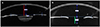
Figure 2
Associations between axial length and difference of anterior chamber parameters (ACD-Pentacam®, [A], ACD-IOL Master®, [B], ACA [C], ACV [D], Epithelium-Iris distance [E] and iris-lens [IOL] distance [F]). ACD = anterior chamber depth; ACA = anterior chamber angle; ACV = anterior chamber volume; IOL = intraocular lens. Statistically significant p-value <0.01 using Pearson's correlation analysis.
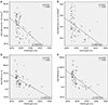
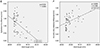
Figure 3
Changes in anteror segment before and after phacoemulsification in a short and a long eye, measured with Scheimpflug images of Pentacam®,. Change in anteror segment of a short eye (axial length [AL] 21.23), before phacoemulsification (A) and after phacoemulsificaion (B), change in anteror segment of a long eye (AL 27.34) before phacoemulsification (C) and after phacoemulsificaion (D).
References
1. Lee AC, Qazi MA, Pepose JS. Biometry and intraocular lens power calculation. Curr Opin Ophthalmol. 2008; 19:13–17.


2. Schröder S, Langenbucher A. Relationship between effective lens position and axial position of a thick intraocular lens. PLoS One. 2018; 13:e0198824.
3. Haigis W, Lege B, Miller N, Schneider B. Comparison of immersion ultrasound biometry and partial coherence interferometry for intraocular lens calculation according to Haigis. Graefes Arch Clin Exp Ophthalmol. 2000; 238:765–773.
4. Dinc UA, Gorgun E, Oncel B, et al. Assessment of anterior chamber depth using Visante optical coherence tomography, slitlamp optical coherence tomography, IOL Master, Pentacam and Orbscan IIz. Ophthalmologica. 2010; 224:341–346.
5. Shankar H, Taranath D, Santhirathelagan CT, Pesudovs K. Anterior segment biometry with the Pentacam: comprehensive assessment of repeatability of automated measurements. J Cataract Refract Surg. 2008; 34:103–113.
6. McAlinden C, Khadka J, Pesudovs K. A comprehensive evaluation of the precision (repeatability and reproducibility) of the Oculus Pentacam HR. Invest Ophthalmol Vis Sci. 2011; 52:7731–7737.
7. Sanders DR, Retzlaff J, Kraff MC. Comparison of the SRK II formula and other second generation formulas. J Cataract Refract Surg. 1988; 14:136–141.
8. Retzlaff JA, Sanders DR, Kraff MC. Development of the SRK/T intraocular lens implant power calculation formula. J Cataract Refract Surg. 1990; 16:333–340.
9. Holladay JT, Prager TC, Chandler TY, et al. A three-part system for refining intraocular lens power calculations. J Cataract Refract Surg. 1988; 14:17–24.
10. Olsen T. Calculation of intraocular lens power: a review. Acta Ophthalmol Scand. 2007; 85:472–485.
11. Kucumen RB, Yenerel NM, Gorgun E, et al. Anterior segment optical coherence tomography measurement of anterior chamber depth and angle changes after phacoemulsification and intraocular lens implantation. J Cataract Refract Surg. 2008; 34:1694–1698.
12. Nolan WP, See JL, Aung T, et al. Changes in angle configuration after phacoemulsification measured by anterior segment optical coherence tomography. J Glaucoma. 2008; 17:455–459.
13. Memarzadeh F, Tang M, Li Y, et al. Optical coherence tomography assessment of angle anatomy changes after cataract surgery. Am J Ophthalmol. 2007; 144:464–465.

14. Pereira FA, Cronemberger S. Ultrasound biomicroscopic study of anterior segment changes after phacoemulsification and foldable intraocular lens implantation. Ophthalmology. 2003; 110:1799–1806.
15. Abulafia A, Barrett GD, Kleinmann G, et al. Prediction of refractive outcomes with toric intraocular lens implantation. J Cataract Refract Surg. 2015; 41:936–944.
16. Ribeiro F, Castanheira-Dinis A, Dias JM. Refractive error assessment: influence of different optical elements and current limits of biometric techniques. J Refract Surg. 2013; 29:206–212.
17. Praveen MR, Shah GD, Vasavada AR, et al. A study to explore the risk factors for the early onset of cataract in India. Eye (Lond). 2010; 24:686–694.
18. Chang JS, Lau SY. Correlation between axial length and anterior chamber depth in normal eyes, long eyes, and extremely long eyes. Asia Pac J Ophthalmol (Phila). 2012; 1:213–215.
19. Sedaghat MR, Azimi A, Arasteh P, et al. The relationship between anterior chamber depth, axial length and intraocular lens power among candidates for cataract surgery. Electron Physician. 2016; 8:3127–3131.
20. Su PF, Lo AY, Hu CY, Chang SW. Anterior chamber depth measurement in phakic and pseudophakic eyes. Optom Vis Sci. 2008; 85:1193–1200.
21. Kim M, Park KH, Kim TW, Kim DM. Changes in anterior chamber configuration after cataract surgery as measured by anterior segment optical coherence tomography. Korean J Ophthalmol. 2011; 25:77–83.
22. Kasai K, Takahashi G, Kumegawa K, Dogru M. Measurement of early changes in anterior chamber morphology after cataract extraction measured by anterior segment optical coherence tomography. Graefes Arch Clin Exp Ophthalmol. 2015; 253:1751–1756.
23. Muzyka-Woźniak M, Ogar A. Anterior chamber depth and iris and lens position before and after phacoemulsification in eyes with a short or long axial length. J Cataract Refract Surg. 2016; 42:563–568.
24. Donoso R, Mura JJ, López M, Papic A. Emmetropization at cataract surgery. Looking for the best IOL power calculation formula according to the eye length. Arch Soc Esp Oftalmol. 2003; 78:477–480.
25. Yang S, Whang WJ, Joo CK. Effect of anterior chamber depth on the choice of intraocular lens calculation formula. PLoS One. 2017; 12:e0189868.
26. Ascaso FJ, Huerva V, Grzybowski A. Epidemiology, etiology, and prevention of late IOL-Capsular bag complex dislocation: review of the literature. J Ophthalmol. 2015; 2015:805706. .
 XML Download