

 PDF
PDF ePub
ePub Citation
 Print
Print


Abstract
Purpose
To evaluate clinical findings in phlyctenular keratoconjunctivitis patients and assess the function and morphology of Meibomian glands using an interferometer (LipiView®, TearScience, Morrisville, NC, USA) in such patients.
Methods
This retrospective study included 19 eyes of 13 patients diagnosed with phlyctenular keratoconjunctivitis. The lipid layer thickness (LLT) and meibograph of each eye was quantified by tear interferometry. Tear film break-up time (TBUT) and corneal staining score were measured. Meibomian gland morphology (lid margin vascularity, plugging of gland orifices, lid margin irregularity, lid margin thickening, and partial glands) was evaluated based on anterior photographs and meibographs.
Results
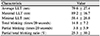
The mean age was 21.3 years (8–44 years). Mean BUT and Oxford corneal staining scores were 2.6 ± 1.2 seconds and 1.9 ± 0.8, respectively. Abnormal findings of the Meibomian glands were observed in all patients. The mean LLT was 79.6 ± 27.4 µm and the incomplete eye blinking frequency was 3.8 ± 5.9 during 20 seconds. The graphs of the tear lipid layer showed various patterns such as flat, up-hill, down-hill, and mixed.
Figures and Tables
Figure 1
Examples of graphs which tear film lipid layer thickness measured by Lipiview® (TearScience Inc., Morrisville, NC, USA). Flat type in case 3, up-hill type in case 10, down-hill type in case 13, mix type in case 5.
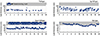
Figure 2
Initial presentation of a 10-year-old female patient (case 3). Telangiectasia, plugging of Meibomian gland orifices, and lid debris can be seen (A, B). The ocular surface is shown, accompanied by superficial neovascularization, corneal infiltration, and mild hyperemia of the bulbar conjunctiva in both eyes (C, D). Fluorescein staining shows superficial punctate keratitis on both corneas, along with infiltration (E, F).
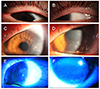
Figure 3
Images obtained by Lipiview® (TearScience Inc., Morrisville, NC, USA) interferometer in 8-year-old (case 1, A), 14-year-old (case 4, B), 10-year-old (case 3, C, D) and 25-year-old (case 10, E, F).Meibomian gland dilatation, partial gland and gland dropout were observed in younger patients.

References
1. Gary C. Phlyctenular keratoconjunctivitis and marginal staphylococcal keratitis. In : Krachmer J, Mannis M, Holland E, editors. Cornea. 3rd ed. St. Louis: Mosby;2010. p. 1143–1148.
2. Suzuki T, Mitsuishi Y, Sano Y, et al. Phlyctenular keratitis associated with meibomitis in young patients. Am J Ophthalmol. 2005; 140:77–82.


3. Smolin G, Okumoto M. Staphylococcal blepharitis. Arch Ophthalmol. 1977; 95:812–816.
4. Suzuki T, Sano Y, Sasaki O, Kinoshita S. Ocular surface inflammation induced by Propionibacterium acnes. Cornea. 2002; 21:812–817.
5. Suzuki T. Meibomitis-related keratoconjunctivitis: implications and clinical significance of meibomian gland inflammation. Cornea. 2012; 31:S41–S44.
6. Farpour B, McClellan KA. Diagnosis and management of chronic blepharokeratoconjunctivitis in children. J Pediatr Ophthalmol Strabismus. 2001; 38:207–212.
7. Suzuki T, Morishige N, Arita R, et al. Morphological changes in the meibomian glands of patients with phlyctenular keratitis: a multicenter cross-sectional study. BMC ophthalmol. 2016; 16:178.

8. Isreb M, Greiner J, Korb D, et al. Correlation of lipid layer thickness measurements with fluorescein tear film break-up time and Schirmer's test. Eye (Lond). 2003; 17:79–83.
9. Olson MC, Korb DR, Greiner JV. Increase in tear film lipid layer thickness following treatment with warm compresses in patients with meibomian gland dysfunction. Eye Contact Lens. 2003; 29:96–99.
10. Eom Y, Lee JS, Kang SY, et al. Correlation between quantitative measurements of tear film lipid layer thickness and meibomian gland loss in patients with obstructive meibomian gland dysfunction and normal controls. Am J Ophthalmol. 2013; 155:1104–1110.e2.
11. Mitra M, Menon G, Casini A, et al. Tear film lipid layer thickness and ocular comfort after meibomian therapy via latent heat with a novel device in normal subjects. Eye (Lond). 2005; 19:657–660.
12. Craig JP, Tomlinson A. Importance of the lipid layer in human tear film stability and evaporation. Optom Vis Sci. 1997; 74:8–13.
13. Arita R, Minoura I, Morishige N, et al. Development of definitive and reliable grading scales for meibomian gland dysfunction. Am J Ophthalmol. 2016; 169:125–137.
14. Jo DH, Kim MK, Wee WR, Lee JH. Analysis of clinical characteristics in phlyctenular keratoconjunctivitis at a tertiary center. J Korean Ophthalmol Soc. 2011; 52:7–13.
15. Suzuki T, Kinoshita Y, Tachibana M, et al. Expression of sex steroid hormone receptors in human cornea. Curr Eye Res. 2001; 22:28–33.
16. Peng CC, Cerretani C, Braun RJ, Radke C. Evaporation-driven instability of the precorneal tear film. Adv Colloid Interface Sci. 2014; 206:250–264.
17. Bron AJ, Tomlinson A, Foulks GN, et al. Rethinking dry eye disease: a perspective on clinical implications. Ocul Surf. 2014; 12:2 Suppl. S1–S31.
18. Foulks GN. The correlation between the tear film lipid layer and dry eye disease. Surv Ophthalmol. 2007; 52:369–374.
19. Finis D, Pischel N, Schrader S, Geerling G. Evaluation of lipid layer thickness measurement of the tear film as a diagnostic tool for meibomian gland dysfunction. Cornea. 2013; 32:1549–1553.
20. Arita R, ltoh K, Inoue K, Amano S. Noncontact infrared meibography to document age-related changes of the meibomian glands ina normal population. Ophthalmology. 2008; 115:911–915.
21. Yin Y, Gong L. The evaluation of meibomian gland function, morphology and related medical history in Asian adult blepharokeratoconjunctivitis patients. Acta Ophthalmol. 2017; 95:634–638.
22. Den S, Shimizu K, Ikeda T, et al. Association between meibomiangland changes and aging, sex, or tear function. Cornea. 2006; 25:651–655.
23. Ozdemir M, Temizdemir H. Age-and gender-related tear function changes in normal population. Eye (Lond). 2010; 24:79–83.
24. Maïssa C, Guillon M. Tear film dynamics and lipid layer characteristics--effect of age and gender. Cont Lens Anterior Eye. 2010; 33:176–182.
25. Yin Y, Gong L. Reversibility of gland dropout and significance of eyelid hygiene treatment in meibomian gland dysfunction. Cornea. 2017; 36:332–337.
26. Mizoguchi T, Arita R, Fukuoka S, Morishige N. Morphology and function of meibomian glands and other tear film parameters in junior high school students. Cornea. 2017; 36:922–926.
27. Tsubota K, Yokoi N, Shimazaki J, et al. New perspectives on dry eye definition and diagnosis: a consensus report by the Asia Dry Eye Society. Ocul Surf. 2017; 15:65–76.
28. King-Smith PE, Reuter KS, Braun RJ, et al. Tear film breakup and structure studied by simultaneous video recording of fluorescence and tear film lipid layer images. Invest Ophthalmol Vis Sci. 2013; 54:4900–4909.
29. Arita R, Morishige N, Fujii T, et al. Tear interferometric patterns reflect clinical tear dynamics in dry eye patients. Invest Ophthalmol Vis Sci. 2016; 57:3928–3934.
30. Hirota M, Uozato H, Kawamorita T, et al. Effect of incomplete blinking on tear film stability. Optom Vis Sci. 2013; 90:650–657.
31. Jie Y, Sella R, Feng J, et al. Evaluation of incomplete blinking as a measurement of dry eye disease. Ocul Surf. 2019; 17:440–446.
32. Culbertson WW, Huang AJ, Mandelbaum SH, et al. Effective treatment of phlyctenular keratoconjunctivitis with oral tetracycline. Ophthalmology. 1993; 100:1358–1366.
 XML Download