

 PDF
PDF ePub
ePub Citation
 Print
Print


HIGHLIGHTS
• Induction therapy played a signifincant role in kidney transplantation improvement.
• Acute rejection and delayed graft functions are related to graft and recipent survival.
• Compared with basiliximab, rabbit anti-thymocyte globulin (rATG) is effective at lowering acute rejection and delayed graft function rates.
• Low-dose rATG may be considered an effective induction therapy in deceased donor kidney transplantation.
INTRODUCTION
In recent years, short- and long-term graft survival rates have improved for recipients of kidneys from both living and deceased donors [12]. Development of induction therapy played a significant role in this improvement, typically rabbit anti-thymocyte globulin (rATG) and basiliximab.
Because rATG targets multiple surface antigens on T-cells, its consumption of T-cells is more effective, as are other effects on NK cells and B-cells [34]. Basiliximab is a monoclonal antibody for the interleukin-2 receptor that can effectively inhibit T-cell proliferation and activation mediated by interleukin-2 [5]. Both have been shown to reduce the incidence of acute rejection (AR) and delayed graft function (DGF) after renal transplantation [678]. These two outcomes are important because in the posttransplantation period, AR significantly reduces long-term graft survival [9], and DGF may increase the incidence of AR, having negative effects on long-term graft and patient survival [1011].
Compared with basiliximab, rATG is effective at lowering AR and DGF rates, but studies have shown that the incidence of infection and malignancy is significantly higher in patients receiving rATG [12]. As a result, rATG and basiliximab have been actively studied, and both rATG dose and basiliximab efficacy are the subject of considerable controversy. Additionally, few studies on low-dose rATG have been published. The purpose of this study is to discuss effective induction treatment strategies through a multidisciplinary analysis of high-dose rATG, low-dose rATG, and basiliximab treatments in deceased donor kidney transplantation (DDKT) at Samsung Medical Center, Korea, from 2003–2016.
METHODS
Study Design
This retrospective, single-center study analyzed Samsung Medical Center's electronic medical records and kidney transplantation database. We screened 542 recipients who underwent DDKT between June 2003 and April 2006. We excluded 66 recipients who underwent kidney retransplantation, 26 who underwent multiorgan transplantation, and 22 pediatric recipients. In induction therapy, four no-induction recipients, 21 added rituximab recipients, and eight alemtuzumab recipients were excluded. The remaining 395 recipients were divided into three groups. Group 1 (n=184) received basiliximab, group 2 (n=98) received high-dose rATG, and group 3 (n=113) received low-dose rATG. This study protocol was reviewed and approved by the Institutional Review Board of Samsung Medical Center, Sungkyunkwan University School of Medicine (IRB No. SMC2019-05-057).
Induction Therapy
For their induction-immunosuppressive agents, recipients in group 1 received an interleukin-2 receptor antagonist induction (Basiliximab, Novartis Pharmaceuticals, Basel, Switzerland; 20 mg/kg, two doses on days 0 and 4), recipients in group 3 received rATG (Genzyme, Cambridge, MA, USA; 1.5 mg/kg, three doses on days 0, 1, and 2), and those in group 2 received rATG for more than 4 days. Standard criteria donor recipients received basiliximab, and expanded criteria donor recipients received rATG. Before 2007, most recipients belonged to group 2, contrary to post 2007 when most belonged to group 3.
Maintenance Therapy
When oral intake was possible, all recipients were initiated on tacrolimus (Prograf, Astellas Pharma US, Deerfield, IL, USA; 0.1 mg/kg/day with a target trough level of 8–10 ng/mL until 1 month after surgery and then at 5–8 ng/mL) and mycophenolate (CellCept, Roche Laboratories, Nutley, NJ, USA; 750 mg twice daily) at the time of admission. All recipients were given 500 mg of intravenous methylprednisolone during the operation until postoperative day 2, followed by a tapered dose of 60 mg per day for 5 days and prednisolone 8 mg, twice per day, for a month (starting on postoperative day 8). Then, recipients received 4 mg of methylprednisolone twice daily for 2 months.
Infection Prophylaxis and Monitoring
All recipients received itraconazole and Bactrim for prophylaxis against fungi and Pneumocystis jirovecii. Cefotaxime was used for bacterial prophylaxis until postoperative day 2. Cytomegalovirus (CMV) antigenemia test was started in March 1997 and the BKV PCR test was started in October 2007 in our center. When a donor was positive for CMV immunoglobulin G but the recipient was negative, the recipient was intravenously administered ganciclovir for 2 weeks while in the hospital and then valganciclovir for 10 weeks for insurance coverage. In the rATG groups, ganciclovir was administered as a CMV prophylaxis for 2 weeks. Otherwise, we followed a preemptive treatment strategy. CMV infection was monitored weekly in the first postoperative month and monthly thereafter via the CMV antigenemia test. When patients had a viral count above 50/400,000 white blood cells or had confirmed tissue-invasive CMV disease, intravenous ganciclovir was used as preemptive therapy. Polyomavirus BK virus (BKV) infection was monitored weekly in the first postoperative month, and then monthly via urine cytology or BKV DNA detection using the polymerase chain reaction. If urine BK virus DNA was >107 copies/mL, blood BKV DNA load was assessed regularly. If the blood BKV DNA load was >104 copies/mL with serum creatinine (Cr) elevation, we performed allograft biopsy. If BKV replication disappeared from the blood, urine BKV DNA level was used for follow-up [13].
End Points
The primary end points were rates of graft failure, patient survival, AR, and DGF. DGF refers to the acute kidney injury that occurs in the first week of kidney transplantation, which necessitates dialysis. AR is defined according to the Banff classification and is determined by a pathologist. Acute cellular rejection and antibody mediated rejection can be classified by histological examination, but in this study, they didn't need to be distinguished. The secondary end points were CMV infection, BKV infection, and other infections. Graft function represented by serum Cr level and the estimated glomerulus filtration rate (eGFR) of the three groups were also compared.
Statistical Analysis
Differences among the three groups were analyzed using Fisher's exact test for categorical variables and Kruskal-Wallis test for continuous variables. Post-hoc analyses were also performed. Pairwise comparisons between groups were performed using Fisher's exact test for categorical variables and a Wilcoxon rank-sum test for continuous variables. Graft and patient survival rates were obtained by Kaplan-Meier analysis. Risk-factor analysis was performed by Cox proportional-hazards regression analysis and logistic regression analysis. Variables with a P-value <0.1 in the univariate analysis were included in the multivariable analysis. The generalized estimating equation was applied to analyze repeated measurements for serum Cr and eGFR levels. Statistical analysis was executed using SAS ver. 9.4 (SAS Institute Inc., Cary, NC, USA).
RESULTS
Recipient and Donor Characteristics
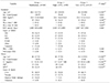
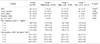
Recipient and donor characteristics are summarized in Table 1. The median recipient age and recipient diabetes mellitus proportion of group 3 were 56 years and 29.2%, respectively; the highest values among groups (P<0.001 and P=0.049, respectively). Human leukocyte antigen (HLA) class 1 and 2 mismatches were significantly different among the three groups (P=0.019 and P=0.007, respectively), with group 2 mismatches significantly higher than those of group 1 (P=0.012 and P=0.003, respectively). There was no significant difference in the other comparisons. The median age of donors in group 3 was 54 years, which was significantly higher than that of the other two groups (P<0.001). Group 2 and 3 serum Cr levels were 1.8 and 1.78 mg/dL, respectively, which were significantly higher than that of group 1 (1.1 mg/dL; P<0.001 and P<0.001, respectively).
Clinical Outcomes
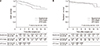
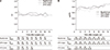
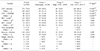
Graft function outcomes are summarized in Table 2. The DGF rate was lowest in group 1 (1.6%) and highest in group 3 (21.2%; P=0.004). However, there was no significant difference in DGF between group 2 and 3 (P=0.942). The graft failure rate was lowest in group 3 (4.4%) and highest in group 2 (27.6%; P=0.007), and it was not significantly different between group 3 and 1 (P=0.08). In a graft survival curve produced by Kaplan-Meier analysis, graft survival of group 3 and 1 was significantly higher than that of group 2 (group 3 vs. group 2: P=0.012, group 1 vs. group 2: P=0.043) (Fig. 1A). However, AR rate and recipient death were not statistically different in all three groups (Table 2, Fig. 1B). Graft function estimated using serum Cr level and eGFR is shown in Fig. 2. There was no significant difference between the three groups at any point in time. Infection Outcomes Infection outcomes were divided into posttransplant and within 1 year of posttransplant events. The results are summarized in Table 3. For CMV infection, the cases requiring preemptive therapy (viral count above 50/400,000 white blood cells) were further subdivided. In overall CMV infection, the infection rate was lowest in group 1 (59.2%) and highest in group 3 (88.5%; P<0.001). Within a year of the operation, the CMV infection rate in group 1 (51.1%) was statistically significantly lower than that in group 2 (67.3%) and 3 (87.6%; P<0.001 and P<0.001, respectively). The same results were obtained for CMV infection requiring treatment (CMV ≥ 50/400,000).
There were no statistically significant differences in BK virus, bacterial, and fungal infections among the three groups. In addition, viral, pneumocystis pneumonia, and tuberculosis infections were difficult to statistically analyze because of the small number of events.
DISCUSSION
While rATG is known to benefit from basiliximab at AR and DGF rates, basiliximab benefits from cancer and infection rates [1415]; thus, which drug is more useful remains a subject of discussion. Our study attempted to explore effective induction treatments by analyzing graft survival, recipient survival, AR rate, DGF rate, and infection rates.
DGF is related to graft survival, and its risk factors include obesity, high donor serum Cr level, a positive panel reactive antibody (PRA) test, old donor, old recipient, and long cold-ischemia time [161718]. In a study by Chen et al. [14], low-dose rATG (1 mg/kg on days 0, 1, and 2) significantly reduced the rates of DGF and AR compared with basiliximab in high-risk recipients (DGF, P=0.035; AR, P=0.004). Gavela et al. [19] reported that AR rate was significantly reduced in a low-dose rATG group (1.25 mg/kg on days 0 and 2) compared with a basiliximab group (P<0.001). The DGF rate was not statistically significant in older donors (P=0.08), but the authors suspected that low-dose rATG could reduce the DGF rate (low-dose rATG group, 33%; basiliximab group, 55.6%). In our study, group 1 had a lower rate of DGF than the other groups, and there was no significant difference in AR rate. This may be related to recipient and donor characteristics. In the studies mentioned above, there was no statistically significant difference in recipient and donor characteristics between groups. However, in our study, there were differences in recipient and donor characteristics between groups. Compared to group 1, group 3 had older recipients (P<0.001) and donors (P<0.001), and higher donor serum Cr levels (P<0.001). Moreover, according to our riskfactor analysis, donor serum Cr level was the risk factor for DGF (P<0.001), and donor age was the risk factor for AR (P=0.029) (Supplementary Tables 1 and 2). Therefore, our results were different those of previous studies.
In graft failure, the rate of group 2 was higher than that of group 1 (P=0.040). Based on the characteristics of our study, we predicted that donor serum Cr level and HLA mismatches were related, and some studies have found that donor serum Cr level and HLA mismatches can affect graft failure [2021]. However, in our graft failure risk factor analysis, neither donor serum Cr level nor HLA mismatches was statistically significant (serum Cr, P=0.318; HLA class 1 mismatch, P=0.504; HLA class 2 mismatch, P=0.390) (Supplementary Table 3). Group 3 donors were older and had higher donor serum Cr levels than the other groups. Additionally, donor age was a significant risk factor, according to graft failure risk factor analysis (P<0.001) (Supplementary Table 3). Group 3 also had older recipients and a relatively large number of diabetic patients (recipient age, P<0.001; diabetes, P=0.049). However, its graft failure rate (4.4%) was significantly lower than that of group 2 (27.6%; P=0.004) and lower than that of group 1 (12.5%), although there was no significant difference between the two groups (P=0.080). Although group 3 had a short-term follow-up, which made predictions difficult, it is expected that its recipients will experience better graft survival than those of group 2, and will be comparable to those of group 1 at long-term follow-up (Fig. 1).
De novo donor-specific antibody (DSA) production after kidney transplantation is a risk factor for AR and graft failure [2223], and rATG is known to produce less de novo DSA than basiliximab [24]. However, in our study, there was no significant difference in DSA incidence among the three groups (P=0.802). The risk factors for de novo DSA are retransplantations, preformed DSA, and higher PRA [25]. In our study, patients with kidney retransplantation were excluded, the preformed DSA recipient rate was only 3.4% (group 1, 2.81%; group 2, 2.2%; and group 3, 5.45%), and the PRA >50% recipient rate was only 6.3% (group 1, 4.5%; group 2, 6.5%; and group 3, 9.2%). Recipients in the Brokhof et al.'s study [24] were moderately sensitized recipients, while most of ours were low-risk recipients. We therefore concluded that the low possibility of production of de novo DSA did affect the incidence of de novo DSA in the three groups.
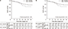
A previous study reported that CMV infection directly or indirectly affects graft survival and recipient survival [26], and another study suggests that CMV infection is an independent risk factor for AR [27]. However, in the current era of prophylaxis/preemptive antiviral treatment, factors other than CMV infection are known to be related to long-term graft and recipient survival [28]. In our study, the rATG groups had significantly higher CMV infection rates than the basiliximab group (P<0.001), especially the low-dose rATG group (P=0.001). However, similar to a recent study, our study shows that graft survival rates were not significantly different according to CMV infection that required preemptive ganciclovir treatment (CMV ≥50/400,000) (Fig. 3).
There are some limitations to our study. As a retrospective study, patients were not randomly assigned to groups. As rATG is frequently used in high-risk patients, selection bias was unavoidable. The short-term follow-up of the low-dose rATG group limited accurate analysis. Our study has some strengths as well. While most studies have compared two groups (basiliximab vs. low-dose rATG, high-dose rATG vs. low-dose rATG), ours compared three. We also analyzed various complications of kidney transplantation. An additional prospective and randomized study may reveal the utility of induction therapy.
Graft survival and patient survival rates of the low-dose rATG group were comparable with those of the basiliximab group even though the donors were older and had higher serum Cr levels, and the recipients in the low-dose rATG group were older and more likely to have diabetes. The CMV infection rate of the low-dose rATG group was higher than that of the basiliximab group. However, with ganciclovir preemptive treatment, CMV infection did not influence graft outcome. Therefore, low-dose rATG may be considered an effective induction therapy.
 XML Download