Journal List > Korean J Transplant > v.33(4) > 1141367

Kim: Macrophages in xenotransplantation

Abstract

Xenotransplantation refers to organ transplantation across species. Immune rejection of xenografts is stronger and faster than that of allografts because of significant molecular differences between species. Recent studies have revealed the involvement of macrophages in xenograft and allograft rejections. Macrophages have been shown to play a critical role in inflammation, coagulation, and phagocytosis in xenograft rejection. This review presents a recent understanding of the role of macrophages in xenograft rejection and possible strategies to control macrophage-mediated xenograft rejection.

HIGHLIGHTS

• Damage-associated molecular pattern (DAMP) release during ischemia reperfusion injury is one of the main causes of activation of macrophages, which play a critical role in inflammation and coagulation in xenograft rejection.
• Cross-talk between macrophages, hepatocytes, and vascular endothelial cells by producing immune mediators, such as monocyte chemoattractant protein 1 (MCP-1), interleukin (IL)-6, and creactive protein (CRP) may play a critical role in inflammatory responses and coagulation in pig-to-baboon organ transplantation.
• Early generation of MCP-1, IL-6, and CRP as well as DAMPs needs to be controlled to avoid inflammation and coagulation.

INTRODUCTION

Xenotransplantation is the transplantation of organs, tissues, or cells across species. Pigs are considered to be an ideal organ source for xenotransplantation due to their physiological similarity to humans and the feasibility of pig breeding. However, immune rejection of xenografts is believed to be stronger and faster than that of allografts. This can be attributed to the significant molecular differences between species. Xenograft rejection can be temporally divided into four interlinked immune rejection types: hyperacute rejection (HAR), acute vascular and cellular rejections, and chronic rejection [1]. HAR is mediated by human antibodies against carbohydrate moieties, such as galactose α1,3-galactose (α-gal) and N-glycolylneuraminic acid, present on the surfaces of pig endothelial cells [2]. Binding of pre-existing antibodies to these antigens and subsequent complement activation destroy pig endothelial cells, resulting in xenograft rejection within a few minutes [3]. During the last two decades, outcomes of pig-to-non-human primate organ transplantation have been markedly improved owing to development of genetically-engineered pigs; transplants from such pigs help avoid HAR [4]. However, many aspects of immune responses to xenografts still pose a critical problem for successful transplantation.
Macrophages are phagocytic innate immune cells that play a crucial role in host defense. Recent studies have revealed the involvement of macrophages in immune rejection of organ transplants. In animal allotransplant models, macrophages recognize allogeneic antigens, induce immune responses, and thus contribute to graft rejection [56]. In addition, they are able to kill allogeneic cells by phagocytosis [7]. Clinical studies exhibit a positive correlation between macrophage infiltration and graft rejection [891011]. The recently arising role of macrophages in allograft rejection must be one of the interesting topics in the field of transplant immunology. Therefore, the purpose of this paper is to review the recent understanding of the role of macrophages in xenograft rejection and possible strategies to control macrophage-mediated xenograft rejection.

ROLE OF MACROPHAGES IN XENOGRAFT REJECTION

Inflammation and Coagulation

Inflammation, triggered by innate immune cells as a defense mechanism against infectious agents or tissue damage, is a major problem in organ transplantation. Damaged host cells release or secrete various damage-associated molecular patterns (DAMPs) [12]. In organ transplantation, DAMP release from injured tissues is inevitable during ischemia reperfusion injury (IRI) and levels of the released DAMPs increase following IRI. DAMPs are recognized by cellular receptors of various cell types including, the Toll-like receptors (TLRs) of macrophages, and the inflammasomes, which trigger intracellular signaling that leads to inflammatory responses and subsequent initiation of rejection of the transplanted organ [56]. As shown in Table 1, IRI-induced release of various DAMPs, such as high-mobility group box 1 (HMGB1) [13141516], nuclear DNAs or mitochondrial DNAs [171819], heat shock proteins (HSPs) [20212223], and histones [24], was detected in different types of solid organs after transplantation.
Unlike other DAMPs, adenosine triphosphate (ATP) is a relatively small molecule and is recognized by specific cell surface receptors, such as P2X and P2Y. Binding of extracellular ATP to these receptors induces inflammatory responses of macrophages [25]. A study using a murine liver allotransplantation model has suggested extracellular ATP involvement in increased graft dysfunction and the involvement of the reduction of regulatory T cell frequency in overall graft survival [26].
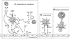
The role of the coagulation pathway in IRI and its crosstalk with the inflammatory pathway have been recently proposed [27]. Tissue factor (TF), which is the primary initiator of coagulation and is expressed on both, monocytes/macrophages and endothelial cells, is a central player in providing a bridge between these pathways [28]. Macrophages play a critical role in coagulation as well as inflammation in xenograft rejection (Fig. 1). During IRI, DAMPs activate monocytes/macrophages, which then, produce proinflammatory cytokines. In response to DAMPs and the pro-inflammatory cytokines, TF is rapidly induced by these cells in the graft recipients, and becomes exposed to blood [2930]. Cell surface TF can complex with factor VIIa, and thereby trigger coagulation by activating factor X and subsequent coagulating factors. TF can also induce protease-activated receptor-mediated signaling, which leads to the production of more inflammatory cytokines, upregulation of adhesion molecules, and suppression of thrombomodulin (Fig. 1A) [31].
One of the reasons for the stronger immune rejection of xenografts than that of allografts is associated with vascular endothelial cells. In the early phase of organ xenotransplantation, xenoantigen-specific innate antibodies can bind to and activate the endothelial cells, causing the release of proinflammatory immune mediators, as well as the activation of the complement mediated destruction of the endothelial layer. Monokines, such as interleukin (IL)-1β, IL-6, and tumor necrosis factor-α, secreted by monocytes/macrophages, can also activate vascular endothelial cells across pig and human species [32]. Activated endothelial cells release CD62, von Willebrand Factor, platelet factor 4, and CD40 ligand (Fig. 1A) in addition to proinflammatory cytokines. All these mediators eventually lead to the recruitment of more inflammatory cells and activation of coagulation [31]. The interplay between the inflammatory responses and the coagulation system plays a significant role in xenograft rejection [33343536].

Phagocytosis

The mechanism by which macrophages distinguish between self and allogeneic non-self organs, tissues, cells, or antigens and promote organ rejection has been recently clarified [37]. Mice that lack T, B, and natural killer cells could distinguish allogeneic antigens from those of self-tissues and induce an innate response. This innate allo-activation is triggered by mismatch between donor and recipient signal regulatory protein α (SIRPα), which is a cell surface molecule interacting with CD47. Similarly, macrophages are able to recognize and destroy xenografts through their cell surface interactions between CD47 and SIRP 1α (Fig. 1B). Due to their molecular incompatibility, an impaired interaction between pig CD47 and human SIRP 1α can result in the phagocytic killing of pig endothelial cells by human macrophages [38]. However, pig hematopoietic cells expressing human CD47 could be protected from phagocytic killing by human macrophages in hematopoietic cell engraftment experiments [41].

Chemotaxis and Acute Phase Responses

Macrophages are the major cells infiltrating into an allograft during severe rejection [42]. Similarly, macrophage infiltration occurs just after IRI of a xenograft and persists until graft rejection [4344]. The infiltration level of macrophages was significantly higher in α-gal knockout xenogeneic islets than in allogeneic islets [45]. The mechanism of monocyte accumulation within a xenograft is thought to be associated with the production of chemokines, such as monocyte chemoattractant protein 1 (MCP-1), in the graft [3546].
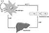
In pig-to-baboon heart and kidney transplantation, it was observed that early elevated serum levels of MCP-1, IL-6, and C-reactive protein (CRP), which is an acute phase protein synthesized by hepatocytes in response to proinflammatory cytokines [47], precede consumptive coagulopathy [35]. In addition, increased numbers of monocytes were associated with enhanced expression of TF [35]. These results, taken together with those of previous reports indicate that IL-6 provokes liver cells to produce CRP [48], which stimulates endothelial cells to produce MCP-1 [49], and both IL-6 [50] and CRP [51] promote TF expression, suggesting the occurrence of cross-talk between macrophages, hepatocytes, and vascular endothelial cells through the production of these mediators. MCP-1, produced by the activated vascular endothelial cells, recruit monocytes/macrophages, which are eventually activated to produce IL-6, which stimulates liver cells to release CRP, which in turn activates vascular endothelial cells. This positive feedback loop may play a critical role in inflammatory responses and coagulation in pig-to-baboon organ transplantation (Fig. 2). Therefore, early upregulation of CRP, IL-6, and MCP-1 levels needs to be suppressed to avoid inflammation-induced coagulopathy.

STRATEGIES TO CONTROL MACROPHAGE-MEDIATED XENOGRAFT REJECTION

A recent study has suggested that there is increasing evidence for sustained inflammatory response in pig-to-baboon xenograft recipients, and this systemic inflammation is a critical hurdle for successful xenotransplantation [52]. Therefore, therapeutic prevention of inflammation is necessary to achieve successful pig organ xenotransplantation.

Anti-inflammatory Drugs

A commonly used immunosuppressive regimen in recent studies of pig-to-non-human primate xenotransplantation includes antithymocyte globulin, cobra venom factor (CVF), corticosteroids, and an immunophilin inhibitor, such as cyclosporine A, tacrolimus, or rapamycin. In addition to this regimen, several other selective biological drugs aimed at suppressing immune responses after xenotransplantation have been tested in separate studies, such as anti-CD154 monoclonal antibody (mAb), CTLA-4 fusion proteins, anti-CD40 mAb, anti-CD20 mAb, anti-CD25 mAb, anti-IL-6 receptor mAb, and anti-LFA1 mAb [45,53]. Among them, mAbs for blocking the interaction between CD40 and CD154, and CTLA-4 fusion protein appear to be essential for achieving long term xenograft survival. CD40–CD154 interactions stimulate the inflammatory response. CD40 expression is induced in activated monocytes/macrophages while CD154 is also expressed on monocytes/macrophages during inflammation [54]. Blockade of the IL-6 receptor with the anti-IL-6 receptor mAb, tocilizumab, resulted in a reduction in the levels of CRP [36] and serum histones [55] upon pig-to-non-human primate xenotransplantation.
Other inflammatory drugs were also tested. Nuclear factor kappa B inhibitor, parthenolide, significantly suppressed histone-induced pig endothelial cell death in in vitro study [55]. CVF, which had been originally used to deplete complements causing HAR following xenotransplantation, was found to reverse the increased IL-6 and MCP-1 levels in pig-to-baboon heart and artery patch transplantation [56].
Since the effective treatment of an established inflammatory response to DAMPs is relatively difficult, selective and rapid blocking or scavenging of released DAMPs would be a more promising therapeutic strategy. Indeed, anti-histone therapy was found to prevent histone-induced inflammation in xenotransplantation [55]. Mice treated with HMGB1 antibody were protected against pulmonary dysfunction and had improved lung allograft outcomes [16]. Blockade of HMGB1 secretion by small molecule inhibitor was found to be beneficial to prevent the loss of islet grafts and to reverse diabetes in murine syngeneic islet transplantation [57]. Administration of ATP antagonist to a recipient mouse for 2 weeks led to prolonged survival of the transplanted allogeneic heart [58].
Laboratory studies have suggested possible manipulation of the inflammatory response by using a DAMP antigen rather than using an antibody or antagonist. Ischemic preconditioning with HMGB1 protected grafts from IRI through TLR4 signaling in renal and hepatic allotransplantation [5960]. In addition, genetic overexpression of HSP27 could reduce IRI-induced apoptosis of graft cells and delay the onset of acute rejection in murine heart allotransplantation [61].

Targeting Macrophages

Deletion or inhibition of macrophages can attenuate graft injury and prolong graft survival [62]. In recent animal and clinical studies, some macrophage subsets have been reported to act as regulatory cells, and the adoptive transfer of these macrophages significantly prolonged graft survival. A subset of macrophages was found to suppress allogeneic T cell proliferation and inhibit dendritic cell maturation [6364]. Furthermore, adoptive transfer of these macrophages promotes graft survival and minimizes immunosuppression [6465].
Although immunological memory has long been thought to be driven exclusively by adaptive immunity, new evidence suggests that various tissue-derived factors can induce epigenetic changes, leading to the formation of innate memory of macrophages [66]. Further understanding of the mechanisms of innate memory could allow us to dampen the response to DAMPs induced during the xenograft rejection.

CONCLUSIONS

Although T cells are major players of organ transplant rejection, early inflammation after transplantation is critical for T-cell mediated immune rejection. Thus, prevention of inflammation following transplantation might help avoid acute graft rejection. Monocytes/macrophages are critical immune mediators of inflammation, and thus are important targets for immune modulation. Owing to phenotypical and functional heterogeneity of macrophages, the identification and/or isolation of distinct subsets of macrophages involved in graft rejection or tolerance is essential to develop macrophage-based therapeutic strategies. Compared to allotransplantation, xenotransplantation has some advantages, including feasibility of genetic modification and preconditioning for transplantation. Thus, understanding precise mechanisms of macrophage-mediated immune responses in organ xenotransplantation will enable establishment of strategies to modulate macrophage function, which can improve the outcomes of xenotransplantation in future clinical practice.

Figures and Tables

Fig. 1

(A–C) Central role of activated macrophages in inflammation, coagulation, phagocytosis, and antigen presentation [31]. TNF, tumor necros; IL, interleukin; MCP-1, monocyte chemoattractant protein 1; DAMP, damage-associated molecular pattern; TLR, Toll-like receptor; TF, tissue factor; PLT, platelet; VWF, von Willebrand factor; PF4, platelet factor 4; TM, thrombomodulin; PAR, protease-activated receptor; ICAM-1, intercellular adhesion molecule-1; VCAM-1, vascular cell adhesion molecule-1; SIRPα, signal regulatory protein α.

kjt-33-74-g001
Fig. 2

Possible cross-talk between macrophages, hepatocytes, and vascular endothelial cells through the production of interleukin (IL)-6, monocyte chemoattractant protein 1 (MCP-1), and C-reactive protein (CRP) in inflammatory responses and coagulation in pig-to-baboon organ transplantation. TF, tissue factor.

kjt-33-74-g002
Table 1

DAMPs identified in inflammatory diseases and upon clinical organ transplantation [3940]

kjt-33-74-i001

DAMP, damage-associated molecular pattern; K, kidney; H, heart; Lu, lung; ATP, adenosine triphosphate; Li, liver; HSP, heat shock protein; IL, interleukin; mtDNA, mitochondrial DNA; mtROS, mitochondrial reactive oxygen species; mtTFA, mitochondrial transcription factor A; EDN, eosinophil-derived neurotoxin.

a)Designated organs where indicated DAMPs have been identified and studied in clinical solid organ transplantation.

Notes

Conflict of Interest No potential conflict of interest relevant to this article was reported.

Funding/Support This study was supported by research grant from the Korean Society for Transplantation (2019-04-03001-002).

References

1. Samstein B, Platt JL. Physiologic and immunologic hurdles to xenotransplantation. J Am Soc Nephrol. 2001; 12:182–193.
crossref
2. Galili U. The alpha-gal epitope and the anti-Gal antibody in xenotransplantation and in cancer immunotherapy. Immunol Cell Biol. 2005; 83:674–686.
crossref
3. Cooper DK, Ekser B, Tector AJ. Immunobiological barriers to xenotransplantation. Int J Surg. 2015; 23(Pt B):211–216.
crossref
4. Denner J. Xenotransplantation-progress and problems: a review. J Transplant Technol Res. 2014; 4:1000133.
crossref
5. Zecher D, van Rooijen N, Rothstein DM, Shlomchik WD, Lakkis FG. An innate response to allogeneic nonself mediated by monocytes. J Immunol. 2009; 183:7810–7816.
crossref
6. Oberbarnscheidt MH, Zeng Q, Li Q, Dai H, Williams AL, Shlomchik WD, et al. Non-self recognition by monocytes initiates allograft rejection. J Clin Invest. 2014; 124:3579–3589.
crossref
7. Liu W, Xiao X, Demirci G, Madsen J, Li XC. Innate NK cells and macrophages recognize and reject allogeneic nonself in vivo via different mechanisms. J Immunol. 2012; 188:2703–2711.
crossref
8. Matheson PJ, Dittmer ID, Beaumont BW, Merrilees MJ, Pilmore HL. The macrophage is the predominant inflammatory cell in renal allograft intimal arteritis. Transplantation. 2005; 79:1658–1662.
crossref
9. Xu L, Collins J, Drachenberg C, Kukuruga D, Burke A. Increased macrophage density of cardiac allograft biopsies is associated with antibody-mediated rejection and alloantibodies to HLA antigens. Clin Transplant. 2014; 28:554–560.
crossref
10. Bergler T, Jung B, Bourier F, Kühne L, Banas MC, Rümmele P, et al. Infiltration of macrophages correlates with severity of allograft rejection and outcome in human kidney transplantation. PLoS One. 2016; 11:e0156900.
crossref
11. Bräsen JH, Khalifa A, Schmitz J, Dai W, Einecke G, Schwarz A, et al. Macrophage density in early surveillance biopsies predicts future renal transplant function. Kidney Int. 2017; 92:479–489.
crossref
12. Matzinger P. Tolerance, danger, and the extended family. Annu Rev Immunol. 1994; 12:991–1045.
crossref
13. Wu H, Chen G, Wyburn KR, Yin J, Bertolino P, Eris JM, et al. TLR4 activation mediates kidney ischemia/reperfusion injury. J Clin Invest. 2007; 117:2847–2859.
crossref
14. Kaczorowski DJ, Nakao A, Vallabhaneni R, Mollen KP, Sugimoto R, Kohmoto J, et al. Mechanisms of Toll-like receptor 4 (TLR4)-mediated inflammation after cold ischemia/reperfusion in the heart. Transplantation. 2009; 87:1455–1463.
crossref
15. Yang Z, Deng Y, Su D, Tian J, Gao Y, He Z, et al. TLR4 as receptor for HMGB1-mediated acute lung injury after liver ischemia/reperfusion injury. Lab Invest. 2013; 93:792–800.
crossref
16. Weber DJ, Gracon AS, Ripsch MS, Fisher AJ, Cheon BM, Pandya PH, et al. The HMGB1-RAGE axis mediates traumatic brain injury-induced pulmonary dysfunction in lung transplantation. Sci Transl Med. 2014; 6:252ra124.
crossref
17. Hu Q, Wood CR, Cimen S, Venkatachalam AB, Alwayn IP. Mitochondrial damage-associated molecular patterns (MTDs) are released during hepatic ischemia reperfusion and induce inflammatory responses. PLoS One. 2015; 10:e0140105.
crossref
18. De Vlaminck I, Martin L, Kertesz M, Patel K, Kowarsky M, Strehl C, et al. Noninvasive monitoring of infection and rejection after lung transplantation. Proc Natl Acad Sci U S A. 2015; 112:13336–13341.
crossref
19. Gielis EM, Ledeganck KJ, De Winter BY, Del Favero J, Bosmans JL, Claas FH, et al. Cell-free DNA: an upcoming biomarker in transplantation. Am J Transplant. 2015; 15:2541–2551.
crossref
20. Baba HA, Schmid KW, Schmid C, Blasius S, Heinecke A, Kerber S, et al. Possible relationship between heat shock protein 70, cardiac hemodynamics, and survival in the early period after heart transplantation. Transplantation. 1998; 65:799–804.
crossref
21. Schimke I, Lutsch G, Schernes U, Kruse I, Dübel HP, Pregla R, et al. Increased level of HSP27 but not of HSP72 in human heart allografts in relation to acute rejection. Transplantation. 2000; 70:1694–1697.
crossref
22. Sarri S, Shaw SM, Gieschen-Krische MA, Archer L, Yonan N, Fildes JE. Myocardial heat shock protein 60 expression is upregulated following acute cardiac rejection. Transpl Immunol. 2009; 21:140–142.
crossref
23. Maehana T, Tanaka T, Kitamura H, Fukuzawa N, Ishida H, Harada H, et al. Heat shock protein 90α is a potential serological biomarker of acute rejection after renal transplantation. PLoS One. 2016; 11:e0162942.
crossref
24. Esmon CT. Molecular circuits in thrombosis and inflammation. Thromb Haemost. 2013; 109:416–420.
crossref
25. Idzko M, Ferrari D, Eltzschig HK. Nucleotide signalling during inflammation. Nature. 2014; 509:310–317.
crossref
26. Yoshida O, Dou L, Kimura S, Yokota S, Isse K, Robson SC, et al. CD39 deficiency in murine liver allografts promotes inflammatory injury and immune-mediated rejection. Transpl Immunol. 2015; 32:76–83.
crossref
27. Jennewein C, Paulus P, Zacharowski K. Linking inflammation and coagulation: novel drug targets to treat organ ischemia. Curr Opin Anaesthesiol. 2011; 24:375–380.
28. Osterud B, Olsen JO, Bjørklid E. What is blood borne tissue factor? Thromb Res. 2009; 124:640–641.
crossref
29. Ezzelarab M, Garcia B, Azimzadeh A, Sun H, Lin CC, Hara H, et al. The innate immune response and activation of coagulation in alpha1,3-galactosyltransferase gene-knockout xenograft recipients. Transplantation. 2009; 87:805–812.
crossref
30. Lin CC, Ezzelarab M, Shapiro R, Ekser B, Long C, Hara H, et al. Recipient tissue factor expression is associated with consumptive coagulopathy in pig-to-primate kidney xenotransplantation. Am J Transplant. 2010; 10:1556–1568.
crossref
31. Foley JH, Conway EM. Cross talk pathways between coagulation and inflammation. Circ Res. 2016; 118:1392–1408.
crossref
32. Gao H, Liu L, Zhao Y, Hara H, Chen P, Xu J, et al. Human IL-6, IL-17, IL-1β, and TNF-α differently regulate the expression of pro-inflammatory related genes, tissue factor, and swine leukocyte antigen class I in porcine aortic endothelial cells. Xenotransplantation. 2017; 24:e12291.
crossref
33. Iwase H, Ekser B, Zhou H, Dons EM, Cooper DK, Ezzelarab MB. Platelet aggregation in humans and nonhuman primates: relevance to xenotransplantation. Xenotransplantation. 2012; 19:233–243.
crossref
34. Ezzelarab MB, Cooper DK. Systemic inflammation in xenograft recipients (SIXR): a new paradigm in pig- to-primate xenotransplantation? Int J Surg. 2015; 23(Pt B):301–305.
crossref
35. Ezzelarab MB, Ekser B, Azimzadeh A, Lin CC, Zhao Y, Rodriguez R, et al. Systemic inflammation in xenograft recipients precedes activation of coagulation. Xenotransplantation. 2015; 22:32–47.
crossref
36. Iwase H, Ekser B, Zhou H, Liu H, Satyananda V, Humar R, et al. Further evidence for sustained systemic inflammation in xenograft recipients (SIXR). Xenotransplantation. 2015; 22:399–405.
crossref
37. Dai H, Friday AJ, Abou-Daya KI, Williams AL, Mortin-Toth S, Nicotra ML, et al. Donor SIRPa polymorphism modulates the innate immune response to allogeneic grafts. Sci Immunol. 2017; 2:eaam6202.
crossref
38. Ide K, Wang H, Tahara H, Liu J, Wang X, Asahara T, et al. Role for CD47-SIRPalpha signaling in xenograft rejection by macrophages. Proc Natl Acad Sci U S A. 2007; 104:5062–5066.
39. Todd JL, Palmer SM. Danger signals in regulating the immune response to solid organ transplantation. J Clin Invest. 2017; 127:2464–2472.
crossref
40. Roh JS, Sohn DH. Damage-associated molecular patterns in inflammatory diseases. Immune Netw. 2018; 18:e27.
crossref
41. Tena A, Kurtz J, Leonard DA, Dobrinsky JR, Terlouw SL, Mtango N, et al. Transgenic expression of human CD47 markedly increases engraftment in a murine model of pig-to-human hematopoietic cell transplantation. Am J Transplant. 2014; 14:2713–2722.
crossref
42. Hancock WW, Thomson NM, Atkins RC. Composition of interstitial cellular infiltrate identified by monoclonal antibodies in renal biopsies of rejecting human renal allografts. Transplantation. 1983; 35:458–463.
crossref
43. Candinas D, Lesnikoski BA, Robson SC, Miyatake T, Scesney SM, Marsh HC Jr, et al. Effect of repetitive high-dose treatment with soluble complement receptor type 1 and cobra venom factor on discordant xenograft survival. Transplantation. 1996; 62:336–342.
crossref
44. Lin Y, Vandeputte M, Waer M. Natural killer cell- and macrophage-mediated rejection of concordant xenografts in the absence of T and B cell responses. J Immunol. 1997; 158:5658–5667.
45. Samy KP, Davis RP, Gao Q, Martin BM, Song M, Cano J, et al. Early barriers to neonatal porcine islet engraftment in a dual transplant model. Am J Transplant. 2018; 18:998–1006.
crossref
46. Ehrnfelt C, Kumagai-Braesch M, Uzunel M, Holgersson J. Adult porcine islets produce MCP-1 and recruit human monocytes in vitro. Xenotransplantation. 2004; 11:184–194.
crossref
47. An HJ, Jang JW, Bae SH, Choi JY, Yoon SK, Lee MA, et al. Serum C-reactive protein is a useful biomarker for predicting outcomes after liver transplantation in patients with hepatocellular carcinoma. Liver Transpl. 2012; 18:1406–1414.
crossref
48. Boras E, Slevin M, Alexander MY, Aljohi A, Gilmore W, Ashworth J, et al. Monomeric C-reactive protein and Notch-3 co-operatively increase angiogenesis through PI3K signalling pathway. Cytokine. 2014; 69:165–179.
crossref
49. Han KH, Hong KH, Park JH, Ko J, Kang DH, Choi KJ, et al. C-reactive protein promotes monocyte chemoattractant protein-1—mediated chemotaxis through upregulating CC chemokine receptor 2 expression in human monocytes. Circulation. 2004; 109:2566–2571.
crossref
50. Kruithof EK, Mestries JC, Gascon MP, Ythier A. The coagulation and fibrinolytic responses of baboons after in vivo thrombin generation: effect of interleukin 6. Thromb Haemost. 1997; 77:905–910.
51. Hage FG, Szalai AJ. C-reactive protein gene polymorphisms, C-reactive protein blood levels, and cardiovascular disease risk. J Am Coll Cardiol. 2007; 50:1115–1122.
crossref
52. Li J, Hara H, Wang Y, Esmon C, Cooper DK, Iwase H. Evidence for the important role of inflammation in xenotransplantation. J Inflamm (Lond). 2019; 16:10.
crossref
53. Cooper DK, Dou KF, Tao KS, Yang ZX, Tector AJ, Ekser B. Pig liver xenotransplantation: a review of progress toward the clinic. Transplantation. 2016; 100:2039–2047.
54. Engel D, Seijkens T, Poggi M, Sanati M, Thevissen L, Beckers L, et al. The immunobiology of CD154-CD40-TRAF interactions in atherosclerosis. Semin Immunol. 2009; 21:308–312.
crossref
55. Li T, Lee W, Hara H, Long C, Ezzelarab M, Ayares D, et al. An investigation of extracellular histones in pig-to-baboon organ xenotransplantation. Transplantation. 2017; 101:2330–2339.
crossref
56. Iwase H, Liu H, Li T, Zhang Z, Gao B, Hara H, et al. Therapeutic regulation of systemic inflammation in xenograft recipients. Xenotransplantation. 2017; 24:e12296.
crossref
57. Chung H, Hong SJ, Choi SW, Koo JY, Kim M, Kim HJ, et al. High mobility group box 1 secretion blockade results in the reduction of early pancreatic islet graft loss. Biochem Biophys Res Commun. 2019; 514:1081–1086.
crossref
58. Vergani A, Tezza S, D'Addio F, Fotino C, Liu K, Niewczas M, et al. Long-term heart transplant survival by targeting the ionotropic purinergic receptor P2X7. Circulation. 2013; 127:463–475.
crossref
59. Wu H, Steenstra R, de Boer EC, Zhao CY, Ma J, van der Stelt JM, et al. Preconditioning with recombinant high-mobility group box 1 protein protects the kidney against ischemia-reperfusion injury in mice. Kidney Int. 2014; 85:824–832.
crossref
60. Izuishi K, Tsung A, Jeyabalan G, Critchlow ND, Li J, Tracey KJ, et al. Cutting edge: high-mobility group box 1 preconditioning protects against liver ischemia-reperfusion injury. J Immunol. 2006; 176:7154–7158.
crossref
61. Seemampillai B, Germack R, Felkin LE, McCormack A, Rose ML. Heat shock protein-27 delays acute rejection after cardiac transplantation: an experimental model. Transplantation. 2014; 98:29–38.
crossref
62. Qi F, Adair A, Ferenbach D, Vass DG, Mylonas KJ, Kipari T, et al. Depletion of cells of monocyte lineage prevents loss of renal microvasculature in murine kidney transplantation. Transplantation. 2008; 86:1267–1274.
crossref
63. Hutchinson JA, Riquelme P, Sawitzki B, Tomiuk S, Miqueu P, Zuhayra M, et al. Cutting edge: immunological consequences and trafficking of human regulatory macrophages administered to renal transplant recipients. J Immunol. 2011; 187:2072–2078.
crossref
64. Riquelme P, Haarer J, Kammler A, Walter L, Tomiuk S, Ahrens N, et al. TIGIT(+) iTregs elicited by human regulatory macrophages control T cell immunity. Nat Commun. 2018; 9:2858.
crossref
65. Hutchinson JA, Brem-Exner BG, Riquelme P, Roelen D, Schulze M, Ivens K, et al. A cell-based approach to the minimization of immunosuppression in renal transplantation. Transpl Int. 2008; 21:742–754.
crossref
66. Netea MG, Latz E, Mills KH, O'Neill LA. Innate immune memory: a paradigm shift in understanding host defense. Nat Immunol. 2015; 16:675–679.
crossref
TOOLS
ORCID iDs

Jae Young Kim
https://orcid.org/0000-0001-9323-3580

Similar articles