

 PDF
PDF ePub
ePub Citation
 Print
Print


HIGHLIGHTS
• Small-for-size graft expected graft recipient weight ratio (GRWR) cutoff of 0.8.
• Volume-related factors may significantly affect graft survival in living donor liver transplantation (LDLT).
• Early allograft dysfunction characterized by prolonged cholestasis and coagulopathy on day 7 after LDLT may fit the clinical presentations of post-LDLT graft dysfunction better than small-for-size syndrome.
• Graft selection to maintain an expected GRWR >0.8, and full venous drainage and inflow modulation using splenic artery ligation may become the standard.
INTRODUCTION
Living donor liver transplantation (LDLT) was first introduced for pediatric patients in Brazil in 1989 [1], the positive outcomes of which resulted in the gradual recognition of LDLT as a treatment of choice for pediatric patients with end-stage liver diseases [2]. In 1993, Kawasaki et al. [3] reported in their pediatric LDLT series that a graft volume standard liver volume ratio (GV/SLV) of 46% was within the safe limit. They were the first to use the term “small-for-size (SFS)” graft to describe grafts with GV/SLV <1.0. In 1994, the same Shinshu group [4] performed the first LDLT in adults using a left lobe graft with a GV/SLV of 45% in a small adult female recipient. In 1996, a Hong Kong group [5] reported successful adult LDLT using a left lobe graft with 25% of the ideal liver size in a patient with acute liver failure, thus demonstrating that an SLV of 25% was within the limit for successful LDLT.
However, Emond et al. [6] reviewed pediatric LDLTs using lateral segment grafts (n=23) and adult LDLT using left lobe grafts (n=2), reporting delayed functional recoveries in five recipients receiving a SFS graft defined as <50% of the expected total liver size and non-function as a case with 23% of the expected total liver size. They reported that an SFS graft <50% of the expected total liver size showed characteristic pathological presentations including ischemia, cholestasis, and regeneration after LDLT. Under such circumstances, the Hong Kong group [7] in turn reported that right lobe grafts with the middle hepatic vein should be used in adult LDLT to prevent SFS-related graft dysfunction, demonstrating the safety of removing the extended right lobe in donors.
DEFINING SFSG
In 1997, the Kyoto group also showed negative outcomes of left lobe grafts with graft recipient weight ratio (GRWR) <1.0, especially in older donors [8] and revised their strategy in 2000 to use mainly right lobe grafts without the middle hepatic vein in adult LDLT with GRWR ≥0.8 [9]. Recently, the group decreased the lower limit of graft weight to a GRWR of 0.7 for under extensive inflow modulation using splenectomy [10]. The Kyushu group initially started the LDT program with a lower limit of GV/SLV of 30% with successful outcomes in the initial series [11]. However, the initial 50 cases performed with this strategy resulted in prolonged cholestasis and ascites output in 20% of cases. Therefore, to decrease the incidence of graft dysfunction and maximize the success rate, the Kyushu group instead used more right lobe grafts, not less than 35% of the lower limit of GV/SLV, corresponding to a GRWR of 0.7% under aggressive graft inflow modulation [12]. In 2001, the Tokyo group reported prolonged cholestasis and elongation of prothrombin time for GV/SLV <40%, resulting in poor graft survival (80%); thus, a GV/SLV of 40% has been used as the lower limit for graft selection since [13]. A literature review showed that the major LDLT transplant centers in Eastern and Western countries apply a GRWR of 0.8 or GV/SLV of 40% of the expected graft size as the safe limit for successful LDLT without inflow modulation [1415161718]. Currently, SFSG can be defined as a GRWR of 0.8 or GV/SLV of 40%, although small-for-size syndrome (SFSS) after an LDLT graft could be attributed to multiple factors including not only graft size but also donor age, graft steatosis, recipient condition, and portal hypertension [1920212223].
DIFFERENCES IN EXPECTED AND ACTUAL GRAFT VOLUMES
When debating the graft volume (GV), we should consider the differences between expected and actual GVs. Urata et al. [24] showed that the conversion ratio for liver weight (g) and liver volume (cm3) was not 1.0 and should instead be 1.12 g/mL, with a possible error of 12%. In 2001, Marcos et al. [25] described differences between hepatic venous-oriented radiological anatomy and portal-oriented surgical anatomy regarding the boundaries between the right and left lobes, possibly resulting in volume errors of 100 g with a 1-cm deviation in the cutting line. However, three-dimensional (3D) volumetry software with possible portal-oriented 3D reconstruction of LDLT grafts and portal demarcation line-oriented donor hepatectomy technique, which was recently reported by Suh et al. [15], might address this bias. Hiroshige et al. [26] performed an experimental study in small animals to evaluate the changes in preserved graft weights, reporting that procured grafts lost 4% of their weight in 15 minutes during preservation due to the high osmolarity of the preservation solution. Radtke et al. [27] evaluated changes in liver volume by infusing contrast medium during computed tomography (CT) scans, reporting enlargement by as much as 7% during the venous phase from the plain phase and reduction of as much as 11% during procurement. Because 3D-CT volumetry is usually performed using the venous CT phase, the differences between expected and actual volumes could be 18%.
Because of these innate errors between the expected and actual GV, Yoneyama et al. [28] established coefficient factors of 0.84 and 0.86 for the right and left lobe grafts, respectively, to determine the expected and actual GVs. They also reported no difference in actual and expected cirrhotic liver volume for a coefficient factor of 1.01, suggesting that a soft and normal liver graft with good elasticity might have larger differences in predicted and actual graft weight and volume. Kayashima et al. [29] found higher graft shrinkage (14%) during preservation in LDLT grafts procured from younger donors compared to those from older donors (4.4%). Thus, most surgical papers might be describing actual GV, as highlighting good graft function even with small graft size. However, strategic descriptions of the graft selection may refer to the expected volume; the actual GV may be 15%–20% smaller. A transplant surgeon might have a chance to have an LDLT graft with enough predicted GV with a largely reduced GV on a back-table. Thus, the definition of an SFS graft could be 0.8 for the “expected” GRWR or 40% for the expected GV/SLV rather than for the actual GRWR or GV/SLV.
DEFINING SFSS
Ben-Haim et al. [30] first used the term “small-for-size syndrome (SFSS)” to describe an LDLT graft with cholestasis and coagulopathy in disparity with normalization of transaminase levels together with graft blood flow, although no objective definitions were described. They mentioned that recipient factors could contribute to SFSS and that GRWR as low as 0.6% can be used without SFSS in patients within Child A level, whereas GRWR ≥0.85% was necessary to avoid SFSS in those with Child-Pugh class B or C. Soejima et al. [31] proposed the first objective definition of SFSS in 2003 as total bilirubin >5 mg/dL and daily ascites output exceeding 1 L at 14 days after LDLT. They revised the definition in 2005 as prolonged functional cholestasis, with total bilirubin >10 mg/dL and daily ascites production >1 L at postoperative day 14 with a graft survival rate of 90% [12]. In contrast, Dahm et al. [32] defined SFSS as GRWR <0.8 and the presence of two of the following for 3 consecutive days during the first postoperative week: total bilirubin >5 mg/dL, international normalized ratio (INR) >2 and encephalopathy grade 3 or 4, excluding technical, immunological and infectious factors. Their definition focused on primary non-function after deceased donor liver transplantation [33] with a prompt presentation of non-functional liver. It also had problems including the exclusion of GRWR ≥0.8 and focusing only on the very early period after LDLT without prolonged cholestasis. Thereafter, Hill et al. [34] defined SFSS in 2009 as the presence of significant cholestasis with serum bilirubin >10 mg/dL after postoperative day 7, coagulopathy with an INR >1.5, and daily ascites output >2 L in the absence of technical problems. They used grafts with an expected GRWR ≥0.8 and compared graft outcomes between the cases with actual GRWR ≥0.8 and those <0.8. They concluded that graft size was not the only determinant of outcome after LDLT and that inflow modification may help to prevent certain problems associated with LDLT. Since the early 2000s, LDLT centers have adopted their own institutional policies regarding the lower cutoff for graft selection as 0.8 for GRWR and 40% of GV/SLV; thus, there is significant selection bias in terms of GV in the reports since that time. In the 2010s, factors including prolonged cholestasis and coagulopathy, in addition to GV, become the focus in describing graft dysfunction in LDLT caused by multiple factors besides GV.
EARLY ALLOGRAFT DYSFUNCTION RATHER THAN SFSS
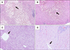
In 2012, Ikegami et al. [35] advocated a conceptual shift from SFSS to primary graft dysfunction after LDLT. Their review of adult LDLT cases showed a high early graft mortality rate (50%) in patients with functional cholestasis, with total bilirubin >20 mg/dL after post-LDLT day 7, with very high sensitivity and specificity. Huge ascites as a manifestation of graft dysfunction did not negatively impact graft survival. Although GV did not affect the occurrence of primary graft dysfunction, model for end-stage liver disease (MELD) score >15, the presence of portosystemic shunt, donor age >45 years, portal venous pressure >20 mmHg at the end of surgery, and operative blood loss >10 L were associated with primary graft dysfunction in LDLT. The proposed typical pathological findings of primary graft dysfunction including SFSS were ballooned hepatocytes with cholestasis in the pericentral venous zone forming a demarcation line with healthy hepatocytes in the periportal zone. Higher serum peak total bilirubin level in primary graft dysfunction was also associated with the shift in the ballooned hepatocyte borderline with cholestasis from the central venous to portal areas (Fig. 1A and B). Pathological findings in primary graft dysfunction can be distinguished from cholestatic lobular hepatitis C by pan-lobular hepatocyte ballooning (Fig. 1C) and acute rejection with mixed cellular infiltrations around the periportal area (Fig. 1D).
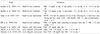
In the meantime, the concept of early allograft dysfunction (EAD) characterized by high early aminotransferase levels, persistent cholestasis, and coagulopathy in the first week after LT in the use of extended-criteria donors for deceased donor liver transplantation has been accepted in Western countries [363738]. Olthoff et al. [39] proposed a definition of EAD in LDLT as the presence of jaundice with bilirubin >10 mg/dL or coagulopathy with an INR >1.6 on day 7 without technical complications. The following A2ALL study [40] showed that EAD, with a five-fold risk of short-term graft loss, was associated with a left lobe graft, smaller graft weight, higher preoperative bilirubin, higher portal pressure, higher donor age, and higher donor body mass index. The reasons for graft size for EAD in this study might be attributed to nation-wide studies including LDLT centers with small case numbers. The Kyoto group [41] evaluated the impact of EAD criteria, reporting that 22 patients (8.5%) satisfied both criteria. Graft survival was significantly decreased by the coexistence of both factors (68.2%), compared to only bilirubin >10 mg/dL (24.3%), INR >1.6 (37.5%), and neither (11.9%); however, SFS grafts with GRWR <0.8 did not negatively impact graft survival rates. The Kyoto EAD criteria in LDLT might be reasonable for graft mortality in LDLT; therefore, cholestasis and coagulopathy should be the primary manifestations related to graft mortality in EAD including SFSS. The various definitions for EAD including SFSS are listed in Table 1.
HOW TO OVERCOME EAD AFTER LDLT
The basic concept of EAD including SFSS is represented by the absence of technical complications, and inflow and outflow issues as previously described [424344]. The major donor and recipient factors associated with EAD after DLLT include GRWR <0.8, donor age >45 years, MELD score >20, recipient with sarcopenia and unable to walk independently, posttransplant portal pressure >15 mmHg, and post-LDLT complications including sepsis [17181920303132333435424344]. Because management of recipients with end-stage liver disease is difficult, donor and graft selection with inflow modulation to control portal pressure are the only options to overcome EAD.
Among inflow modulation methods, splenic artery ligation is widely recognized as the safest intervention with optimal outcomes [34454647]. The benefit of splenic artery ligation is attributed to increased hepatic arterial inflow due to obstruction of the splenic artery and decreased portal inflow via splenic outflow. The possible challenge in splenic artery ligation is the potential for bleeding and pancreas injury due to a deeply located splenic artery beneath the pancreas. The Kyushu group has performed simultaneous splenectomy during LDLT with the use of vessel sealing devices and endo- stapling devices to perform tie-less and blood-less splenectomy, with more potent decompression of portal inflow than that of splenic artery ligation [484950]. The rationale for splenectomy is based on the fact that spleen is a major supplier of vasoconstrictive molecules, including endothelin, to the liver; thus, splenectomy results in hepatic vasodilatation [5152]. Ikegami et al. [50] usually performed splenic artery ligation before splenectomy for easier and safer handling of enlarged spleens in end-stage liver disease. However, splenectomy is not globally recognized as a standard inflow modulation procedure in LDLT, except for the Kyushu and Kyoto groups, due to bleeding complications, portal and splenic venous thrombus, and post-splenectomy sepsis. Recently, the Asan group reported innovative splenic devascularization in LDLT, with sufficient portal decompression and fewer complications compared to those for splenectomy [53].
The creation of portosystemic shunts for portal decompression was first reported in the early to middle 2000s, with optimal outcomes in LDLT using extremely small grafts [545556]. However, because of unstable portal inflow due to liver regeneration in LDLT and portal steal phenomena leading to graft mortality, no Japanese institute has recently performed this procedure [44]. We experienced a case requiring an extra-small graft with expected and actual GV/SLV of 36% and 23%, respectively, from a 20-year-old donor into a patient with hepatocellular carcinoma, in which an end-to-side portocaval shut after splenectomy was created to modulate excessive portal inflow to the small graft [57]. The graft showed hepatofugal portal flow 4 days after surgery, followed by immediate re-laparotomy for shunt ligation, resulting in a good postoperative course. We should have recognized that this young and soft graft had an expected GV/SLV of 36% to accommodate sufficient portal flow and that the GV/SLV of 23% was a temporary presentation due to dehydration [26272829].
The generally recommended strategies for graft selection and inflow modulation might include the use of a modified right lobe graft with GRWR >0.8 and full venous drainage with splenic artery ligation. However, special techniques in LDLT could be performed in experienced LDLT centers for future development including the use of smaller grafts including the left lobe or posterior segment, the use of much larger grafts including dual grafts or extended right lobe graft, and technically demanding portal inflow modulations including tie-less splenectomy and creation of portosystemic shunts.
CONCLUSION
The historical changes over more than 25 years in LDLT include those from pediatric to adult patients, from a possible chance to generally accepted stable outcomes, and right lobe grafts with GRWR >0.8 becoming mainstream in LDLT, with worldwide expansion. We should recognize a SFS graft expected GRWR cutoff of 0.8. Because volume-related factors may significantly affect graft survival in LDLT due to of institutional guidelines based on a GRWR >0.8, EAD characterized by prolonged cholestasis and coagulopathy on day 7 after LDLT may fit the clinical presentations of post-LDLT graft dysfunction better than SFSS. Graft selection to maintain an expected GRWR >0.8, and full venous drainage and inflow modulation using splenic artery ligation may become the standard.
 XML Download