

 PDF
PDF ePub
ePub Citation
 Print
Print


SUMMARY OF PRESENTATION
Sacral spinal schwannomas are very rare and constitute only 1–5% of all schwannomas.1 Local recurrence and malignant transformation of schwannoma are very rare.1 The aim of this study was to review a case of giant sacral schwannoma focusing on its diagnosis and surgical management.
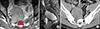
A 42-year old man was admitted to our hospital complaining of buttock and right inguinal pain. Magnetic Resonance Image (MRI) revealed 10.5 cm×7.5 cm×6.5 cm solid mass along ventral aspect of S1-S4 (Fig. 1A, B). The tumor arose from the right S1 sacral foramen compressing the right S1 nerve root and extended into pelvic cavity (Fig. 1A). The tumor showed low signal intensity on T2-weighted imaging and iso to high signal intensity on T1-weighted imaging with homogenous enhancement. Computed tomography scans of the abdomen showed bony erosion of the right S1 foramen (Fig. 1C).
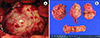
Tumor resection was performed using a retroperitoneal approach with assistance from a urologist. After careful dissection of the surrounding structures, we encountered a large pelvic mass deep in the pelvis. The pelvic mass was bordered by the internal iliac artery, bladder and rectum. The longest diameter of the tumor was 10 cm and had a yellowish color, and a overall firm consistency (Fig. 2A, B). Gross complete removal of the mass was achieved without any retained mass around the sacral foramen. On the basis of a pathologic exam, the lesion was diagnosed as schwannoma. The postoperative period was uneventful. Preoperative symptoms improved, there were no postoperative complications, and the patient was discharged within a week.
Sacral schwannoma is classified into four stages according to the involved structures and many authors have been performing anterior, posterior, combined approach based on the stage of tumor.2 Tumors with large presacral growths (type II, IV) can be resected via anterior approach, however, there are several things to keep in mind when preparing for an anterior approach.2 First, because of the slow growing nature of the tumor, the border of the tumor can be adherent to visceral organs, vascular structures, ureter, which can be injured during dissection.1 Second, a spinal surgeon can diagnose this tumor but a multidisciplinary approach involving a general surgeon or urologist when deciding the appropriate surgical management is highly advised.1
A retroperitoneal approach for sacral schwannoma is a challenging and rewarding method. This approach can provide a wide corridor to access the entire tumor as opposed to the posterior approach. In this case, the patient showed favorable outcomes as compared with the anterior approach without complications such as sphincter disturbance or neurologic deterioration. We suggest that when the tumor is mainly in a presacral space, the anterior approach with help of urologist can be a good alternative option.
 XML Download