

 PDF
PDF ePub
ePub Citation
 Print
Print


INTRODUCTION
Patients with end-stage renal disease (ESRD) who are on maintenance hemodialysis experience a decrease in blood pressure (BP) during their dialysis treatment, because the fluid retained during the interdialytic period is removed by ultrafiltration.1 Intradialytic BP patterns are also influenced by antihypertensive medications, the sympathetic nervous system, cardiac and vascular diseases, and the hemodialysis treatment.12 Deviations from the typical course, known as intradialytic hypotension and hypertension, can complicate dialysis and result in adverse outcomes such as all-cause mortality and cardiovascular events.2 Therefore, efforts to understand the pathophysiology of intradialytic BP abnormalities and prevent these complications are warranted. Intradialytic hypotension occurs when the rate of fluid removal exceeds that of plasma refill, conversely, intradialytic hypertension is associated with volume overload.2 However, volume factors may not be sufficient to explain the pathophysiology of intradialytic BP abnormalities, and an investigation of other causes including cardiovascular abnormalities is needed.2
Cardiovascular disease is the major cause of death in patients with ESRD.3 Various traditional and uremia-related factors have been established as causative for the disproportionate burden of cardiovascular disease in the ESRD population.4 One of the common uremia-related factors is vascular calcification, which is caused by abnormal endocrine and mineral metabolism.5 Various studies have reported that vascular calcification can be a predictor of increased cardiovascular morbidity and mortality.567 Moreover, recent studies have shown a significant relationship between the vascular calcification that was assessed by plain radiograph and intradialytic hypotension in patients with ESRD on hemodialysis.89 These studies indicated that vascular calcification could result in a predisposition for intradialytic hypotension. Therefore, the assessment of vascular calcification may be useful for predicting the occurrence of intradialytic hypotension as well as the probability of cardiovascular events in patients with ESRD on maintenance hemodialysis.
In this study, we assessed the degree of coronary artery calcification as an indicator for the burden of cardiovascular disease using images obtained from nongated chest computed tomography (CT), and examined the correlation between calcification and intradialytic BP patterns in patients with ESRD on maintenance hemodialysis. We evaluated the impacts of coronary artery calcification on intradialytic BP abnormalities and further investigated their independent impacts on cardiovascular events and all-cause mortality.
MATERIALS AND METHODS
1. Patients
This study recruited adult patients with ESRD who underwent outpatient maintenance hemodialysis three times a week between October 2011 and April 2016. Of the 141 outpatients, we enrolled 40 who had chest CT images. The baseline was defined as the time when body composition was assessed, and we allowed 3 to 12 months to elapse after the CT conductance in order to avoid the acute ill periods needing CT. This study included a total of 36 patients who had clinical and body composition data, and who had follow-up periods of at least one month.
The included patients were reviewed from the baseline until death, loss to follow-up, or the study end point (August 2017). This study was approved by the Institutional Review Board (IRB) of Chung-Ang University Hospital (IRB number: C2016146[1889]). The IRB waived the need for written consent from the patients, because this study had a retrospective design, and the subjects were de-identified.
2. Coronary artery calcification imaging and scoring
The included 36 patients received CT scans for the following reasons: 12 for identification of pneumonia; six for evaluation of new lesions such as infiltration or effusion on chest X-ray; six for differential diagnosis of malignancy due to a lung nodule; six for evaluation of chronic respiratory symptoms such as cough, sputum, or dyspnea; three for monitoring of cancer recurrence after treatment; and three for surveillance of health examination. The CT images revealed the following: 12 normal or nonspecific findings; seven cases of pneumonia; six chronic stable lesions including chronic bronchitis, emphysema, bronchiectasis, and tuberculosis sequelae; five fluid-retention related lesions; three bases of acute bronchitis; two of active tuberculosis; and one of diffuse alveolar hemorrhage. After the CT scans, the patients received either outpatient or inpatient management, and were followed-up on for at least four months or until death, loss to follow-up, or the study end point.
Chest CT images were acquired on 3 mm standard nongated CT scanners (Brilliance 64 and Brilliance iCT 256, Philips Healthcare, Cleveland, OH, USA; or LightSpeed Pro 16 and Optima 660, GE Medical systems, Milwaukee, WI, USA) with or without contrast. The coronary artery calcium score (CACS) was measured using the scoring system (in units) developed by Agatson et al.10 Thereafter, patients were categorized into two groups: CACS <400 or ≥400.
3. BP measurements
The enrolled patients underwent hemodialysis using either a Fresenius 5008s (Fresenius Medical Care, Homburg, Germany) or Artis (Gambro Dasco SpA, Medolla, Italy) machine. Transition of machines did not occur. The patients were treated with bicarbonate dialysis fluid at a normal temperature (36.0–36.5℃). Dialysis was conducted three times a week for four hours per session with a 200–350 mL/min blood flow. The intradialytic BP was obtained in the supine position using an automated device built into the hemodialysis machine that was calibrated regularly.
Four-week BP measurements were obtained before the body composition examination. Intradialytic hypotension was defined as either the requirement for fluid administration or the following criteria11: minimum intradialytic systolic BP <100 mmHg (Nadir100) or pre-dialytic systolic BP − minimum intradialytic systolic BP ≥30 mmHg (Fall30). Conversely, intradialytic hypertension was defined as an increase in systolic BP ≥10 mmHg during hemodialysis.12 Patients that had a ≥30% exposure period during their hemodialysis sessions and who met the defined intradialytic hypotension or hypertension during their 12 treatment sessions were classified as having these complications.
4. Body composition analysis
Body composition was assessed using a multifrequency BIA device (InBody S10, Biospace, Seoul, South Korea). A total of eight electrodes were placed on the surfaces of the thumbs, fingers of the hands, and balls of the feet and heels in the supine position. The measurement was made within 30 minutes of starting the first dialysis session after the weekend. The BIA-derived parameters included intracellular water, extracellular water (ECW), and total body water (TBW). The ECW/TBW was used for the volume status estimation.13 All BIA tests were performed using the same technique by nursing staff trained in the manufacturer's protocol.
5. Data collection
Demographic and clinical data were collected from the patients' electronic medical records and included age, sex, height, cause of ESRD, duration of renal replacement therapy, dialysis access type, and medications used such as antihypertensive agents and erythropoiesis-stimulating agents. In addition, dialysate sodium levels, interdialytic weight gain, and ultrafiltration rates were obtained. The comorbidity burden was assessed using the modified Charlson Comorbidity Index.14 Age was not included in the modified Charlson Comorbidity Index b,ut was used for the multivariate analysis adjustment.
All of the patients' blood samples were drawn under fasting conditions before the first-in-week dialysis sessions, except for postdialysis blood urea nitrogen. The laboratory data included hemoglobin, albumin, blood urea nitrogen, calcium, phosphate, intact parathyroid hormone, uric acid, sodium, total carbon dioxide, and C-reactive protein levels. Dialysis adequacy (Kt/Vurea) and normalized protein catabolic rates were estimated using a single-pool urea kinetic model.15
6. Statistical analysis
Continuous variables were expressed as a median (25th, 75th percentile) and were compared using the Wilcoxon rank-sum test. Categorical variables were expressed as a number (percentage) and were compared between groups using the chi-square test. Pearson correlation coefficients were calculated to find the associations among CACS and the mean values of intradialytic systolic BP fall (pre-dialytic systolic BP – minimum intradialytic systolic BP) and nadir systolic BP (minimum intradialytic systolic BP). Univariate and multivariate Cox proportional hazard models were used to determine the hazard ratio (HR) for cardiovascular events and all-cause mortality. Cardiovascular events referred to cardiac death, acute coronary syndrome, cerebrovascular accident, acute exacerbation of heart failure, or acute peripheral artery occlusion. Variables with a p<0.10 in the univariate analyses were included in the multivariate models. All statistical analyses were performed using SPSS Statistics version 18.0 (IBM Corp., Armonk, NY, USA). A two-sided p value <0.05 was considered significant.
RESULTS
1. Baseline characteristics according to CACS
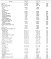
The 36 patients in this study were followed up for a median of 25 (13, 47) months, and their median CACS was 236 (82, 1316). The patients were classified based on their CACS into a group of 22 (61.1%) with a CACS <400 and a group of 14 (38.9%) with a CACS ≥400. Table 1 shows the differences of baseline characteristics between the two groups. The patients with a CACS ≥400 had higher dialysis duration, lower prescription rates of diuretics, and lower levels of albumin than those with a CACS <400 (p=0.002, 0.006, and 0.035, respectively). However, interdialytic weight gain, ultrafiltration rate, and ECW/TBW showed no difference between the two groups.
2. Intradialytic BP patterns according to the CACS groups
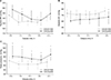
BP measurements over the course of dialysis treatment were compared between the patients with a CACS <400 and ≥400 (Fig. 1). The median systolic and diastolic BP differed at some time points. However, the pulse pressure was higher in the CACS ≥400 group than in the CACS <400 group during treatment (p=0.010, 0.002, 0.011, 0.011, and 0.002). The median systolic BP fall was −23 (−31, −12) mmHg in the CACS <400 group and −19 (−40, −16) mmHg in the CACS ≥400 group (p=0.619), and the median nadir systolic BP was 120 (113, 127) mmHg and 124 (112, 140) mmHg, respectively (p=0.377).
3. Relationship between CACS and intradialytic BP abnormalities
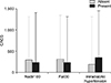
We explored the relationship between CACS and intradialytic BP abnormalities. No difference in the prevalence of intradialytic BP abnormalities was observed between the two CACS groups (Table 1). We further assessed CACS according to the type of intradialytic BP abnormalities (Fig. 2). The median CACS was comparable, irrespective of both intradialytic hypotension (Nadir100 and Fall30) and intradialytic hypertension (p=1.000, 0.661, and 0.626, respectively).
In addition, the relationships among CACS, intradialytic systolic BP fall, and nadir systolic BP were evaluated (Fig. 3). An association between systolic BP fall and nadir systolic BP (r=−0.4; p=0.010) was observed; however, CACS showed no association with the others (p=0.277 and 0.415).
4. Impacts of CACS and intradialytic BP abnormalities on cardiovascular events and mortality
We investigated the association of CACS and intradialytic BP abnormalities with the occurrence of cardiovascular events and all-cause mortality, and if they affected each other. Cardiovascular events occurred in seven patients, and deaths occurred in six patients during the study period. Tables 2 and 3 show HRs for cardiovascular events and all-cause mortality. CACS was associated with cardiovascular events, but this association disappeared after adjustments for age and pulse pressure (HR 1.001; p=0.058). Conversely, CACS was a predictor for all-cause mortality and was independent of both age and the comorbidity index (HR 1.001; p=0.010). We did not find any association between intradialytic BP abnormalities and outcomes.
DISCUSSION
We retrospectively investigated the association between CACS and intradialytic BP patterns. Patients with a high CACS had wider intradialytic pulse pressure than those with a low CACS. However, we observed no difference in the prevalence of intradialytic hypotension and hypertension between the two CACS groups. Patients with intradialytic hypotension or hypertension had comparable CACS to those without intradialytic BP abnormalities. In addition, we found no correlation between CACS and intra dialytic systolic BP fall and nadir systolic BP. We further explored the impacts of CACS and intradialytic BP abnormalities on outcomes and identified that CACS predicted the occurrence of cardiovascular events and all-cause mortality, but intradialytic hypotension (Nadir100 and Fall30) and hypertension were not associated with outcomes.
CACS is a strong predictor of coronary artery disease, cardiovascular events, and all-cause mortality regardless of chronic kidney disease status.16 CACS is usually quantified using electrocardiographic-gated CT in order to minimize motion artifacts from the beating heart and provide fine cuts through the coronary arteries. However, CACS can be detected and quantified on nongated chest CT scans, and this method has been shown to correlate well with CACS obtained from electrocardiographic-gated scans.1718 Given this evidence, we hypothesized that CACS quantified from nongated chest CT scans could be predictive for cardiovascular events also in patients with ESRD on maintenance hemodialysis. In this study, patients with a high CACS had longer dialysis vintages and slightly higher comorbidity burdens compared to those with a low CACS, which demonstrates that the extent of calcification increases with longer times on hemodialysis, as shown in previous studies.519 In addition, we found that a high CACS from nongated chest CT scans was associated with an increased rate of cardiovascular events and all-cause mortality in this retrospective ESRD population. As far as we know, our study is the first to use nongated chest CT scans to measure CACS to predict outcomes in patients with ESRD as well as the general population.2021
Pulse pressure is influenced by vascular elasticity, which decreases as calcium accumulates within the vessel wall. Previous studies demonstrated strong correlations between calcification and arterial stiffening.2223 Arterial stiffness is an important determinant of pulse pressure, and its measurement is valuable for the evaluation of cardiovascular risk. This study shows that the presence of coronary artery calcification is related to widened pulse pressure, and we deduced that CACS could be a marker for arterial stiffness. A previous study by Haydar et al.24 also reported that pulse wave velocity, which is a measure of arterial stiffness, was related to the degree of CT-derived CACS in patients with chronic kidney disease; moreover, they found that the correlation was independent of BP. We extrapolated that CACS is a measure of arterial stiffness and is predictive for atherosclerotic cardiovascular events independent of pulse pressure, although the statistical significance on cardiovascular events is weakened after the adjustment.
This study discovered that coronary artery calcification is not associated with either intradialytic hypotension or hypertension. Intradialytic hypotension occurs when the rate of fluid removal exceeds that of plasma refill, which results in a decrease of the effective arterial blood volume. Based on this mechanism, a reduction in the ultrafiltration rate during dialysis treatments can prevent this complication.25 Although the study could not estimate whether the impacts of calcification on intradialytic BP abnormalities depend on volume factors, the patients had comparable ECW/TBW, interdialytic weight gain and ultrafiltration rate between the groups. However, reduction of the ultrafiltration rate may not be enough to prevent intradialytic hypotension. Our previous study showed that patients with a considerable decrease in systolic BP during dialysis had a similar volume status and prescribed ultrafiltration rates to those with normal BP patterns.26 Thus, an investigation of other causes, such as autonomic dysfunction or cardiovascular abnormalities, is needed to understand the pathophysiology of intradialytic hypotension.2 On the other hand, intradialytic hypertension is associated with volume overload and other causal factors such as endothelial dysfunction.22627 Vascular calcification may be a predisposing factor for intradialytic BP abnormalities because of its role in cardiovascular events and mortality.567 Previous studies on Korean patients with ESRD receiving hemodialysis found a relationship between vascular calcification and intradialytic hypotension,89 which was in contrast to our study's findings. These previous studies scored aortic arch and abdominal aortic vascular calcification using a plain-chest radiograph and lateral radiograph of the abdomen, respectively. The current study had a smaller sample size than these two previous studies, but we used the more accurate CT-image measure to assess vascular calcification. Although the conflicting results are noteworthy, it may be the result of differences in the arteries used. The previous studies used he aorta, but we used the coronary arteries where calcification of atherosclerotic plaques is more prominent, even in dialysis patients.28 In other words, CACS from the coronary arteries mainly quantifies intimal calcification, but aortic calcification is dependent on both intimal and medial calcification. Nevertheless, how intimal or medial calcification induces a drop in BP during hemodialysis remains unclear. Until further studies determine the pathophysiologic mechanism, we cannot conclude whether the presence of vascular calcification is a risk factor for intradialytic hypotension or if it is an independent condition that occurs in distressed patients with ESRD on hemodialysis. Likewise, some studies have established that patients with intradialytic hypertension had stiff arteries assessed by pulse wave velocity,2930 but the relationship of either arterial stiffness or vascular calcification with intradialytic hypertension remains unclear. In summary, this study concludes that calcified vessels may be unconnected to intradialytic BP abnormalities, so further research is essential to clarify the vascular factors that cause a considerable BP drop or rise over the course of dialysis treatment. The current study is the first to evaluate the relationship between CACS and intradialytic BP abnormalities in patients with ESRD.
The current study has several limitations. First, we used a small sample size from a single center, which limits the power of the results and may ignore some differences. However, we could deduce that CACS was irrelevant to both intradialytic hypotension and hypertension using three different analyses. In addition, we could deduce that CACS has an independent impact on outcomes, regardless of either intradialytic hypotension or hypertension. Second, we used retrospective data, which may result in an information bias. However, errors of classification or outcome measurement did not occur, because our data was obtained from electronic medical records and did not include the subjects' recall. Third, most patients receive chest CT scans in acute ill states, which could influence the occurrence of outcomes. As a result, we looked into the reasons why the chest CTs were carried out, and the baseline time was elapsed to at least three months after the CT conductance. Fourth, we need to comment on the technique used for scoring CACS; this study did not conduct standardized measurements of CACS, and therefore problems with accuracy may exist. However, we could generate significant findings by our technique, which corresponded to previous trials.2021 Furthermore CACS obtained from nongated CT scans for lung cancer screening has been demonstrated as a valuable biomarker in diagnosis and risk prediction for coronary heart disease.31
In conclusion, we investigated the impact of coronary artery calcification on intradialytic BP patterns in patients with ESRD on maintenance hemodialysis. CACS measured using nongated chest CT scans was associated with widened pulse pressure, but was not related to either intradialytic hypotension or hypertension. The current study shows that coronary artery calcification might be unrelated to intradialytic BP abnormalities.


 XML Download