

 PDF
PDF ePub
ePub Citation
 Print
Print


INTRODUCTION
Cerebral cortical vein thrombosis (CVT) can be presented as an isolated form or combined with venous sinus thrombosis (1). Isolated CVT is difficult to diagnose because there are many anatomical variations in the cerebral venous system (23). Susceptibility-weighted imaging (SWI) is a considerably sensitive MR sequence for the detection of intravascular deoxygenated blood as well as extravascular blood products. It has been reported SWI is valuable regarding the detection of intravascular thrombus(4).
CASE REPORT
An age 26 female was presented with a headache since two weeks ago. She complained of occipital headache, nausea, and vomiting when upright, which were completely relieved when lying down. Neurologic examination showed a mild reduction in motor power in both of her lower extremities. The cerebrospinal fluid (CSF) pressure was measured as 0 cm H2O on the lumbar puncture. Laboratory testing including CSF study was unremarkable. She denied recent history of trauma, lumbar puncture, acupuncture, hematologic disease, pregnancy, or oral contraceptive use.
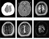
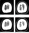
CT and CT venography (7) were obtained in the emergency department. A cortical vein of the left cerebral vertex showed high attenuation on non-contrast image, which was not opacified on CT venography (Figs. 1, 2). On the second day of admission, brain MR examination was performed with a 3T scanner (Ingenia; Philips Healthcare, Best, the Netherlands). There was a small amount of subdural fluid collection. The pachymeninges were thickened and strongly enhanced after the administration of the Gadolinium contrast agent. There was a hyperintense lesion in a cortical vein of the left cerebral vertex on T1-weighted image, not detected on T2-weighted image. SWI showed a blooming artifact due to thrombus in and beyond the cortical vein (Fig. 1). MR myelography was performed on day four of hospitalization, but CSF leakage was not detected. Ten days after the initial CT venography, follow-up CT venography was obtained. The cortical vein thrombus showed iso-attenuation as the cerebral cortex on non-contrast image and remained a filling defect on CT venography (Fig. 2).
The patient received anticoagulation therapy from the second day of admission and her symptoms slowly improved. She was discharged a month later without neurological deficits.
DISCUSSION
Our patient showed typical clinical and MR findings of SIH such as orthostatic headache, low CSF pressure, subdural fluid collection, and diffuse pachymeningeal thickening (5). SIH is a rare risk factor of cerebral venous thrombosis. Approximately 2.1% of cases are complicated by venous thrombosis (6). The relationship between SIH and cerebral venous thrombosis has not been clearly established. SIH is commonly caused by CSF leakage into the spinal epidural space. The reduced intracranial CSF spaces are compensated by subdural fluid collection and enlargement of the cerebral venous spaces, leading to a reduction in the flow velocity of the cerebral venous system. Additionally, the loss of intracranial CSF volume may cause stretching of the cerebral venous system and damage to the venous lining. These changes facilitate the formation of thrombi in the cerebral cortical veins and venous sinuses (89).
CVT is usually associated with venous sinus thrombosis. SIH is a risk factor of cerebral venous sinus thrombosis, a certain case as in our patient may present as isolated CVT (1011). Thus, it is crucial to detect imaging evidence of isolated CVT as well as venous sinus thrombosis in the case of SIH. However, isolated CVT is rare and difficult to diagnose because there are many anatomical variations in the cerebral cortical veins and the CT attenuation and MR signal intensity of CVT change according to the age of thrombus (23) (Fig. 2). Even if digital subtraction angiography (DSA), CT venography or MR venography is performed, it is sometimes difficult to detect intravenous filling defects due to thrombi since the thrombosed cortical veins are not often opacified (Fig. 2b).
In this case, we could easily detect the isolated CVT with SWI (Fig. 1f). It has been reported that SWI is valuable regarding the detection of intravascular thrombus. In acute stroke patients with large vessel occlusion, SWI can detect a blooming artifact due to thrombus in and beyond the cerebral arterial lumen. Additionally, it can also be valuable in assessing the burden of thrombus, which may facilitate establishing a therapeutic strategy for thrombolysis (4). On SWI, the cerebral veins usually appear as dark signal intensity structures since they have relatively high content of deoxy-hemoglobin that produces a susceptibility effect (12). In our patient, the thrombosed cortical vein was significantly larger in diameter than the adjacent normal veins. Thus, it was not difficult to judge that it was caused by cortical vein thrombosis. The high signal intensity of the corresponding vein on T1-weighted image was also valuable regarding the diagnosis (Fig. 1d).
In summary, SIH can be a risk factor for isolated CVT. SWI is valuable regarding the diagnosis of isolated CVT.
 XML Download