

 PDF
PDF ePub
ePub Citation
 Print
Print


A 42 year-old-woman presented with acute dyspnea. The patient had a history of resection of glioblastoma multiforme involving both frontal lobes 3 weeks previously.
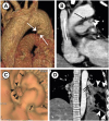
Computed tomography (CT) showed an 8–9 mm filling defect (arrow in Figure 1B and C) with low attenuation and lobulated contour at the junction of the left main pulmonary artery and patent ductus arteriosus (PDA) (arrows in Figure 1A and arrowheads in Figure 1B and C), suggesting endarteritis. In addition, a pulmonary embolus (arrows in the Figure 1D) was noted in the left lower pulmonary artery resulting in pulmonary infarction (arrowheads in Figure 1D) in the corresponding lung. Because of the patient's poor general condition, conservative management was elected using intravenous broad-spectrum antibiotics (vancomycin 500 mg every [q] 12 hours and doripenem monohydrate 250 mg q 8 hours) and intravenous heparin 20,000 IUy. The patient expired 14 days later due to septic shock.
The major complications of PDA are heart failure and endarteritis.1) Although the risk of endarteritis is relatively low in patients with silent PDA (i.e., small size of PDA without audible murmur), any PDA has a risk for endarteritis.1) There is increasing opportunity to initially identify a silent PDA using CT because of its increased usage. However, a small PDA can be missed if interpreting physicians review only the axial images due to its small size. Thus, careful inspection on coronal and sagittal views as well as 3-dimensional images is critical to making the diagnosis.
 XML Download